Abstract
Background Triangular fibrocartilage complex (TFCC) injury often results in distal radioulnar joint (DRUJ) instability. However, not all patients with a ruptured TFCC have an unstable DRUJ as in these patients a distal oblique bundle (DOB) may be present. We assumed that augmentation of the DOB leads to a more stable situation following reinsertion of the TFCC. We present the clinical results of a new surgical technique using the TightRope system as a DOB augmentation.
Description of Technique All cases were treated under regional anesthesia with the TightRope implant for which a tunnel was drilled from the distal ulna through the radius along the path of the DOB. The TightRope was passed through the tunnel and secured with buttons on either side. X-rays were made during surgery to confirm correct positioning.
Methods A retrospective study was performed analyzing 21 cases treated with a TightRope augmentation of the DOB. The primary outcome was measured using the patient-rated wrist evaluation (PRWE) score at least 12 months after surgery.
Results Postoperatively, the DRUJ was stable in all patients. The median PRWE score was 16 for the injured side compared to zero for the uninjured side ( p -value: < 0.001). The median pronation and supination were not statistically significant when we compared the injured side to the uninjured side. The median grip strength was 31 kg for the injured side compared to 38 kg for the uninjured side ( p -value: 0.015). There were two minor postoperative complications (10%).
Conclusion This technique is capable of restoring DRUJ stability with a short immobilization period resulting in good patient-related outcomes and a low complication rate.
Keywords: distal radioulnar joint, triangular fibrocartilage complex, wrist stabilization, TightRope
Distal radioulnar joint (DRUJ) instability usually results from a disruption of the triangular fibrocartilage complex (TFCC), and the extrinsic stabilizers, such as the extensor carpi ulnaris and its subsheath or the distal oblique bundle (DOB). 1 Okada et al published a cadaveric study in which 40% of the specimens had a DOB. Moreover, the absence of the DOB was correlated with an unstable DRUJ. 2 This may explain why not all patients with a ruptured TFCC have an unstable DRUJ. In these patients a DOB may be present.
Clinical diagnosis of DRUJ instability relies on physical examination and could be supported with radiographical images or a diagnostic arthroscopy. Symptomatic DRUJ instability may be present if the patient has a history of wrist trauma, persisting pain, and decreased function of the wrist. 3 4 5 6 7 The ballottement test seems to be the most reliable test with a sensitivity of 66% and a specificity of 68%. 8 This test assesses the dorsopalmar instability between the ulna and radius in both the neutral, pronated, and supinated position. 9
Radiographical signs that are associated with DRUJ instability are widening of the DRUJ gap, an ulna plus variance, and ulnar styloid fractures through the fovea or base. 3 10 11 A positive hook test during wrist arthroscopy may identify a rupture of the foveal insertion of the TFCC. 12 13 14
Numerous surgical techniques are described for treating DRUJ instability but none of them have shown superiority. Many of these techniques require a prolonged immobilization in a long arm or elbow cast. In our cadaveric study, using a suture-button expansion system that is placed in the direction of the DOB stabilizing the DRUJ, we found that the suture-button expansion system reduced dorsal displacement of the radius in an unstable DRUJ. 1
We assumed that this suture-button technique leads to a more stable situation with less recurrent DRUJ instability and requiring shorter immobilization in a short arm cast following foveal reinsertion of the TFCC. The purpose of this study is to describe the clinical results of this new surgical technique using the TightRope system as a DOB augmentation in patients with an unstable DRUJ.
Surgical technique : The suture-button expansion system used was the TightRope implant system according to the manufacturer's guidelines (Arthrex, Naples, FL).
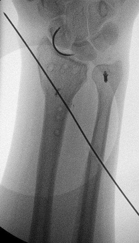
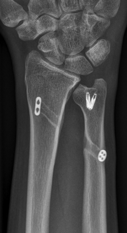
Surgery was performed under regional anesthesia in all patients. A tourniquet was used to create a bloodless field. A curved ulnar incision was made centered over the fifth extensor compartment of the wrist under regional anesthesia. The extensor digiti quinti tendon was released from the fifth extensor compartment and was held to the radial side. From proximal to distal the DRUJ was opened carefully not to injure the TFCC. A curettage of the scar tissue in the fovea was performed followed by reinsertion of the deep palmar and dorsal limbs of the TFCC to the fovea using a suture anchor technique (Mitek, Depuy Synthes, Raynham, MA). The extensor retinaculum was closed in a reefed fashion. After reinsertion of the TFCC, a bone tunnel was drilled under fluoroscopic guidance using a Kirschner wire (K-wire) at the lateral part of the distal ulna through the radius with the wrist in a neutral position in a 45-degree angle aiming just proximal of the sigmoid notch ( Fig. 1 ). At the point where the K-wire exited the radius an additional incision was made and a cannulated drill was used to drill the bone tunnels protecting the soft tissues. The TightRope was then passed through both tunnels starting from the ulnar side. The buttons were securely engaged on both cortices ( Fig. 2 ). The arm was pronated and supinated a few times making sure that there was a correct reduction and full range of motion (ROM) before tightening the knot. Postoperatively, patients received a short forearm cast for 4 weeks. After cast treatment the patient was instructed to start pro- and supination exercises under supervision of a specialized hand physiotherapist. After 6 weeks full weight bearing was allowed.
Fig. 1.

Bone tunnel drilled using a Kirschner wire (K-wire).
Fig. 2.

( A ) Tunnel with the TightRope implant secured with suture-buttons on both ends. ( B ) Mitek anchor at the ulnar fovea where the triangular fibrocartilage complex (TFCC) was reinserted.
Methods
Study Design and Study Population
A retrospective study was conducted of all adult patients that were treated with the suture-button expansion system between June 2016 and October 2020 and was approved by the institutional review board. All patients were treated in a level II trauma center in the Netherlands with an expertise in hand and wrist surgery.
Inclusion Criteria
All consecutive adult patients with a grossly unstable DRUJ due to TFCC lesion were included. Grossly unstable was defined as: an unstable DRUJ ballottement test, where the ulna could be dislocated completely out of the sigmoid notch and/or a positive hook test seen during diagnostic arthroscopy.
Outcomes Measures
The primary outcome measure of this study was the patient-rated wrist evaluation (PRWE) score. 15 The PRWE is a 15-item questionnaire designed to measure wrist pain and disability in activities of daily living. The PRWE allows patients to rate their levels of wrist pain and disability from 0 to 10, and consists of two subscales: (1) pain subscale and (2) function subscale. The total score per subscale ranges from 0 to 50, making a total score range from 0 to 100 in which the lower the score the better the wrist function.
Secondary outcome measurements were active ROM, grip strength, and any reported complications. The active ROM was measured according to the guidelines of the neutral-0-method using a goniometer. Flexion and extension and pro- and supination were measured. Grip strength was measured with a hand dynamometer (Baseline, White Plains, NY) in both injured and uninjured sides. Complications were defined as postoperative infections, failure of the repair leading to DRUJ instability, and persisting pain. The ballottement test was performed as described above.
Statistical Analysis
Baseline characteristics and outcome measures were categorized using descriptive statistics. For continuous data, the mean and standard deviation (parametric data) or the median and percentiles (nonparametric data) were reported. For categorical data numbers and frequencies were reported. Normality of continuous data was tested with the Shapiro–Wilk test and homogeneity of variances was tested using the Levene's test. A p -value of < 0.05 was taken as a threshold of statistical significance in all statistical tests and all tests were two-sided. Missing values were not replaced. Data were analyzed using the IBM Statistical Package for the Social Sciences (SPSS) (Version 25, SPSS Inc., Chicago, IL).
Results
Demographics
A total of 21 patients were treated with the TightRope system between June 2016 and October 2020. In Table 1 the baseline demographics are presented. The median age was 36 years (interquartile range [IQR]: 26–53) and 11 patients (52%) were male. The main cause of trauma to the wrist was either a fall from standing height or higher or during sports activities. Half of the patients had injured their dominant hand. Eight patients (38%) were secondary referrals from elsewhere. Median time until presentation in our hospital was 173 days (IQR: 55–547).
Table 1. Patient demographics.
| Total number of patients, N = 21 | |
|---|---|
| Age (in years), median (IQR) | 36 (26–53) |
| Gender, N (%) | |
| Male | 11 (52) |
| Female | 10 (48) |
| Trauma mechanism, N (%) | |
| Fall from standing height | 4 (19) |
| Fall from height (> 2 meter) | 4 (19) |
| Sports | 5 (24) |
| Motorized vehicle | 3 (14) |
| Heavy lifting | 3 (14) |
| Missing | 2 (10) |
| Affected hand, N (%) | |
| Right | 11 (52) |
| Left | 10 (48) |
| Affected hand is dominant hand, N (%) | 10 (48) |
| Second opinion, N (%) | 8 (41) |
| Days until presentation in our hospital, | |
| Median (IQR) | 173 (55–547) |
| ASA classification, N (%) | |
| 1 | 15 (71) |
| 2 | 6 (29) |
| Ulnar sided pain, N (%) | 14 (67) |
| Pain during wringing, N () | 9 (43%) |
| Pain while lifting, N (%) | 8 (38) |
| Snapping sensation in wrist, N (%) | 5 (24) |
| Positive fovea sign, N (%) | 9 (43) |
| Unstable DRUJ ballottement test, N (%) | 19 (91) |
| Widened DRUJ on X-ray, N (%) | 4 (29) |
| PSU fracture on X-ray, N (%) | 7 (33) |
| Ulna plus on X-ray, N (%) | 3 (14) |
| Diagnostic arthroscopy, N (%) | 10 (48) |
| Days until surgery, median (IQR) | 92 (69–143) |
Abbreviations: ASA, American Society of Anesthesiologists; DRUJ, distal radioulnar joint; IQR, interquartile range; N, number; PSU, ulnar styloid process.
Most patients experienced ulnar-sided pain (67%). Other symptoms or complaints were: pain during wringing (43%), pain during lifting objects (38%), and a clicking sensation of the wrist (24%). Nineteen patients had an unstable DRUJ ballottement test (91%). There were two patients of which the DRUJ ballottement test was not documented but they did have a positive hook test during diagnostic arthroscopy. Forty-three percent of the patients had a positive fovea sign during physical examination. Radiographically, there was a widened DRUJ in four patients (19%), an old fracture of the ulnar styloid process in seven patients (33%), and an ulna plus variation in three patients (14%). In 10 (48%) out of 21 patients a diagnostic arthroscopy was performed before the definitive TightRope surgery was planned. Median time until definitive TightRope surgery was 92 days (IQR: 69–143) after injury in which 16 (76%) patients had intensive hand therapy prior to surgery.
Diagnostic Arthroscopy
A total of 10 patients had a diagnostic arthroscopy prior to their definitive surgery. During arthroscopy several injuries were identified. These consisted of a TFCC lesion classified according to Palmer, a positive ghost sign, a positive trampoline test, any signs of synovitis, and scapholunate and lunotriquetral ligament injuries classified according to Geissler ( Table 2 ).
Table 2. Diagnostic arthroscopy.
| Total number of patients, N = 10 | |
|---|---|
| Palmer classification, N (%) | |
| 1A | 1 (10) |
| 1B | 8 (80) |
| 1C | 0 (0) |
| 1D | 0 (0) |
| 2A | 0 (0) |
| 2B | 0 (0) |
| 2C | 1 (10) |
| 2D | 0 (0) |
| 2E | 0 (0) |
| Positive ghost sign, N (%) | 10 (100) |
| Signs of synovitis, N (%) | 4 (19) |
| Positive hook test, N (%) | 8 (80) |
| SL ligament injury | |
| None | 8 (80) |
| 1 | 0 (0) |
| 2 | 1 (10) |
| 3 | 1 (10) |
| 4 | 0 (0) |
| LT ligament injury | |
| None | 7 (70) |
| 1 | 0 (0) |
| 2 | 1 (10) |
| 3 | 2 (20) |
Abbreviations: LT, lunotriquetral; SL, scapholunate.
Postsurgery
The median duration of surgery was 60 minutes (IQR: 42–71). Postoperatively, all patients were treated with a circular forearm cast for a median of 29 days (IQR: 27–34). All patients received hand therapy and were followed for at least 12 months. At 3 months all patients had a stable DRUJ ballottement test. The median pronation was 85 degrees (IQR: 84–90) and the median supination was 70 degrees (IQR: 49–86).
The median PRWE score of the injured hand was 16 (IQR: 11–23) at a median follow-up of 35 months (IQR: 20–50). Table 3 provides an overview of the demographics and PRWE score per included patient. The DRUJ ballottement test showed a stable DRUJ for all patients, both for the injured and uninjured side. The median pronation was 90 degrees (IQR: 89–90) and the median supination was 90 degrees (IQR: 84–90) at a median follow-up of 35 months. The median grip strength was 31 (IQR: 25–47) kg for the injured side and 38 kg (IQR: 26–55) for the noninjured side ( Table 4 ).
Table 3. Overview of patient demographics and outcome measures.
| Patient | Trauma mechanism | Initial injury | Injury-TightRope interval | Pronation/supination 3 mo postop |
Pronation/supination > 12 mo postop | Long-term PRWE score |
|---|---|---|---|---|---|---|
| ♀ 48 y | Fall from height | DRF (AO-A) | 5 mo | 70/70 | 45/70 | 18 |
| ♂ 19 y | Sports | DRF (AO-A) | 11 mo | 70/20 | 85/80 | 9 |
| ♂ 36 y | Heavy lifting | Contusion wrist | 9 mo | 90/60 | 90/90 | 6 |
| ♀ 56 y | Fall from standing height | DRF (AO-C) | 3 mo | 85/70 | 90/90 | 6 |
| ♀ 18 y | Heavy lifting | TFCC tear | 3 mo | 85/70 | 85/90 | 33 |
| ♀ 20 y | Fall from standing height | DRF (AO-A) | 130 mo | 85/35 | 90/90 | 51 |
| ♂ 51 y | Fall from standing height | TFCC tear, Mason type II radial head fracture | 10 mo | 90/60 | 85/70 | 18 |
| ♂ 30 y | Motorized vehicle accident | Contusion wrist | 6 mo | 90/90 | 90/90 | 13 |
| ♂ 25 y | Fall from standing height | DRF (AO-C) | 28 mo | 85/75 | 90/90 | 11 |
| ♀ 65 y | Fall from standing height | Transolecranon fracture- dislocation | 20 mo | 80/35 | 90/70 | 32 |
| ♂ 29 y | Motorized vehicle accident | TFCC tear | 25 mo | 85/45 | 90/90 | 5 |
| ♀ 63 y | Fall from height | TFCC tear | 6 mo | 85/85 | Unknown | Unknown |
| ♂ 44 y | Fall from standing height | TFCC tear | 80 mo | 60/50 | Unknown | Unknown |
| ♂ 39 y | Sports | Triquetral fracture | 4 mo | 90/90 | 90/90 | 15 |
| ♀ 33 y | Sports | Contusion wrist | 12 mo | 85/60 | 90/85 | 62 |
| ♀ 55 y | Sports | Distortion wrist | 4 mo | 85/50 | 90/90 | 12 |
| ♂ 17 y | Sports | Epiphysiolysis wrist | 10 mo | 90/90 | 90/90 | 18 |
| ♀ 57 y | Fall from height | DRF (AO-A) | 8 mo | 90/80 | 90/90 | 13 |
| ♂ 32 y | Heavy lifting | Distortion wrist | 28 mo | 90/90 | 90/90 | 18 |
| ♀ 46 y | Fall from height | DRF (AO-C) | 29 mo | 85/70 | Unknown | Unknown |
| ♂ 27 y | Motorized vehicle accident | DRF (AO-A) | 8 mo | 90/90 | 90/90 | 20 |
Abbreviations: ♂, male; ♀, female; DRF, distal radius fracture; PRWE, patient-rated wrist evaluation; TFCC, triangular fibrocartilage complex.
Table 4. Long-term follow-up data.
| Total number of patients, N = 18 | p -Value a | |
|---|---|---|
| PRWE score, median (IQR) | ||
| Injured side | 16 (11–23) | < 0.001 |
| Noninjured side | 0 (0–0) | |
| DRUJ ballottement test, N (%) | ||
| Grade 0 | 18 (100) | |
| Grip strength (kg), median (IQR) | ||
| Injured side | 31 (25–47) | 0.015 |
| Noninjured side | 38 (26–55) | |
| Palmar flexion (degrees), median (IQR) | ||
| Injured side | 67 (66–87) | 0.018 |
| Noninjured side | 84 (70–90) | |
| Dorsal flexion (degrees), median (IQR) | ||
| Injured side | 59 (51–68) | 0.046 |
| Noninjured side | 62 (56–72) | |
| Ulnar deviation (degrees), median (IQR) | ||
| Injured side | 49 (40–52) | 0.059 |
| Noninjured side | 50 (43–57) | |
| Radial deviation (degrees), median (IQR) | ||
| Injured side | 26 (20–30) | 0.154 |
| Noninjured side | 31 (23–32) | |
| Pronation (degrees), median (IQR) | ||
| Injured side | 90 (89–90) | 0.059 |
| Noninjured side | 90 (90–90) | |
| Supination (degrees), median (IQR) | ||
| Injured side | 90 (84–90) | 0.059 |
| Noninjured side | 90 (90–90) | |
Abbreviations: DRUJ, distal radioulnar joint; IQR, interquartile range; PRWE, patient-rated wrist evaluation.
p -Value was tested using the Wilcoxon signed rank test.
Complications
Postoperatively, one patient experienced pain caused by the ulnar suture-button, which was removed. Another patient experienced persisting ulnar-sided wrist pain for which he received a corticosteroid (CS) injection at the posterior interosseous nerve to settle the symptoms permanently.
Radiographic Results
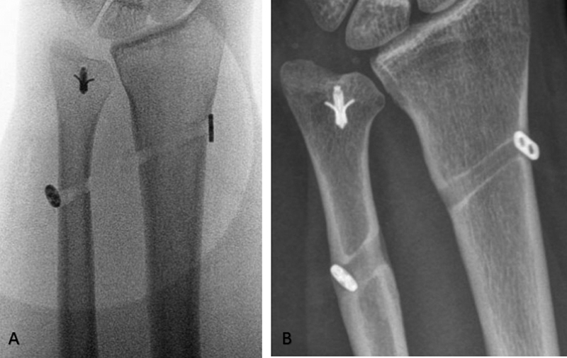
In our hospital we do not routinely perform X-ray imaging postoperatively. X-ray images were made in eight cases postoperatively because of complaints the patients experienced, either due to new injures or slow onset symptoms without a clear new injury. In two cases the complaints were explained by the complications as described in the previous paragraph. None of the postoperative images showed osteoarthritic changes. Five patients had a new injury to the same wrist for which all symptoms vanished in time without any intervention. One patient had a ganglion cyst in the same wrist. After removal the patient was cleared from any pain. We did see widening of tightrope holes in three cases but it did not seem to be of any importance since all symptoms improved spontaneously ( Fig. 3 ).
Fig. 3.

( A ) Peroperative X-ray imaging. ( B ) Postoperative X-ray imaging showing widening of the suture-button hole on the ulnar side.
Discussion
Symptomatic DRUJ instability often requires surgical treatment. However, not all patients with a TFCC tear have an unstable DRUJ as in these patients the secondary stabilizers and an intact DOB may be present.
Our results show that TFCC reinsertion with augmentation of the DOB with the TightRope system leads to good patient-related outcomes. The median PRWE score was 16 (IQR: 11–23) and all patients had a stable DRUJ after 3 months of follow-up. Two patients (10%) had a postoperative complication of which one was successfully treated with a CS injection and this patient completed the long-term follow-up with a full pro- and supination and a PRWE score of 20. The other patient needed a second surgery in which the ulnar placed suture-button was removed. After this reintervention the patient still had a stable DRUJ, gained full ROM but had persisting pain. A computed tomography scan showed signs of degenerative changes at the DRUJ which were treated conservatively. This patient reported a PRWE score of 62 at final follow-up. One other patient reported a significantly higher PRWE score of 51. This patient was a young, morbidly obese woman of 20 years who presented late following a conservatively treated distal radial fracture in the past. Despite full pro- and supination at final follow-up she persisted with pain and decreased strength in her wrist. There were no other identifiable reasons for her ongoing pain.
The TightRope implant is a suture-button expansion system and provides a relatively simple, reproducible, and minimally invasive procedure for the stabilization of the DRUJ as shown in our previously published cadaveric study. 1 Riggenbach et al published another cadaveric study testing the same principle of DOB reconstruction to stabilize the DRUJ with success. 16 A similar treatment has been described by Brink and Hannemann in which the palmaris tendon was used with the DOB reconstruction. 17 They observed a mean PRWE score of 33.6 at a mean follow-up of 25 months after surgery. Our surgical procedure is similar and only differs from the fact that the TightRope implant uses a synthetic graft instead of the palmaris tendon making the procedure less invasive.
The DOB originates from the distal ulna and runs distally to insert on the dorsal inferior rim of the sigmoid notch of the radius. It has an anatomical relationship with the TFCC and both act as DRUJ stabilizers. The origin of the DOB is situated proximal of the ulnar head, whereas TFCC ligaments insert right on top of the ulnar head. In the case of ulnar shortening its clinical relevance becomes more important as the DOB seems to take over as primary DRUJ stabilizer. 18 By augmenting the DOB in the wrist following TFCC trauma we indeed observed a stable end construct with good functional gain comparable, if not superior, to the existing published results. 1 16 17 18
Noteworthy is that only 40 to 60% of patients have a DOB. However, for this treatment we did not consider it important. We augmented the DOB in combination with a foveal reinsertion of the TFCC with an anchor to ensure a more stable situation and a shorter period of immobilization in a forearm cast. None of our patients presented with recurrent DRUJ instability. Moloney et al described their 20-year results of TFCC repair which entailed a reoperation in 34% and a recurrence of DRUJ instability in 17% of their patients. 19 This suggests that only TFCC repair may be insufficient.
We acknowledge the limitations of this study, such as its retrospective nature, its small sample size, and the lack of a comparative cohort. Additionally, there is the lack of preoperative PRWE scores to safely demonstrate improvement after surgery. However, this study does introduce a new technique that could be applied in patients with DRUJ instability. We would advise for further research to conduct a prospective comparative study between the available surgical techniques with a calculated sample size.
Conclusion
DOB augmentation for DRUJ stabilization using the TightRope system results in satisfactory patient-reported outcomes, a low complication rate, and no recurrence of DRUJ instability at a median follow-up of 35 months.
Conflict of Interest None declared.
Ethical Approval
An ethical approval for this study was obtained from our local ethics committee in advance.
References
- 1.de Vries E N, Walenkamp M MJ, Mulders M AM, Dijkman C D, Strackee S D, Schep N WL. Minimally invasive stabilization of the distal radioulnar joint: a cadaveric study. J Hand Surg Eur Vol. 2017;42(04):363–369. doi: 10.1177/1753193416656773. [DOI] [PubMed] [Google Scholar]
- 2.Okada K, Moritomo H, Miyake J et al. Morphological evaluation of the distal interosseous membrane using ultrasound. Eur J Orthop Surg Traumatol. 2014;24(07):1095–1100. doi: 10.1007/s00590-013-1388-6. [DOI] [PubMed] [Google Scholar]
- 3.Fujitani R, Omokawa S, Akahane M, Iida A, Ono H, Tanaka Y. Predictors of distal radioulnar joint instability in distal radius fractures. J Hand Surg Am. 2011;36(12):1919–1925. doi: 10.1016/j.jhsa.2011.09.004. [DOI] [PubMed] [Google Scholar]
- 4.Lindau T, Hagberg L, Adlercreutz C, Jonsson K, Aspenberg P. Distal radioulnar instability is an independent worsening factor in distal radial fractures. Clin Orthop Relat Res. 2000;(376):229–235. doi: 10.1097/00003086-200007000-00031. [DOI] [PubMed] [Google Scholar]
- 5.Lindau T, Adlercreutz C, Aspenberg P. Peripheral tears of the triangular fibrocartilage complex cause distal radioulnar joint instability after distal radial fractures. J Hand Surg Am. 2000;25(03):464–468. doi: 10.1053/jhsu.2000.6467. [DOI] [PubMed] [Google Scholar]
- 6.Andersson J K, Axelsson P, Strömberg J, Karlsson J, Fridén J. Patients with triangular fibrocartilage complex injuries and distal radioulnar joint instability have reduced rotational torque in the forearm. J Hand Surg Eur Vol. 2016;41(07):732–738. doi: 10.1177/1753193415622342. [DOI] [PubMed] [Google Scholar]
- 7.Nicolaidis S C, Hildreth D H, Lichtman D M. Acute injuries of the distal radioulnar joint. Hand Clin. 2000;16(03):449–459. [PubMed] [Google Scholar]
- 8.Wijffels M, Brink P, Schipper I. Clinical and non-clinical aspects of distal radioulnar joint instability. Open Orthop J. 2012;6(01):204–210. doi: 10.2174/1874325001206010204. [DOI] [PMC free article] [PubMed] [Google Scholar]
- 9.Moriya T, Aoki M, Iba K, Ozasa Y, Wada T, Yamashita T. Effect of triangular ligament tears on distal radioulnar joint instability and evaluation of three clinical tests: a biomechanical study. J Hand Surg Eur Vol. 2009;34(02):219–223. doi: 10.1177/1753193408098482. [DOI] [PubMed] [Google Scholar]
- 10.Omokawa S, Iida A, Fujitani R, Onishi T, Tanaka Y. Radiographic predictors of DRUJ instability with distal radius fractures. J Wrist Surg. 2014;3(01):2–6. doi: 10.1055/s-0034-1365825. [DOI] [PMC free article] [PubMed] [Google Scholar]
- 11.Kwon B C, Seo B K, Im H J, Baek G H. Clinical and radiographic factors associated with distal radioulnar joint instability in distal radius fractures. Clin Orthop Relat Res. 2012;470(11):3171–3179. doi: 10.1007/s11999-012-2406-4. [DOI] [PMC free article] [PubMed] [Google Scholar]
- 12.Nakamura T, Sato K, Okazaki M, Toyama Y, Ikegami H. Repair of foveal detachment of the triangular fibrocartilage complex: open and arthroscopic transosseous techniques. Hand Clin. 2011;27(03):281–290. doi: 10.1016/j.hcl.2011.05.002. [DOI] [PubMed] [Google Scholar]
- 13.Adams B D, Berger R A. An anatomic reconstruction of the distal radioulnar ligaments for posttraumatic distal radioulnar joint instability. J Hand Surg Am. 2002;27(02):243–251. doi: 10.1053/jhsu.2002.31731. [DOI] [PubMed] [Google Scholar]
- 14.Atzei A, Luchetti R, Carletti D, Marcovici L L, Cazzoletti L, Barbon S. The hook test is more accurate than the trampoline test to detect foveal tears of the triangular fibrocartilage complex of the wrist. Arthroscopy. 2021;37(06):1800–1807. doi: 10.1016/j.arthro.2021.03.005. [DOI] [PubMed] [Google Scholar]
- 15.MacDermid J C, Turgeon T, Richards R S, Beadle M, Roth J H. Patient rating of wrist pain and disability: a reliable and valid measurement tool. J Orthop Trauma. 1998;12(08):577–586. doi: 10.1097/00005131-199811000-00009. [DOI] [PubMed] [Google Scholar]
- 16.Riggenbach M D, Conrad B P, Wright T W, Dell P C. Distal oblique bundle reconstruction and distal radioulnar joint instability. J Wrist Surg. 2013;2(04):330–336. doi: 10.1055/s-0033-1358546. [DOI] [PMC free article] [PubMed] [Google Scholar]
- 17.Brink P RG, Hannemann P FW. Distal oblique bundle reinforcement for treatment of druj instability. J Wrist Surg. 2015;4(03):221–228. doi: 10.1055/s-0035-1556856. [DOI] [PMC free article] [PubMed] [Google Scholar]
- 18.Moritomo H. The distal oblique bundle of the distal interosseous membrane of the forearm. J Wrist Surg. 2013;2(01):93–94. doi: 10.1055/s-0032-1333428. [DOI] [PMC free article] [PubMed] [Google Scholar]
- 19.Moloney M, Farnebo S, Adolfsson L. 20-year outcome of TFCC repairs. J Plast Surg Hand Surg. 2018;52(03):193–197. doi: 10.1080/2000656X.2017.1415914. [DOI] [PubMed] [Google Scholar]


