Abstract
Long-acting injectable medications, such as atovaquone, offer the prospect of a “chemical vaccine” for malaria, combining drug efficacy with vaccine durability. However, selection and transmission of drug-resistant parasites is of concern. Laboratory studies have indicated that atovaquone resistance disadvantages parasites in mosquitoes, but lack of data on clinically relevant Plasmodium falciparum has hampered integration of these variable findings into drug development decisions. Here we generate atovaquone-resistant parasites that differ from wild type parent by only a Y268S mutation in cytochrome b, a modification associated with atovaquone treatment failure in humans. Relative to wild type, Y268S parasites evidence multiple defects, most marked in their development in mosquitoes, whether from Southeast Asia (Anopheles stephensi) or Africa (An. gambiae). Growth of asexual Y268S P. falciparum in human red cells is impaired, but parasite loss in the mosquito is progressive, from reduced gametocyte exflagellation, to smaller number and size of oocysts, and finally to absence of sporozoites. The Y268S mutant fails to transmit from mosquitoes to mice engrafted with human liver cells and erythrocytes. The severe-to-lethal fitness cost of clinically relevant atovaquone resistance to P. falciparum in the mosquito substantially lessens the likelihood of its transmission in the field.
Subject terms: Parasitology, Translational research, Malaria, Antiparasitic agents
Malaria parasites from patients who fail atovaquone therapies are highly drug-resistant, with mutations at Y268 in cytochrome b. Here the authors show that this mutation results in multiple defects in the parasite’s development and prevents transmission from mosquitoes to mice.
Introduction
Despite concerted efforts, that over several decades successfully cut by half the world’s malaria burden, in recent years progress has stalled and appears to be reversing. Estimates in 2021 were of 247 million infections and 619,000 deaths, attesting to malaria’s ongoing threat to public health and productivity1. Of greatest concern are infections with Plasmodium falciparum, which comprise 90% of cases and may be lethal, though geographically widespread P. vivax also causes significant morbidity. Bednets, vector reduction, various drug regimens, and, most recently, the P. falciparum RTS,S vaccine, are means of control, but new strategies are clearly needed. Among the candidates are long-acting injectable antimalarials2, which for chemoprophylaxis have aptly been termed “chemical vaccines”3.
Although long-acting injectable drugs are mainstays in contraceptive and antipsychotic therapy, new advances in drug formulation have brought this modality into use against infectious diseases4–6. Every-two-month injection of viral integrase inhibitor cabotegravir was recently approved as monoprophylaxis against sexually acquired HIV infection7. Chief among the benefits of long-acting injectable drugs is that they circumvent the commonplace problem of ensuring, over months to years of time, compliance with the frequent dosing required by oral regimens. In previous studies with P. berghei ANKA in mice, we found that an oral dose of atovaquone protected for one day, but when the same dose was given as a single injection of atovaquone nanoparticles in aqueous suspension, the protection lasted four weeks, a duration expected to be longer in humans8.
Reflecting the complex lifecycle of malaria parasites within humans (each stage with its distinctive biochemistry and pharmacology), conventional antimalarial drugs have limited scope in human infections. Although it is the sporozoite form that is transmitted by female anopheline mosquitoes, the parasites of critical importance for chemoprophylaxis against symptomatic disease have been the initial (and entirely asymptomatic) liver stage, and the subsequent asexual (pathogenic) erythrocytic stages – targets for causal and suppressive prophylaxis, respectively. Most agents are suppressive (e.g., chloroquine, mefloquine, doxycycline) but the synergistic fixed combination of atovaquone plus proguanil has both causal and suppressive action. Key among the advantages of causal prophylaxis is the substantially smaller number of parasites (difference ≥4 logs) that must be overcome, which reduces the likelihood of failure or resistance.
Atovaquone was devised as a structural mimic of coenzyme Q (or ubiquinone, an essential component in mitochondrial electron transfer) that binds to cytochrome b, collapses the transmitochondrial membrane potential, and impairs the malaria parasite’s obligatory synthesis of pyrimidines9,10. Atovaquone has garnered a reputation for being safe and well-tolerated when used alone for prophylaxis or treatment of Pneumocystis jirovecii or Toxoplasma gondii infection, which entails months to years of high dose therapy and plasma levels logs greater than those required for causal malaria prophylaxis9. Development of atovaquone alone to treat established erythrocytic malaria infections was abruptly paused by the unexpectedly ready appearance of drug resistance, mediated by mutations in cytB, the mitochondrial gene encoding cytochrome b, a finding that confirmed the mechanism of action11,12. In an effort to salvage atovaquone, selected from among several possible partners was proguanil, itself also active against liver-stage parasites and substantially synergistic with atovaquone. Clear reductions in requisite dose of each partner drug against erythrocytic parasites was shown in laboratory and clinical studies11–13. Field studies with atovaquone + proguanil confirmed reliable cures14, and the causal prophylactic activity of atovaquone alone was demonstrated in humans15. Accordingly, fixed combination atovaquone + proguanil was FDA approved in 2000 for the treatment of malaria. Although the impact of co-administered proguanil on efficacy and resistance against liver stages was not assessed, the same formulation was also approved for causal prophylaxis, making this a notable exception to the usual single drug malaria prophylaxis.
Numerous studies have focused on the issue of atovaquone resistance, pinpointing mutations in loops of cytochrome b that extend into the intermembrane space, where reduced coenzyme Q binds. Mutants have been obtained in vivo (from clinical drug treatment failures or murine models) and from in vitro selection. Curiously, there are marked differences in the mutants obtained clinically versus in vitro. The several dozen literature reports of atovaquone + proguanil treatment failures for falciparum malaria all document highly resistant Y268N/S/C mutants16. (Reported without drug sensitivities are I258M17 and no mutation in cytB18,19). By contrast, in vitro experiments with P. falciparum have generated at least 11 different mutations, some multiple, all in the intermembrane space or transmembrane regions of the protein, and with just one exception20, none at Y26812,20,21. Furthermore, atovaquone resistance of in vitro-generated mutants is substantially lower (typically 100- to 1000-fold) than that of the clinically relevant Y268N/S/C. The dichotomy between in vitro and clinical mutants assumed greater importance with intriguing reports that the development of atovaquone-resistant parasites in mosquitoes is impaired or halted22–24, suggesting their transmission may be limited in the field. Unfortunately, these studies have not included an evaluation of the Y268 mutants of P. falciparum that pertain to clinical drug resistance.
Transmissibility, or not, of atovaquone resistance is clearly critical to the use of long-acting injectable atovaquone as a chemical vaccine against malaria. In this study we generated a clinically relevant Y268S mutant of P. falciparum NF54, and, in comparison to isogenic wild type (WT) control parasites, monitored its growth and differentiation in anopheline mosquitoes and its transmissibility to mice engrafted with human hepatocytes and erythrocytes.
Results
Genesis and in vitro characterization of atovaquone-resistant P. falciparum
We were unable to obtain from other sources atovaquone-resistant P. falciparum with a Y268 mutation in cytochrome b, that retained the ability to infect mosquitoes. Accordingly, they were generated in vitro by applying atovaquone pressure to mosquito-competent P. falciparum NF54 lines, minimally passaged after isolation from clinical trial volunteers.
Over a 21-month period, efforts by several investigators, entailing a total of 7.8 × 10⁹ gametocyte-competent parasites, yielded five independent isolates of atovaquone-resistant cells, three of which were Y268S mutants. To select these mutants, continuous drug pressure was applied to various parasite densities and hematocrit levels. Mutants were detected by microscopy 25–47 days after start of pressure (Supplementary Fig. S1a, b). Reasoning that the mutation rate in mitochondrially-encoded cytB may be greater in cells cultured in a relatively high oxygen atmosphere25, selections (and parallel no drug controls) were conducted with cells acclimated to conventional incubator conditions of 5% CO2 in air (i.e., 19% O2)26. This is substantially higher than the 2–3% O2 in classic cultivation of P. falciparum27, or the ~13% O2 in our routine candle jar conditions, but it nevertheless falls within the range experienced by parasites in vivo26. Below-described studies in mosquitoes and mice revealed no difference between parasites maintained continuously in candle jar conditions versus those in 5% CO2 in the air for the ~two-month period of resistance selection.
Parasites that emerged from drug pressure were first evaluated for changes in cytB, by PCR amplification and sequencing of the entire open reading frame (Supplementary Fig. S2a, b). Three independent mutants encoded a Y268S amino acid change (Supplementary Fig. S1b, c). Whole genome sequence analysis of mutant cells was obtained at a coverage of 44- to 57-fold and compared to the reference P. falciparum NF54 gene sequence in PlasmoDB. FreeBayes variant caller in Partek Flow identified the Y268S mutation with a Variant Quality (VarQual) score of 49,191. All other genetic differences were of low relevance with VarQual scores of 721 or less (Supplementary Data File 1).
These collective efforts provided a mutation rate of 6 × 10⁻¹⁰. This is somewhat less than the previously reported 4.6 × 10⁻⁹ for atovaquone against 3D728, and substantially lower than 10−5 reported for atovaquone against multi-drug resistant W2 from Indochina29, perhaps reflecting our use of high drug concentrations for selection, or perhaps there is some impact of the retained (and likely costly to the cell) ability of these parasites to generate infectious gametocytes. Interestingly, all three Y268 mutations were Y-to-S (TAT-to-TCT), as was the only other previously reported in vitro-selected Y268 mutant20. We did not obtain the Y268N (to-AAT) or Y268C (to-TGT) mutants that are also seen clinically. Although all four known in vitro-selected mutants, from two laboratories, are thus Y268S, and this may have happened by chance, failure to obtain Y268N/C may also reflect a biological bias that operates for selection in vivo but not in vitro.
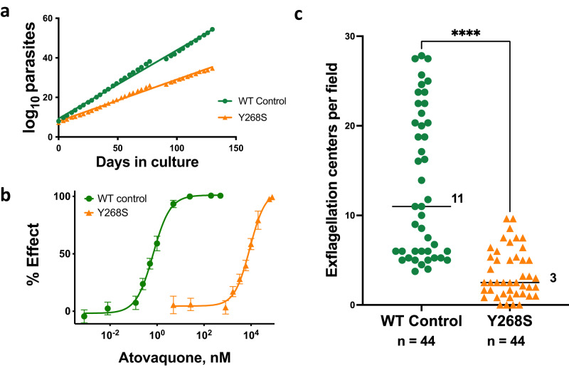
Asexual erythrocytic Y268S parasites had several phenotypic changes. Their growth rate in vitro was only 71% that of isogenic parent cells (Fig. 1a), and they reached lower peak parasitemia (Supplementary Fig. S3a), findings consistent with a fitness cost for this mutation. Compared to WT, mutant parasites had significantly fewer progeny within late schizonts, a difference that almost certainly contributes to slower growth (Supplementary Fig. S3b). However, the cytB mutation and atovaquone resistance remained stable for at least four months in the absence of drug pressure, indicating the fitness cost is sustainable for asexual erythrocytic parasites in vitro.
Fig. 1. Phenotype characterization in vitro of isogenic wild type (WT) and cytochrome b Y268S mutant P. falciparum.
a Cumulative number of asexual parasites in continuous culture. Growth rate of wild type parasites (y = 0.35x + 9.3) was 1.4-fold greater than that of mutant (y = 0.21x + 7.9); n timepoints = 36, R2 > 0.99, data from one continuous experiment. b Atovaquone activity against WT or Y268S asexual erythrocytic parasites (EC50 0.68 nM or 9.0 µM, respectively) was obtained by assaying [3H]hypoxanthine incorporation as a function of atovaquone concentration. Depicted are mean ± SD of quadruplicate determinations from each of three biological replicate experiments (for each data point n = 12; some SD are too small to extend outside the symbols); R2 ≥ 0.994. c Male gametocyte exflagellation, adjusted to 1.5% gametocytemia. Bars, median number of centers across four independent biological replicates (ranges 4 to 28 for WT, and 0 to 10 for mutant); n, the total number of fields examined; ****P < 0.0001, two-sided Mann–Whitney test, single comparison. For WT cells, all 44 microscopy fields had at least one exflagellation center (100%); Y268S evidenced exflagellation in 40 of 44 fields (91%). Source data are provided as a Source Data file.
As expected, the Y268S mutation was associated with substantial atovaquone resistance (Fig. 1b). Baseline EC50 for wild type control parasites was 0.68 nM, in line with published values of less than 1 nM30, but was 9.0 µM for the Y268S mutant, an impressive change in drug susceptibility also in keeping with previous reports12, 31. Preliminary experiments in our hands indicated that EC50 values for atovaquone can vary two- to ten-fold depending on the protein source in the medium (albumax versus human serum) or differential count of the starting culture. These, in conjunction with atovaquone’s limited aqueous solubility at the highest assay concentrations, likely contribute to the 75-fold range of EC50 values reported by different labs for Y268S clinical isolates12,31,32. An ambient atmosphere (3% O2, candle jar, or 5% CO2 in air) had no significant effect on EC50 values. Susceptibilities to artemisinin (9.8 versus 12 nM) or chloroquine (8.2 versus 11 nM) were not significantly affected by the cytochrome b Y268S mutation.
The final in vitro assay we conducted was an assessment of male gametocyte exflagellation (Fig. 1c). This phenomenon normally occurs in the midgut of anopheline vectors after ingestion of an infected bloodmeal, during which male gametocytes release up to eight motile gametes33. This process, induced in vitro by incubating mature gametocytes at room temperature34, revealed a highly significant nearly four-fold decrease in exflagellation centers in Y268S mutants as compared to isogenic controls, likely reflecting an increased demand for mitochondrial respiration35.
Characterization of parasites in anopheline mosquitoes
In all experiments, stage V gametocytemia in infecting bloodmeals was adjusted to be the same, at 0.5%. Ookinetes (both immature and mature) were evaluated in the midguts of blood-fed Anopheles stephensi at 20–26 h after infection. The count in mosquitoes infected with WT parasites was nearly eight-fold greater than that in Y268S mutant-fed insects (Supplementary Fig. S4a, c, d).
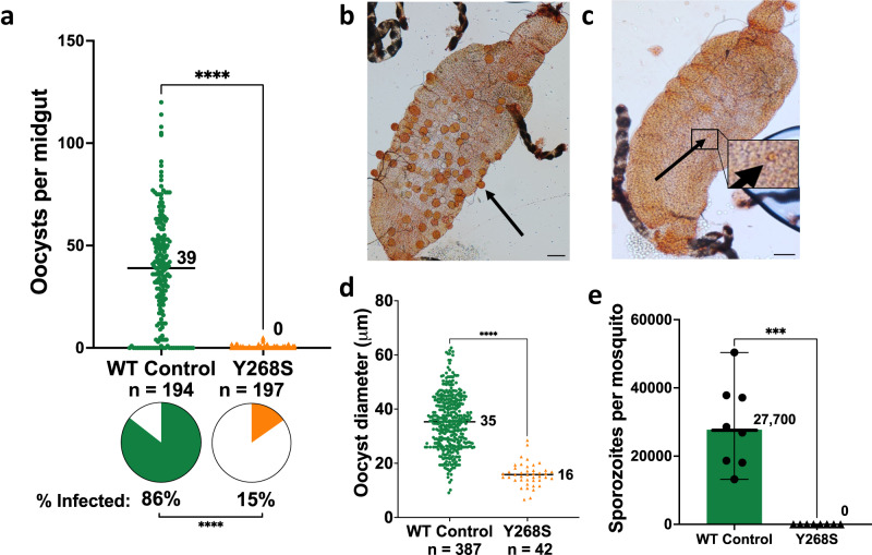
Oocyst numbers on An. stephensi midguts, evaluated in nine independent experiments (Fig. 2a), revealed a highly significant difference in the number of wild type or isogenic Y268S mutants (medians 39 or 0 per midgut, respectively). Prevalence of mutants was similarly reduced (Fig. 2a, pie charts). Furthermore, although drug-resistant Y268S parasites generated up to five oocysts/midgut in 15% of samples, mercurochrome-stained images revealed that their quality was uniformly poor, appearing smaller and malformed relative to their WT parents (Fig. 2b, c), with median diameter less than half that of WT (Fig. 2d).
Fig. 2. Evaluation of wild type and Y268S P. falciparum in An. stephensi tissues.
All feeds were adjusted to 0.5% stage V gametocytemia. a Oocysts on mosquito midguts were counted at 9-10 d after membrane feed. Indicated is the median number of oocysts per midgut in nine biological replicate experiments (ranges 0 to 120 for WT, and 0 to 5 for Y268S); n, the total number of mosquitoes dissected, two-sided Mann–Whitney test, single comparison; pie charts, percent of mosquitoes infected; ****P < 0.0001, two-sided Fisher’s exact test. b, c Photomicrographs of representative midguts infected with WT or Y268S parasites, respectively. Bars, 100 µm; arrows, representative oocysts; inset, magnification of Y268S oocyst; banded black structures are Malpighian tubules. d Oocyst diameters were measured in six biological replicates for wild type, and seven biological replicates for Y268S parasites. Median diameter is indicated (WT range 9 to 63 µm, mutant range 7 to 28 µm); n, the total number of oocysts measured; ****P < 0.0001, two-sided Mann–Whitney test, single comparison. e Salivary glands were harvested at 17–20 d after feed in eight biological replicate experiments entailing a total of 234 WT-fed or 245 Y268S-fed mosquitoes. Symbols, for each experiment, average number of sporozoites per mosquito. No sporozoites were seen in any Y268S preparation. Bars, median (values indicated) with 95% confidence interval; ***P = 0.0002, two-sided Mann–Whitney test, single comparison. Source data are provided as a Source Data file.
An. stephensi salivary glands were examined for sporozoites in eight biological replicate experiments (Fig. 2e). The difference between WT and Y268S mutant parasite counts was stark: in a total of nearly 250 mosquitoes per group, median counts were 27,700 and zero, respectively. The difference in the maturation time of mutant versus wild-type sporozoites as a possible cause of these discrepant counts was ruled out (Supplementary Fig. S5).
To test whether the phenotypic differences between wild-type and atovaquone-resistant parasites in An. stephensi would apply in another major malaria vector, we repeated these experiments with An. gambiae, and obtained essentially the same results: ookinetes were less (Supplementary Fig. S4b, e, f), and median counts were reduced from 22 to 0 for oocysts and from 2700 to 0 for sporozoites/mosquito (Supplementary Fig. S6). As with An. stephensi, no sporozoites were seen in any of the Y268S salivary gland preparations.
Since malaria infections in the field are frequently polyclonal, we wanted to determine whether the severe impairment in Y268S sporozoite number could be rescued by coinfection with wild-type parasites. An. stephensi were fed gametocyte cultures with WT P. falciparum only or with mixtures of 1:1 or 1:10 WT:drug-resistant parasites. In three biological replicates, median oocyst numbers were 11, 5, and 1 for WT, 1:1, and 1:10 groups, respectively (Supplementary Fig. S7a). Sporozoite counts were similarly patterned (Supplementary Fig. S7b). Thus, although total gametocyte numbers in the feeds were constant, the number of tissue parasites, for both oocysts and sporozoites, correlated strongly with proportion of WT gametocytes in the bloodmeal (Supplementary Fig. S7c). In all three replicate experiments, these finding were further confirmed by two complementary RFLP assays, designed to detect 10% or less mutant cytB DNA in midgut or salivary gland samples from mixed infections (Supplementary Fig. S2d). All samples evidenced WT DNA, but no mutant DNA was detected in any of the mixed infection samples (Supplementary Fig. S7d). These findings suggest not only that the cost of the Y268S mutation in mosquitoes is not relieved by coinfection with WT gametes, but indeed that WT parasites may outcompete the mutants in a mixed population.
Transmissibility of P. falciparum from mosquito to humanized mouse
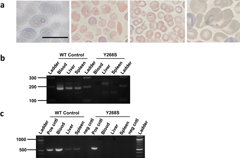
To provide a more biologically relevant, and functional, assay for the transmissibility of the mutant parasite, huHep mice were bitten by An. stephensi infected with either WT or Y268S atovaquone-resistant P. falciparum (Supplementary Fig. S8). In two independent experiments, three mice were bitten by an average of 138 wild-type-infected mosquitoes/mouse and two were bitten by an average of 152 mutant-infected mosquitoes/mouse (median parasite intensities in these mosquitoes were comparable to overall data in Fig. 2a, e: 29-33 WT or zero Y268S oocysts/midgut; 18,000 WT or zero Y268S sporozoites/mosquito). Engraftment of human erythrocytes assayed after infusions on days 5.5 and 6 after infection, was robust (WT and mutant averaged 30 and 29%, respectively). By 7.5 d after infection, all mice bitten by WT-infected mosquitoes were parasitemic (0.18-0.26%) on Giemsa-stained blood smear (Fig. 3a), as confirmed by blood culture. However, no parasites were seen in smears from mice bitten by mutant-infected mosquitoes, and in vitro cultures of ≥1 mL blood samples harvested at necropsy remained parasite-free for at least three weeks. Confirming these results, blood, liver and spleen from mice infected with WT parasites were positive for both STEVOR and cytB, but samples from mice exposed to Y268S mutant P. falciparum were negative by PCRs (Fig. 3b, c).
Fig. 3. Analysis of huHep mouse tissues for evidence of WT or Y268S P. falciparum.
a Giemsa-stained thin smears of peripheral blood from mice bitten 7 to 7.5 d previously by An. stephensi infected with WT parasites. Intracellular ring stage parasites are evident within the engrafted human erythrocytes, which at 6.4 µm are appreciably larger than the endogenous 4.7 µm mouse cells. Samples are from two mice in each of two biological replicate experiments; bar, 10 µm. Analogous peripheral blood from mice bitten by Y268S-infected mosquitoes evidenced no parasites in ≥16,000 RBCs examined per mouse, by at least two independent observers. b Indicated tissues were harvested from mice bitten 7.5 d previously by mosquitoes fed on WT or Y268S 0.5% gametocyte culture; liver and spleen were obtained after exsanguination and saline perfusion. DNA was isolated and analyzed by nested PCR for nuclear DNA-encoded STEVOR sequence (188 bp product). Ladder, indicated size fragments. Tissues were obtained in two biological replicate experiments and PCR assays were done in two or more independent technical replicates. c In a second independent experiment, tissues were harvested as above, and isolated DNA was analyzed by nested PCR for mitochondrial DNA-encoded cytB sequence (538 bp product). Ladder, indicated size markers; pos cntl, template DNA isolated from cognate asexual parasite culture; neg cntl, no template added reaction. (Longer exposures of Panels B and C are in Supplementary Fig. S10.) Tissues were obtained in two biological replicate experiments and PCR assays were done in two or more independent technical replicates.
Discussion
Atovaquone is, in many ways, a uniquely ideal candidate for a long-acting injectable chemical vaccine for malaria. It is potent against both initial liver and pathogenic erythrocytic parasites, for both P. falciparum and P. vivax; it has a decades-long track record of safety in patients whose plasma drug levels are 100 times higher than those required for causal activity; it is not metabolized and has few drug-drug interactions; and it faces no established resistance in the field or recognized cross-resistance with other antimalarials. No other currently available antimalarial even approaches these characteristics. Exciting candidates in development2 still require years of nonclinical study and early clinical trials, none of which can provide the extensive human exposure that bears on safety. More practically, further development as a chemical vaccine is facilitated by the fact that atovaquone (alone as Mepron, or in fixed combination with proguanil as Malarone) is off-patent, which, together with extensive use in humans, streamlines its regulatory requirements and facilitates its more equitable deployment.
Countering these advantages is a concern about atovaquone resistance. Selected readily when symptomatic patients harboring 109-12 erythrocytic parasites are treated with atovaquone alone, or occasionally in such patients treated with atovaquone + proguanil, clinical drug-resistance is mediated almost exclusively by a single point mutation at the Y268 residue in cytochrome b. In the setting of long-acting atovaquone monoprophylaxis, resistance concerns are twofold. First is the possibility that causal therapy may itself select resistant parasites. Several factors mitigate against this, including the fact that drug is present before any parasites are introduced, and that the number of liver stage parasites from which to select a mutant is relatively small (by orders of magnitude, a recognized “bottle neck” in the lifecycle). This analysis is supported by the absence of reported resistance in people prophylaxed with atovaquone + proguanil36, despite its being the most widely prescribed agent for this indication37, and the tens of millions who travel to malarious areas every year38. Second is the concern that mutants occurring in a person dosed with long-acting atovaquone will be transmitted by mosquito to others. Importantly, atovaquone + proguanil is not standard therapy for malaria patients in the field, and there is no cross-resistance with other drugs. In the unlikely event of mosquito-borne resistance, the consequence would not be the loss of available therapies to treat the affected patients but rather the possible loss of atovaquone as a chemical vaccine.
Our studies focused on the transmissibility of the clinically relevant highly atovaquone-resistant cytochrome b Y268S mutant P. falciparum. In 14 independent experiments, including both An. stephensi and An. gambiae mosquitoes, by conventional light microscopy and with multiple experienced observers, we found not one Y268S sporozoite in salivary glands from 345 mosquitoes. Validating this finding, the functional bioassays with huHep mice were especially important. By multiple criteria, including light microscopy (tail snip blood films), exquisitely sensitive PCR for P. falciparum nuclear or mitochondrial genes (liver, blood, spleen), or in vitro culture (of total blood volume), they evidenced no transmission of Y268S parasites. Furthermore, a mixture of mutant and wild-type parasites, to mimic polyclonal infections available to mosquitoes in the field, failed to boost sporozoite numbers above those expected for the WT component alone, and instead suggested that wild type parasites out-compete the mutant. In our hands, the fitness cost of the clinically relevant Y268S mutation in cytochrome b is severe-to-lethal in the mosquito.
At least three other laboratories have evaluated the survival in mosquitoes and in some cases transmissibility, of atovaquone-resistant parasites22–24. None of these reports include a clinically relevant P. falciparum Y268 mutant. Previous studies included P. falciparum with 5 to 20-fold resistant M133I or V259L, and P. berghei with both low and high resistance (M133I, Y268C, Y268N). We also isolated a low-grade L144S mutant, and found that it, like previously reported M133I, generated sporozoites (Supplementary Figs. S1b, S5, S9). The best interpretation for all these studies appears to be that laboratory-generated malaria parasites (including P. falciparum) with low-level atovaquone resistance can produce microscopically-detectable sporozoites in mosquitoes, whereas parasites with clinically relevant high-grade resistance cannot. Of obvious interest is the relevance of these various mutants to the use of atovaquone in humans.
The striking difference in EC50 values seen for atovaquone-resistant parasites obtained in vitro versus those arising in treated humans is almost certainly related to the disparate drug concentrations applied for selection. Atovaquone levels applied in vitro have not exceeded 100 nM, whereas trough plasma concentrations are 5400 nM (2 µg/mL) in subjects dosed daily for malaria prophylaxis39, and are even higher in those treated for infection. This in turn suggests that parasites with low-level resistance will not survive in treated humans. In conjunction with our observation that the clinically relevant, highly resistant Y268S fares poorly, if at all, in mosquitoes, these findings mitigate concern about transmissibility of atovaquone-resistance.
The availability of gametocyte-competent wild-type P. falciparum and isogenic mutants with various degrees of resistance provides a new resource for further studies. For example, with highly resistant Y268S we see fitness costs throughout the lifecycle, starting with a 29% reduced growth rate of asexual erythrocytic stages, and then a progressive loss in the mosquito: 73% reduction in exflagellation, 83% reduction in oocysts, and complete loss of sporozoites (Figs. 1 and 2). By contrast, sporozoite numbers in our L144S mutant are reduced 63%, but not eliminated (Supplementary Fig. S9e). These findings, in parasites that differ by just a single amino acid residue in cytochrome b, suggest that fitness in the mosquito may be related to the degree of atovaquone resistance. On a molecular basis, a relationship between the degree of resistance and fitness implies that mutations in cytochrome b that impair atovaquone binding correspondingly impair catalysis. Highly resistant asexual erythrocytic parasites evidence a mild but detectable growth defect, in keeping with their limited dependence on mitochondrial respiration. However, in the mosquito where mitochondrial function is essential22, even low-level resistance markedly impacts growth, and high-level resistance is lethal. These findings are akin to previous reports of differences in the pattern of P. falciparum drug-resistance polymorphisms in human blood versus mosquito tissues, likely also reflecting dissimilar selective pressures in the hosts, that may impose vector-selective constraints on drug resistance in the field40.
Our studies indicate that atovaquone resistance may not pose as great a threat to long-acting causal prophylaxis as it did historically to the treatment of established erythrocytic infection, and that clinically relevant highly resistant P. falciparum mutants are not transmitted by mosquitoes. In view of resurging malaria numbers, limited efficacy and durability of current biological vaccines, and the need for new control strategies, it would seem sensible to establish proof-of-concept for atovaquone as a chemical vaccine. No amount of work can prove the negative (i.e., resistance is not transmissible) and no laboratory investigations can address the enormous diversity of real-world malaria. Despite the promise of our studies, transmissibility, or not, of atovaquone resistance will best be determined by carefully designed and conducted clinical trials in endemic areas.
Methods
P. falciparum cultivation and mutant selection
Studies were conducted with low passage P. falciparum NF54 (MRA-1000 BEI Resources, NIAID, NIH), grown asynchronously at 37 °C under candle jar atmosphere ( ~ 13% O2, 3% CO2, 84% N2) in RPMI supplemented with 2.1 mM L-glutamine, 25 mM HEPES, 0.25% NaHCO3, and 0.37 mM hypoxanthine, in 10% heat-inactivated human serum (Interstate Blood Bank) and O+ erythrocytes at 2 or 4% hematocrit27. Periodic testing for mycoplasma was negative (MP0025, Sigma-Aldrich). Erythrocytes obtained weekly from healthy donors under a Johns Hopkins Institutional Review Board-approved protocol were provided without identifiers to the laboratories. Prior to atovaquone pressure, parasites were adapted over 14 d to 5% CO2 in air, as described previously26.
For mutant selection, atovaquone (PHR1591, Sigma Aldrich) in DMSO (D128500, ThermoFisher) was further diluted in a medium containing 10% serum such that the final DMSO was ≤0.1%. Cultures were exposed in parallel to atovaquone or no drug (Supplementary Fig. S1a,b). Medium was replaced every other day; cultures were split 1:2 every 7 d; only the first split was retained. Cells were maintained under constant drug pressure for 60 days or until healthy parasites appeared. Upon confirmation of Y268S mutation, atovaquone was removed and drug-resistant lines and their concurrent no-drug controls were returned at once to candle jar conditions, amplified, and aliquots were frozen (Glycerolyte 57, McKesson General Medical). In all subsequent experiments WT and mutants were manipulated and studied identically and in parallel.
To obtain gametocytes, cultures were initiated at 0.5% parasitemia, and 4% hematocrit, and the medium was replaced daily for 15-19 d until gametocytes reached stage V. Mutant parasites are being deposited with ATCC.
In vitro drug sensitivities
Asynchronous P. falciparum (0.25% parasitemia, 1.2% hematocrit) in microtiter plates was exposed to serial dilutions of the drug (in quadruplicate) for 72 h. [3H(G)]-hypoxanthine (16 Ci/mmol, 1 mCi/mL NET177001MC, Perkin Elmer) was added for the final 24 h, and incorporated radiolabel was evaluated in harvested cells, as described previously41. EC50 values were obtained by nonlinear regression analysis (Prism 6; GraphPad).
cytB and whole genome sequences
For cytB sequence in parasites surviving drug pressure, lysates were prepared from cultures at 1% parasitemia (90 °C, 5 min), and an 1848 bp fragment (containing the 1131 bp cytB open reading frame) was amplified with primers (Supplementary Fig. S2a, b) and Phusion DNA polymerase (NEB). PCR product was verified by gel electrophoresis as single band of predicted size, and sequenced. Sequences were aligned to P. falciparum 3D7 cytochrome b (PF3D7_MIT02300) using NCBI blast, and positions on all trace files were assessed in SnapGene 7.0.
For whole genome sequences to identify the causative mutation42, DNA was isolated from 108 asexual parasites; QiAamp DNA Mini Blood kit, Qiagen), quantified (Qubit HS ds DNA assay (Qubit Flex Fluorometer, ThermoFisher)), and quality assessed by Genomic Screentape analysis (TapeStation 2200, Agilent). Barcoded libraries for DNA-Seq were synthesized from 100 ng DNA (Celero EZ-Seq kit with NuQuant, Tecan Genomics), quality assessed by High Sensitivity DNA Lab Chips (BioAnalyzer 2100, Agilent), quantified by Qubit HS dsDNA assay, and sequenced on Illumina’s MiSeq platform, (2 x 300 bp v3 with 5% PhiX; JHMI Synthesis and Sequencing Facility). Using Partek Flow 10.0.22.0410 with sequence defaults, analyses included pre-alignment QA/QC, adaptor/read trimming, reference genome alignment to P. falciparum NF54 (PlasmoDB) using Bowtie2 2.2.5, post-alignment QA/QC, FreeBayes variant calling, variant filtering (VarQual ≥ 30), and variant annotation based on precedence rules. To identify changes unique to atovaquone-resistant parasites, sequences were compared to that of the reference strain P. falciparum NF54 GCA009761475.1 (PlasmoDB).
Gametocyte exflagellation
Just prior to mosquito feed, 200 μL of gametocyte culture was taken to assess male exflagellation43,44. Briefly, cultures were centrifuged in a warm tube (500 x g, 5 min), 2 µL of cell pellet was placed on a glass slide, covered, and incubated (room temperature, 15 min). The number of exflagellation centers in 11 independent 400X fields was counted and normalized to 1.5% gametocytemia. In three of four experiments, counts were obtained in a blinded fashion.
Mosquito maintenance and infection. All experiments were conducted with An. stephensi (Liston, NIH) or An. gambiae (Keele, NIH) reared and maintained in the Johns Hopkins Malaria Research Institute insectary (27 °C, 80% humidity, 12/12-h light/dark cycle), and provided 10% sucrose solution-soaked cotton pads43. For each experimental group, 100–200 mosquitoes at 3–7 d post-emergence were fasted overnight prior to 37 °C water-jacketed glass membrane feeding, for one hour, by established protocols43. All bloodmeals were adjusted to 0.5% stage V gametocytemia. Thereafter, 10% sucrose was withheld for 48 h to eliminate unfed mosquitoes.
Ookinete, oocyst and sporozoite counts
Ookinetes were counted by an adaptation of established protocols45. At 20-26 h post bloodmeal, midguts were dissected from ten visibly blood-fed mosquitoes, placed in 20 μL 3% acetic acid, and vigorously pipetted to lyse midguts. An aliquot (2 μL) was loaded on a hydrophobic slide (VWR, 100488–892), spread to fill a 14 mm circle, dried, fixed with methanol, and stained with Giemsa. For each experimental group, the number of mature and immature ookinetes in sixty 1000X fields was recorded.
Oocyst and sporozoite counts were obtained by adaptation of established protocols43. At 9–10 d post feed, 15–30 midguts per group were dissected into 1X PBS on a hydrophobic slide (VWR, 100488-892). Samples were stained with 0.1% mercurochrome (M7011, Sigma Aldrich), covered, and examined (100X) for oocysts. Oocyst diameters were analyzed in ImageJ version 1.53. At 17–20 d post feed, salivary glands from 20-38 mosquitoes per group were collected, transferred to 100 μL 1X PBS on ice, sedimented (5000 g, 30 sec), and homogenized (sterile pestle, 30 sec). Parasites were counted by hemocytometer to obtain an average sporozoite count per mosquito.
Transmission from An. stephensi to huHep mice
All animal studies were conducted under a protocol approved by the Johns Hopkins Animal Care and Use Committee with huHep mice (FRG KO (Fah–/–, Rag2–/–, Il2rg–/–) on NOD background) engrafted with human hepatocytes and capable of maintaining circulating human erythrocytes (Yecuris46,47). Female mice (27–28 wk old) were housed with sterile water, chow (ad libitum), and bedding, at 20-23 °C and 30-70% humidity, with 12-hr light/dark cycles. Until ≥8 d prior to infection they were maintained on 1 mg/mL 2-(2-nitro-4-trifluoro-methylbenzoyl)1,3-cyclohexedione (CuRx™ Nitisinone, 20-0027) in sterile 10% dextrose. Given the rarity and expense of huHep mice, the subtle, if at all, difference in infectivity of female versus male animals (including humans), these studies were conducted with female mice. Mosquitoes (17–18 d post-infection with WT or isogenic atovaquone-resistant parasites) were fed through a mesh-covered container on mice anesthetized with 4–6 μL/g ip of 14 mg/mL ketamine (67457010810, Myland Pharmaceuticals) plus 1.2 mg/mL xylazine hydrochloride (X1251-1G, Sigma Aldrich) in PBS. Mosquitoes fed for 14 min, with interruption every two min to limit blood loss in mice; over 80% evidenced having taken blood. At 5.5 and 6 d post mosquito probe, mice were injected intravenously with 600 μL human blood (50% red cells, 25% O+ serum, 25% RPMI). Human erythrocyte engraftment was assayed by flow cytometry of peripheral blood incubated 1:50 with APC-conjugated TER-119 rat antimouse monoclonal antibody (BD Biosciences catalog no 557909, lot 9213638), with gates at APC+ and APC− (mouse and human erythrocytes, respectively) (Attune NxT Flow Cytometer, BD Biosciences) (Supplementary Fig. S11).
Starting 6.5 d after infection, Giemsa-stained blood films were monitored twice daily. At patency, all mice were sacrificed. Blood was collected by cardiac puncture into citrate/phosphate/dextrose (Sigma, C7165), for PCR (150 µL aliquot) and in vitro culture (the ≥1 mL residual blood volume). After perfusion with 15 mL 0.9% NaCl, the liver and spleen were harvested, weighed, and stored at −70 °C. Blood cultures were maintained for three weeks or until a robust parasitemia was evident.
PCR assays
Mouse tissues were assayed for STEVOR or cytB by nested PCR. DNA was isolated from blood (QiaAmp DNA Mini Blood Kit, Qiagen; or Monarch Genomic DNA purification kit, NEB) or from perfused organs homogenized (Fisher Bead Mill 4, 2.4 mm metal beads) prior to extraction (DNA Maxi Kit; Qiagen). For STEVOR assay, eluates containing DNA (from 25 µL whole blood, or 2 µg DNA from liver or spleen (Nanodrop 2000, ThermoFisher)) were analyzed as described previously48,49. For cytB analysis, the 25 μL primary amplification reaction contained 50 nM each primer (Supplementary Fig. S2a,c), 200 μM each dNTP (NEB), 1 U Phusion DNA polymerase (NEB), and 10 ng template DNA. The 25 μL nested reaction contained 200 nM of each primer (Supplementary Fig. S2a,c), 200 μM each dNTP, 1 U of Phusion DNA polymerase, and 1 μL of the primary reaction product. Thermocycling conditions for both reactions were: denaturation (98 °C, 3.5 min); 25 cycles of (denaturation (98 °C, 30 sec), annealing (56 °C, 40 sec), and extension (72 °C, 45 sec)); and final extension (72 °C, 5 min). Products were separated by agarose gel electrophoresis and visualized by ethidium fluorescence. Serial dilutions into whole blood of a known number of P. falciparum-infected erythrocytes indicated both methods detect ≥10 parasites per 25 µL blood sample.
Statistical analyses
Data were collected in Excel 16.73. Dose-response data were analyzed as described in detail previously41. Unless indicated otherwise, differences between Y268S mutants and WT controls were assessed by the Mann−Whitney test or Fisher’s exact test, in GraphPad Prism 9.
Reporting summary
Further information on research design is available in the Nature Portfolio Reporting Summary linked to this article.
Supplementary information
Description of Additional Supplementary Files
Source data
Acknowledgements
This work was supported by Unitaid project LONGEVITY (2020-38-LONGEVITY); the Johns Hopkins Malaria Research Institute (JHMRI) and Bloomberg Philanthropies; and the National Institutes of Health (R01AI132359 to PS, R01AI1095453 to TS, and T32AI138953 to DS). RE was supported by a JHMRI postdoctoral fellowship. We thank Chris Kizito, Tassanee Thanakornsombut, and the JHMRI Insectary and Parasitology core facilities for their outstanding support; Dr. Sachie Kanatani for help with mosquito dissections and image development; the Clinical Pharmacology Drug Development Unit for weekly supply of healthy donor blood; Jodie Franklin and the JHMI Synthesis and Sequencing Facility for Illumina sequencing; and Mary Barry for meticulous laboratory maintenance. Kit Carson, Matt Ippolito and Kerry Stuart provided thoughtful help with methods. We thank Marcello Jacobs-Lorena for carefully reading and critiquing the manuscript. P. falciparum, Strain NF54 (Patient Line E), MRA-1000, contributed by Megan G. Dowler, was obtained through BEI Resources, NIAID, and NIH. We are grateful to staff at Yecuris for helpful advice and for generously replacing mice that unexpectedly died.
Author contributions
Research was designed by R.P.B., A.O., S.T.P., T.A.S., P.S., D.S., and A.K.T. Experiments were conducted by R.P.B., V.A.B., M.A.C., A.G.D., R.E., A.E.J., A.M., G.M., E.N., K.R.-R., A.S., T.A.S., P.S., D.S., and A.K.T.. New analytical methods were devised by R.E. and S.T.P. Data were analyzed by R.P.B., V.A.B., A.G.D., R.E., A.E.J., E.N., S.T.P., K.R.-R., T.A.S., P.S., D.J.S., and A.K.T. All authors contributed to writing the manuscript.
Peer review
Peer review information
Nature Communications thanks Geoffrey McFadden and the other, anonymous, reviewer(s) for their contribution to the peer review of this work. A peer review file is available.
Data availability
All data supporting the findings of this study are provided in the paper and its supplementary information. The minimum dataset necessary to interpret, verify and extend the research in this article are provided in the Supplementary Information/Source Data file. Whole genome sequence files for WT parent and Y268S mutant have been deposited in the NCBI Sequence Read Archive (BioProject ID: PRJNA913198; https://www.ncbi.nlm.nih.gov/sra). Source data are provided with this paper.
Competing interests
R.P.B., G.M., A.O., T.A.S., and A.K.T. are coinventors on PCT/GB2017/051746 (Atovaquone long-acting injectable formulation). A.O. is Director of Tandem Nano Ltd; received personal fees from Gilead and Assembly Biosciences; and is co-investigator on funding received by the University of Liverpool from ViiV Healthcare and Gilead Sciences. D. J. S. is Founder, Board Member, and stock/option owner of AliquantumRx; an expert legal consultant for Mabery Legal Firm; coinventor on USP 7,270,948 (Detection of malaria parasites by laser desorption mass spectrometry), USP9,568,471 (Malaria diagnosis in urine), USP 9,642,865 (New angiogenesis inhibitors), and PCT/US2015/046665 (Salts and polymorphs of cethromycin for the treatment of disease); and has received royalties from Binax Inc/D/B/A Inverness Medical for plasmids for HRP aldolase for malaria diagnostic kit. The remaining authors declare no competing interests.
Footnotes
Publisher’s note Springer Nature remains neutral with regard to jurisdictional claims in published maps and institutional affiliations.
Supplementary information
The online version contains supplementary material available at 10.1038/s41467-023-42030-x.
References
- 1.World Health Organization. World Malaria Report 2022 (World Health Organization, 2022).
- 2.Macintyre F, et al. Injectable anti-malarials revisited: Discovery and development of new agents to protect against malaria. Malar. J. 2018;17:402. doi: 10.1186/s12936-018-2549-1. [DOI] [PMC free article] [PubMed] [Google Scholar]
- 3.Phillips MA, Goldberg DE. Toward a chemical vaccine for malaria. Science. 2018;362:1112–1113. doi: 10.1126/science.aav7479. [DOI] [PubMed] [Google Scholar]
- 4.Owen A, Rannard S. Strengths, weaknesses, opportunities and challenges for long acting injectable therapies: Insights for applications in HIV therapy. Adv. Drug. Deliv. Rev. 2016;103:144–156. doi: 10.1016/j.addr.2016.02.003. [DOI] [PMC free article] [PubMed] [Google Scholar]
- 5.Flexner C, Owen A, Siccardi M, Swindells S. Long-acting drugs and formulations for the treatment and prevention of HIV infection. Int. J. Antimicrob. Agents. 2021;57:106220. doi: 10.1016/j.ijantimicag.2020.106220. [DOI] [PMC free article] [PubMed] [Google Scholar]
- 6.Lazarus G, et al. Safety and pharmacokinetic profiles of long-acting injectable antiretroviral drugs for HIV-1 pre-exposure prophylaxis: A systematic review and meta-analysis of randomized trials. Front. Pharmacol. 2021;12:664875. doi: 10.3389/fphar.2021.664875. [DOI] [PMC free article] [PubMed] [Google Scholar]
- 7.U.S. Food and Drug Administration (2021) FDA approves first injectable treatment for HIV pre-exposure prevention. https://www.fda.gov/news-events/press-announcements/fda-approves-first-injectable-treatment-hiv-pre-exposure-prevention. Accessed 20 January 2023.
- 8.Bakshi RP, et al. Long-acting injectable atovaquone nanomedicines for malaria prophylaxis. Nat. Commun. 2018;9:1–8. doi: 10.1038/s41467-017-02603-z. [DOI] [PMC free article] [PubMed] [Google Scholar]
- 9.Nixon GL, et al. Antimalarial pharmacology and therapeutics of atovaquone. J. Antimicrob. Chemother. 2013;68:977–985. doi: 10.1093/jac/dks504. [DOI] [PMC free article] [PubMed] [Google Scholar]
- 10.Vaidya, A. B. “Atovaquone—proguanil combination” in Antimalarial Chemotherapy. (Springer, 2001), pp. 203-218.
- 11.Looareesuwan S, et al. Clinical studies of atovaquone, alone or in combination with other antimalarial drugs, for treatment of acute uncomplicated malaria in Thailand. Am. J. Trop. Med. Hyg. 1996;54:62–66. doi: 10.4269/ajtmh.1996.54.62. [DOI] [PubMed] [Google Scholar]
- 12.Korsinczky M, et al. Mutations in Plasmodium falciparum cytochrome b that are associated with atovaquone resistance are located at a putative drug-binding site. Antimicrob. Agents Chemother. 2000;44:2100–2108. doi: 10.1128/aac.44.8.2100-2108.2000. [DOI] [PMC free article] [PubMed] [Google Scholar]
- 13.Canfield C, Pudney M, Gutteridge W. Interactions of atovaquone with other antimalarial drugs against Plasmodium falciparum in vitro. Exp. Parasitol. 1995;80:373–381. doi: 10.1006/expr.1995.1049. [DOI] [PubMed] [Google Scholar]
- 14.Kremsner PG, Looareesuwan S, Chulay JD. Atovaquone and proguanil hydrochloride for treatment of malaria. J. Travel Med. 1999;6(Suppl 1):S18–S20. [PubMed] [Google Scholar]
- 15.Shapiro TA, Ranasinha CD, Kumar N, Barditch-Crovo P. Prophylactic activity of atovaquone against Plasmodium falciparum in humans. Am. J. Trop. Med. Hyg. 1999;60:831–836. doi: 10.4269/ajtmh.1999.60.831. [DOI] [PubMed] [Google Scholar]
- 16.Staines HM, et al. Clinical implications of Plasmodium resistance to atovaquone/proguanil: A systematic review and meta-analysis. J. Antimicrob. Chemother. 2018;73:581–595. doi: 10.1093/jac/dkx431. [DOI] [PMC free article] [PubMed] [Google Scholar]
- 17.Plucinski, M. M. et al. (2014) Novel mutation in cytochrome B of Plasmodium falciparum in one of two atovaquone-proguanil treatment failures in travelers returning from same site in Nigeria. in Open Forum Infect. Dis. (Oxford University Press), p 59. [DOI] [PMC free article] [PubMed]
- 18.Wichmann O, et al. Malarone treatment failure not associated with previously described mutations in the cytochrome b gene. Malar. J. 2004;3:14. doi: 10.1186/1475-2875-3-14. [DOI] [PMC free article] [PubMed] [Google Scholar]
- 19.Färnert A, et al. Evidence of Plasmodium falciparum malaria resistant to atovaquone and proguanil hydrochloride: Case reports. BMJ. 2003;326:628–629. doi: 10.1136/bmj.326.7390.628. [DOI] [PMC free article] [PubMed] [Google Scholar]
- 20.Lane KD, et al. Selection of Plasmodium falciparum cytochrome B mutants by putative PfNDH2 inhibitors. Proc. Natl Acad. Sci. USA. 2018;115:6285–6290. doi: 10.1073/pnas.1804492115. [DOI] [PMC free article] [PubMed] [Google Scholar]
- 21.Schwöbel B, Alifrangis M, Salanti A, Jelinek T. Different mutation patterns of atovaquone resistance to Plasmodium falciparum in vitro and in vivo: rapid detection of codon 268 polymorphisms in the cytochrome b as potential in vivo resistance marker. Malar. J. 2003;2:5. doi: 10.1186/1475-2875-2-5. [DOI] [PMC free article] [PubMed] [Google Scholar]
- 22.Goodman CD, et al. Parasites resistant to the antimalarial atovaquone fail to transmit by mosquitoes. Science. 2016;352:349–353. doi: 10.1126/science.aad9279. [DOI] [PMC free article] [PubMed] [Google Scholar]
- 23.Blake LD, et al. Menoctone resistance in malaria parasites is conferred by M133I mutations in cytochrome b that are transmissible through mosquitoes. Antimicrob. Agents Chemother. 2017;61:e00689–00617. doi: 10.1128/AAC.00689-17. [DOI] [PMC free article] [PubMed] [Google Scholar]
- 24.Paton, D. G. et al. (2021) Atovaquone-resistant P. falciparum parasites complete sporogony in Anopheles gambiae mosquitoes. in MPM XXXIIp. 465 https://drive.google.com/file/d/1E3Dpod2zXf9WP7WSrxFc1F13Qqii1eUU/view. (Accessed 20 January 2023).
- 25.Lynch M, Koskella B, Schaack S. Mutation pressure and the evolution of organelle genomic architecture. Science. 2006;311:1727–1730. doi: 10.1126/science.1118884. [DOI] [PubMed] [Google Scholar]
- 26.Bakshi RP, Nenortas E, Tripathi AK, Sullivan DJ, Shapiro TA. Model system to define pharmacokinetic requirements for antimalarial drug efficacy. Sci. Transl. Med. 2013;5:205ra135. doi: 10.1126/scitranslmed.3006684. [DOI] [PMC free article] [PubMed] [Google Scholar]
- 27.Trager W, Jensen JB. Human malaria parasites in continuous culture. Science. 1976;193:673–675. doi: 10.1126/science.781840. [DOI] [PubMed] [Google Scholar]
- 28.Bopp SE, et al. Mitotic evolution of Plasmodium falciparum shows a stable core genome but recombination in antigen families. PLoS Genet. 2013;9:e1003293. doi: 10.1371/journal.pgen.1003293. [DOI] [PMC free article] [PubMed] [Google Scholar]
- 29.Gassis S, Rathod PK. Frequency of drug resistance in Plasmodium falciparum: A nonsynergistic combination of 5-fluoroorotate and atovaquone suppresses in vitro resistance. Antimicrob. Agents Chemother. 1996;40:914–919. doi: 10.1128/aac.40.4.914. [DOI] [PMC free article] [PubMed] [Google Scholar]
- 30.Basco LK, Ramiliarisoa O, Le Bras J. In vitro activity of atovaquone against the African isolates and clones of Plasmodium falciparum. Am. J. Trop. Med. Hyg. 1995;53:388–391. doi: 10.4269/ajtmh.1995.53.388. [DOI] [PubMed] [Google Scholar]
- 31.Musset L, Bouchaud O, Matheron S, Massias L, Le Bras J. Clinical atovaquone-proguanil resistance of Plasmodium falciparum associated with cytochrome b codon 268 mutations. Microbes Infect. 2006;8:2599–2604. doi: 10.1016/j.micinf.2006.07.011. [DOI] [PubMed] [Google Scholar]
- 32.Savini H, et al. First case of emergence of atovaquone-proguanil resistance in Plasmodium falciparum during treatment in a traveler in Comoros. Antimicrob. Agents Chemother. 2008;52:2283–2284. doi: 10.1128/AAC.00282-08. [DOI] [PMC free article] [PubMed] [Google Scholar]
- 33.Sinden R, Canning EU, Bray R, Smalley M. Gametocyte and gamete development in Plasmodium falciparum. Proc. R. Soc. B: Biol. Sci. 1978;201:375–399. doi: 10.1098/rspb.1978.0051. [DOI] [PubMed] [Google Scholar]
- 34.Bennink S, Kiesow MJ, Pradel G. The development of malaria parasites in the mosquito midgut. Cell Microbiol. 2016;18:905–918. doi: 10.1111/cmi.12604. [DOI] [PMC free article] [PubMed] [Google Scholar]
- 35.Krungkrai J, Prapunwattana P, Krungkrai S. Ultrastructure and function of mitochondria in gametocytic stage of Plasmodium falciparum. Parasite. 2000;7:19–26. doi: 10.1051/parasite/2000071019. [DOI] [PubMed] [Google Scholar]
- 36.Boggild AK, Lau R, Reynaud D, Kain KC, Gerson M. Failure of atovaquone-proguanil malaria chemoprophylaxis in a traveler to Ghana. Travel Med. Infect. Dis. 2015;13:89–93. doi: 10.1016/j.tmaid.2014.12.010. [DOI] [PubMed] [Google Scholar]
- 37.Freedman DO, Chen LH, Kozarsky PE. Medical considerations before international travel. N. Engl. J. Med. 2016;375:247–260. doi: 10.1056/NEJMra1508815. [DOI] [PubMed] [Google Scholar]
- 38.Angelo KM, et al. Malaria after international travel: a GeoSentinel analysis, 2003-2016. Malar. J. 2017;16:293. doi: 10.1186/s12936-017-1936-3. [DOI] [PMC free article] [PubMed] [Google Scholar]
- 39.Sukwa TY, Mulenga M, Chisdaka N, Roskell NS, Scott TR. A randomized, double-blind, placebo-controlled field trial to determine the efficacy and safety of Malarone (atovaquone/proguanil) for the prophylaxis of malaria in Zambia. Am. J. Trop. Med. Hyg. 1999;60:521–525. doi: 10.4269/ajtmh.1999.60.521. [DOI] [PubMed] [Google Scholar]
- 40.Mharakurwa S, et al. Malaria antifolate resistance with contrasting Plasmodium falciparum dihydrofolate reductase (DHFR) polymorphisms in humans and Anopheles mosquitoes. Proc. Natl Acad. Sci. USA. 2011;108:18796–18801. doi: 10.1073/pnas.1116162108. [DOI] [PMC free article] [PubMed] [Google Scholar]
- 41.Caton E, Nenortas E, Bakshi RP, Shapiro TA. Kinetic driver of antibacterial drugs against Plasmodium falciparum and implications for clinical dosing. Antimicrob. Agents Chemother. 2019;63:e00416–e00419. doi: 10.1128/AAC.00416-19. [DOI] [PMC free article] [PubMed] [Google Scholar]
- 42.Rocamora F, Winzeler EA. Genomic approaches to drug resistance in malaria. Annu. Rev. Microbiol. 2020;74:761–786. doi: 10.1146/annurev-micro-012220-064343. [DOI] [PubMed] [Google Scholar]
- 43.Tripathi, A. K., Mlambo, G., Kanatani, S., Sinnis, P., Dimopoulos, G. Plasmodium falciparum gametocyte culture and mosquito infection through artificial membrane feeding. J. Vis. Exp. 161, e61426 (2020). [DOI] [PMC free article] [PubMed]
- 44.Delves MJ, et al. Routine in vitro culture of P. falciparum gametocytes to evaluate novel transmission-blocking interventions. Nat. Protoc. 2016;11:1668–1680. doi: 10.1038/nprot.2016.096. [DOI] [PubMed] [Google Scholar]
- 45.Zollner GE, Ponsa N, Coleman RE, Sattabongkot J, Vaughan JA. Evaluation of procedures to determine absolute density of Plasmodium vivax ookinetes. J. Parasitol. 2005;91:453–457. doi: 10.1645/GE-391R. [DOI] [PubMed] [Google Scholar]
- 46.Vaughan AM, et al. Complete Plasmodium falciparum liver-stage development in liver-chimeric mice. J. Clin. Invest. 2012;122:3618–3628. doi: 10.1172/JCI62684. [DOI] [PMC free article] [PubMed] [Google Scholar]
- 47.Vaughan AM, et al. Plasmodium falciparum genetic crosses in a humanized mouse model. Nat. Methods. 2015;12:631–633. doi: 10.1038/nmeth.3432. [DOI] [PMC free article] [PubMed] [Google Scholar]
- 48.Cheng Q, et al. Measurement of Plasmodium falciparum growth rates in vivo: a test of malaria vaccines. Am. J. Trop. Med. Hyg. 1997;57:495–500. doi: 10.4269/ajtmh.1997.57.495. [DOI] [PubMed] [Google Scholar]
- 49.Cheng Q, et al. Stevor and rif are Plasmodium falciparum multicopy gene families which potentially encode variant antigens. Mol. Biochem. Parasitol. 1998;97:161–176. doi: 10.1016/s0166-6851(98)00144-3. [DOI] [PubMed] [Google Scholar]
Associated Data
This section collects any data citations, data availability statements, or supplementary materials included in this article.
Supplementary Materials
Description of Additional Supplementary Files
Data Availability Statement
All data supporting the findings of this study are provided in the paper and its supplementary information. The minimum dataset necessary to interpret, verify and extend the research in this article are provided in the Supplementary Information/Source Data file. Whole genome sequence files for WT parent and Y268S mutant have been deposited in the NCBI Sequence Read Archive (BioProject ID: PRJNA913198; https://www.ncbi.nlm.nih.gov/sra). Source data are provided with this paper.


