Abstract
Lateral lumbar interbody fusion (LLIF) is a minimally invasive surgical approach used to treat a variety of degenerative and deformity conditions of the lumbar spine such as advanced degenerative disease, degenerative scoliosis, foraminal and central stenosis. It has emerged as an alternative to the traditional posterior and anterior lumbar approaches with some potential benefits such as lower blood loss and shorter hospital stay. In this article, we provide our single institutional surgical experience including main indications and contraindications, a step-by-step surgical technique description, a detailed preoperative imaging assessment with a focus on magnetic resonance imaging (MRI) psoas anatomy, operative room (OR) setup and patient positioning. A descriptive surgical technical note of the following steps is provided: positioning and fluoroscopic confirmation, incision and intraoperative level confirmation, discectomy and endplate preparation, implant size selection and insertion and final fluoroscopic control, hemostasis check and wound closure along with an instructional surgical video with tips and pearls, postoperative patient care recommendations, common approach-related complications, along with our historical clinical institutional group experience. Finally, we summarize our research experience in this surgical approach with a focus on LLIF as a standalone procedure. Based on our experience, LLIF can be considered an effective surgical technique to treat degenerative lumbar spine conditions. Proper patient selection is mandatory to achieve good outcomes. Our institutional experience shows higher fusion rates with good clinical outcomes and a relatively low rate of complications.
Keywords: Lateral lumbar interbody fusion (LLIF), technical note, lumbar spine
Highlight box.
Surgical highlights
• Lateral lumbar interbody fusion is a suitable alternative to the traditional posterior approach for degenerative and deformity conditions.
• It is associated with lower blood loss and shorter hospital stays compared to the posterior approach.
What is conventional and what is novel/modified?
• The conventional surgical technique is usually a posterior fusion.
• We present our institutional experience using lateral lumbar interbody fusion as a standalone procedure.
What is the implication, and what should change now?
• Standalone lateral interbody fusion provides good outcomes in select cases.
Introduction
Lateral lumbar interbody fusion (LLIF) has become a popular, safe and effective minimally invasive treatment option for a variety of degenerative lumbar spinal conditions (1-3). This surgical technique allows for disc height restoration with subsequent central canal and foraminal indirect decompression. Additionally, it can improve spinal alignment by correcting deformities in both the sagittal and coronal planes (4-7). There is a growing body of literature on LLIF in many aspects such as indications, alignment, outcomes and complications (8-10). The objective of this paper, is to provide a step-by-step technical description of LLIF along with tips and pearls from our institutional, single-center experience. This surgical technique is classified as minimally invasive surgery. We present this article in accordance with the SUPER reporting checklist (available at https://jss.amegroups.com/article/view/10.21037/jss-23-54/rc).
Preoperative preparations and requirements
Indications and contraindications
The main indications for LLIF are low back pain and radiculopathy due to degenerative conditions in the lumbar spine such as advanced degenerative disc disease, degenerative scoliosis, foraminal stenosis, central stenosis, coronal or sagittal deformity, and adjacent segment disease from L1 to L5 levels, based on the iliac crest height. Contraindications for this procedure in our series are tumor or acute vertebral fracture.
Indications for standalone LLIF
We developed a decision-making pathway based on the participating surgeons’ indications with 100% agreement for indicating a standalone LLIF and 95% agreement for not recommending a standalone LLIF. In this regard, we identified favorable factors for standalone LLIF such as the presence of endplate sclerosis/Modic II changes, presence of foraminal stenosis, absence of severe sagittal or coronal malalignment and also some relative contraindications such as segmental hypermobility, smoking, facet joint effusion and osteoporosis (11).
Preoperative imaging considerations
Preoperative X-rays and magnetic resonance imaging (MRI) are critical when considering LLIF surgery. First, confirming the position of the iliac crest on the anterior-posterior X-ray view and the horizontal line between both iliac crests is made to assess the iliac crest height. If the line passes below the target disc, such as L4–L5, there is no need for oblique instrumentation and there is less of a risk of technical complications.
MRI allows visualization of vertebral disc elements as well as paravertebral structures, regarding this technique, proper assessment of psoas muscle anatomy is mandatory. Parameters analyzed by MRI include: identification of the anterior edge of the psoas muscle in relation with the anterior edge of vertebral body, assessment of plexus anatomy relationship with the planned working channel and identification of psoas morphology with special consideration at L4–L5, such as tear drop psoas morphology, identified as being detached anteriorly and laterally, displaying anteroposterior dimensions that were notably larger than their medial-lateral dimensions on axial MRI.
Operative room (OR) set up
Many aspects should be addressed in the OR for LLIF surgery. Fluoroscopy should be positioned facing the ventral aspect of the patient as the surgeon performs the procedure facing the dorsal aspect of the patient. Therefore, selection of the surgical approach side should be considered preoperatively, not only for the surgeon and assistants, but for the anesthesiologist also who usually relies on intravenous access on the side that is free of decubitus pressure.
Patient positioning
Patient positioning is important in any type of surgical procedure and is probably one of the most important aspects in LLIF surgery. Once the approach side has been determined based on accessibility and safety, protective dressings and support surfaces should be prepared to decrease the risk for decubitus related injuries not only to the skin, but also for compressive neurapraxias observed in some cases, especially brachial plexus and common peroneal nerve at the axilla and lateral knee, respectively. Protective dressings are mandatory to prevent these complications. The study was conducted in accordance with the Declaration of Helsinki (as revised in 2013) and approved by the institutional review board of Hospital for Special Surgery (HSS-IRB #2014-097). Written consent was obtained for use of deidentified images in publication.
Step-by-step description
Positioning and fluoroscopic confirmation
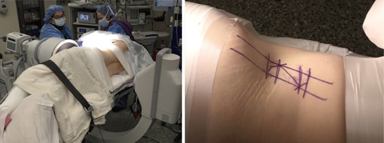
❖ The patient is positioned in lateral decubitus with appropriate padding such as an axillary roll to protect down-sided pressure areas such as the brachial plexus, greater trochanter and common peroneal nerve (Figure 1).
❖ Laterality, decided during preoperative planning, depends on different factors. In coronal plane deformity cases, the concavity side is considered technically better and associated with less vascular damage (12).
❖ The thoracic and pelvic areas are taped to secure the patient in position. The patient’s hips and knees should be partially flexed in a comfortable position. This both increases their stability in the decubitus position, but also releases tension on the psoas muscle and plexus. In addition to taping of the thorax and trochanters, we recommend taping along the thigh and calf to assist with stability and maintain flexion.
❖ The surgical table is flexed to create more space between the iliac crest and rib cage. The patient should be properly positioned so that the greater trochanter is slightly distal to the pivoting area to optimize lateral flexion.
❖ Patient repositioning is mandatory with further taping and revision of pressure points. Patients can easily fall forward or backwards, especially obese patients as this position is less stable that prone or supine.
❖ Radiographic confirmation after proper positioning is advised. The surgical level should be confirmed with anteroposterior and lateral fluoroscopy images. Additionally, the imaging should verify the absence of rotation in the observed area.
❖ Rotation is controlled by operative table hand control. The surgeon asks the radiology technician for fluoroscopy to provide an anterior posterior view. Right and left table tilt changes are made to line up the spinal processes in the midline. Maintaining the orthogonal view is important for proper cage positioning and to avoid contralateral foraminal issues or anterior vessel issues. Fluoroscopy is moved to provide a lateral position view and the table adjusted to provide a proper sagittal view with endplates of the level selected in a parallel position. Careful consideration should be given to table adjustments. Small changes are well tolerated and generally do not result in instability, however, if the table needs to be repositioned in a significant tilt to obtain an orthogonal X-ray, consider restoring the table to normal position and repositioning the patient. Excessive table tilt places the table-based instrumentation in poor ergonomic position, risks further patient translation during the procedure, and ultimately patient safety. In our experience, patient positioning is the key to a successful LLIF. Consider the operative table and fluoroscopy as fixed points and reorient the patient until an orthogonal view is achieved as opposed to reconfiguring the equipment.
❖ Skin marks are drawn and the flank is draped.
❖ At this stage, the anterior and posterior margins of the vertebral body, along with the disc space, are marked. The incision is centered over the disc space in a single-level surgery or between disc spaces in two or more level surgery.
Figure 1.
Lateral decubitus positioning with appropriate proximal and distal taping and skin marks.
Incision and intraoperative level confirmation
❖ The flank area is meticulously prepared and covered with sterile draping to maintain aseptic conditions.
❖ Following a time-out to verify the correct procedure and patient, a surgical blade is used to make a skin incision, precisely centered on the previously marked level. In the case of a single-level procedure, the incision typically spans around 2–3 cm (see Video 1).
❖ After the skin incision, subcutaneous and fat layers are dissected with electrocautery. A self-retaining retractor can easily maintain the open exposure.
❖ The fascia is exposed and bluntly divided with 2 Kelly clamps (Medline, Northfield, IL, USA) in opposite directions.
❖ A muscle-separation technique is recommended. Minimally split the fibers of the abdominal muscles (obliques and transversus abdominis) following the orientation of their respective fibers.
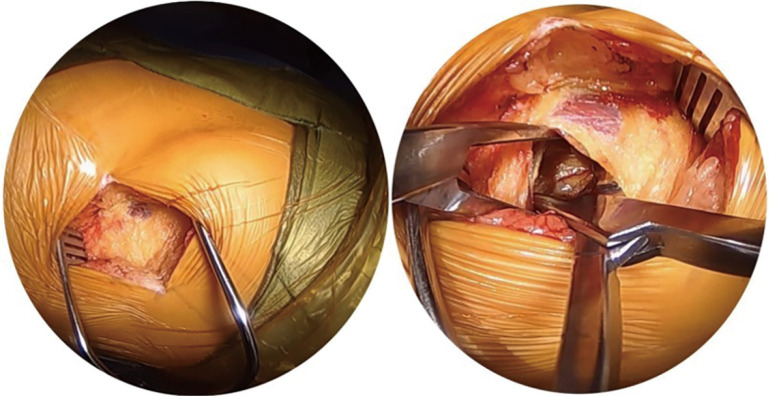
❖ The retroperitoneal area is reached and visualized (Figure 2).
❖ Abdominal and retroperitoneal contents are gently moved from posterior to anterior and the psoas muscle is identified.
❖ Psoas fibers are split along their longitudinal direction and the underlying disc space is exposed using 2 Wylie renal vein retractors (Medline, Northfield, IL, USA).
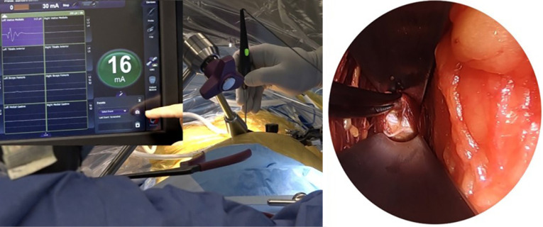
❖ A handheld electromyography (EMG) instrument is utilized to verify the deep positioning of the exiting nerve roots and lumbar plexus at each level. This confirmation is conducted in conjunction with conventional neuromonitoring, which includes somatosensory evoked potentials and spontaneous EMG. During our series, retractor time is meticulously recorded, with particular attention given to the L4-L5 level, aiming for a maximum continuous retraction period of 20 minutes at this specific level.
❖ In the event that a traversing nerve is encountered during the procedure, it is carefully and gently retracted dorsally to ensure its protection and to avoid any potential damage (Figure 3).
❖ Once the level is confirmed through fluoroscopy, a self-retaining retractor system such as MaXcess or NuVasive (San Diego, CA, USA) is employed instead of handheld retraction. This system allows for consistent and continuous exposure of the surgical site throughout the procedure.
Video 1.
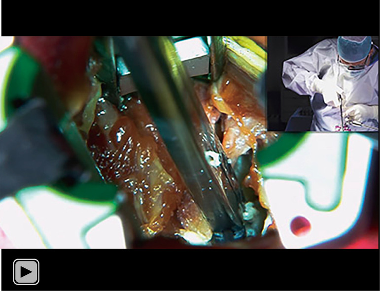
Surgical video demonstrating minimally invasive lateral lumbar interbody fusion technique to the L3-L4 disc space.
Figure 2.
Soft tissue dissection followed by access to the retroperitoneal space.
Figure 3.
Neuromonitoring control and gentle retraction of a traversing nerve.
Discectomy and endplate preparation
❖ After level confirmation, annulotomy with a scalpel and disc material resection are performed using a pituitary rongeur. Curettes and all tools are used carefully so as not to violate the endplates. In cases of severe disc collapse, usually associated with osteophytes, the disc space is difficult to identify. Osteophytes overlying the disc space can be carefully resected with a pituitary rongeur or a similar tool. The disc space may need to be opened by gently malleting a Cobb elevator or osteotome into the space to separate bonded osteophytes. If there is any uncertainty of the local anatomy, positioning should be confirmed with fluoroscopy in a truly orthogonal view to the disc space.
❖ Cobb elevators are used to detach cartilage from the endplates and to carefully release the contralateral annulus.
❖ Placed bullets are used (insert and rotate distraction dilators) and trial components of different sizes. Bullets are inserted slightly over the contralateral edge of the endplates.
The trial position is verified using biplanar fluoroscopy. In the anteroposterior image, the endplates should be symmetrically distracted. In cases where the disc space is significantly collapsed, more frequent fluoroscopic checks are necessary to ensure that the trial is appropriately positioned without any violation of the endplates. This ensures that the trial accurately reflects the desired alignment and helps maintain the structural integrity of the affected area.
Implant size selection and insertion
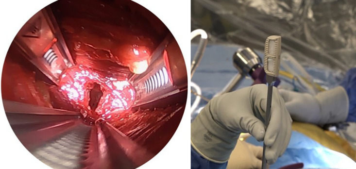
❖ Implant size selection is based on avoiding overstuffing of the disc space to minimize implant subsidence and to prevent endplate fractures, the most common implant height in our experience is size 10 (Figure 4). If sequential disc trials do not provide a good “scratch fit”, serious consideration should be given to possible violation of the anterior longitudinal ligament (ALL) or endplate fracture.
❖ Following irrigation, the implant is filled with the pre-planned graft material, as shown in Figure 5. In this specific case, the implant is packed with recombinant human bone morphogenetic protein-2 (rhBMP-2) on its carrier sponge, which is our preference. However, if bone morphogenetic protein (BMP) is used (off-label), it is crucial to exercise caution to prevent the BMP from passing through the endplates, as this could potentially trigger an osteoclastic reaction in the bone. In cases where the use of BMP might be contraindicated, alternative graft materials like demineralized bone matrix, bone marrow aspirate, or autologous iliac crest graft can be utilized instead of BMP. Proper selection of the graft material is essential to promote successful fusion and minimize the risk of complications.
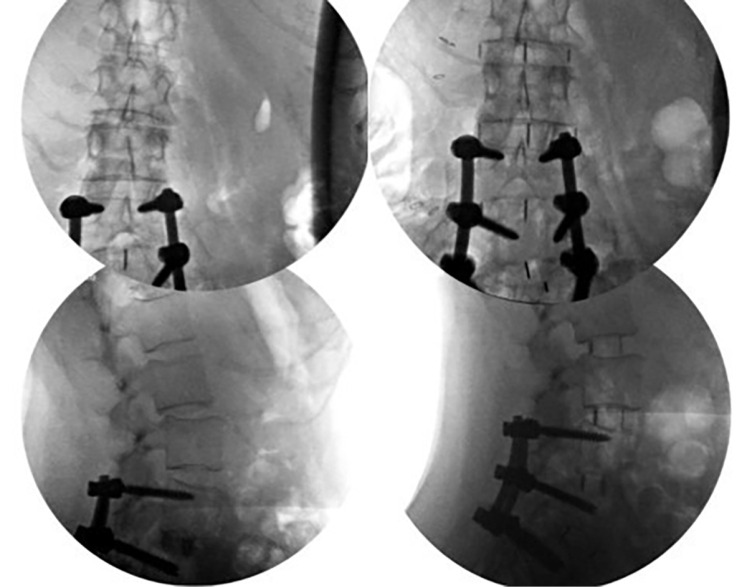
❖ The implant is introduced with the guidance provided by anteroposterior and lateral fluoroscopic imaging. The implant should be positioned to occupy the vertebral body and rest on the dense apophyseal margins bilaterally. Additional segmental stability can be achieved by using a lateral plate in some cases such as when posterior instrumentation is difficult due to small pedicles, or previous instrumentation or endplate violation during the standalone technique.
Figure 4.
Preparation of the disc space and implant selection.
Figure 5.
Anterior-posterior and lateral radioscopic control views showing onsite implant with coronal and sagittal alignment improvement in a patient with previous instrumentation and adjacent segment disease.
Final fluoroscopic control, hemostasis check and wound closure
❖ After placing the implants in all target levels, confirmation of proper realignment and placement of instrumentation with fluoroscopy is performed.
❖ After irrigation and hemostasis check, the wound is closed in a layered fashion.
❖ Upon completion of the procedure, there are two options for patient positioning. The patient can either be turned supine and extubated, or alternatively turned prone for additional fixation. The interbody fusion of the anterior spinal column can be supplemented by posterior pedicle screw fixation, creating a circumferential fusion construct. This additional posterior fixation can be performed either during the same surgical setting or in a staged manner, providing the patient with more time for recovery in between procedures. It is essential to consider the need for additional posterior fixation in cases with high biomechanical stress, such as those involving instability, sagittal imbalance, and spondylolisthesis and/or spondylolysis. By addressing these specific conditions, the surgeon can enhance the overall stability and success of the fusion procedure.
Postoperative considerations and tasks
Postoperatively, patients are transferred to the surgical floor for recovery. Patients who have undergone combined approaches, such as lateral and posterior surgery, should be considered for initial management at a higher level of care for hemodynamic monitoring.
Enhanced recovery after surgery (ERAS) protocols are the standard of care at our institution. This includes pre-, intra-, and post-operative pathways to enhance recovery related to anesthetic and surgical care. Preoperatively, patients are educated on expectations and provided carbohydrate rich beverages up to four hours prior to their operative time. Intraoperatively, multimodal analgesia including the use of regional blocks (e.g., transversus-abdominus plane blocks) and dual anti-nausea therapy are utilized. Postoperatively this includes encouraging early mobilization, nutrition, and multimodal analgesia.
Patients are encouraged to mobilize the day of surgery or first postoperative day after surgery as part of ERAS. This is also good clinical prophylaxis against deep vein thrombosis. Patients who undergo standalone lateral fusions tend to stay 3.3 hospital days on average, according to some reports (13). However, length of stay depends of several factors such as number of levels, revision surgery and patient comorbidities. Thus, single level surgery in a healthy patient could be performed as outpatient. Combined approaches, postoperative pain control, and bowel issues tend to contribute to longer hospital stays.
Common postoperative findings that may influence recovery include anterolateral thigh numbness from lumbar plexus neuropraxia, approach-related pain in the psoas muscle, ileus/constipation, and abdominal wall pseudo-hernia. Femoral nerve neuropraxia, experienced as hip flexion and knee extension weakness, is much less common but can also influence recovery.
Thigh dysesthesia may be present and persistent in 19–30% of patients, with more than half persisting beyond final clinical follow-up (13,14). In patients with significant dysesthesia this can be disconcerting or painful. There is no agreed upon management for this, however our institution tends to treat with one or more doses of intravenous steroid, which may resolve dysesthesia related to plexus irritation and/or inflammation. This is also useful in patients with significant psoas related pain/weakness from direct psoas trauma. Psoas weakness is related to prolonged retractor time and surgeon experience (14). Ileus after LLIF occurs in about 7% of cases. Risk factors include history of gastroesophageal reflux disease (GERD), surgery at the L1-L2 level, and simultaneous posterior instrumentation. In one study, prior abdominal surgery was not found to be an independent risk factor for ileus (15). Management of postoperative ileus includes bowel rest, mobilization, and aperient medications. In rare instances when the ileus does not rapidly resolve, consideration must be given to intraoperative bowel injury and consultation with general surgery is recommended for co-management (16,17). Pseudo-hernia can present as fullness or swelling over the abdominal wall incision. This does not represent a true abdominal wall hernia, but instead this is a result of denervation and relaxation of the abdominal wall musculature. Pseudo-hernia may be present and persistent in up to 2.0–4.2% of patients (18). This can be managed expectantly and with patient reassurance. Femoral nerve palsy is one of the most feared complications after LLIF. Direct injury to the femoral nerve or femoral plexus can result in permanent motor and sensory deficits. Indirect injury via the expandable retractor is more common and related to retraction duration. Femoral nerve palsy that manifests as weakness occurs in up to 1% of cases. Of those patients, the majority will have resolution of the palsy between 6 weeks and 3 months follow-up with expectant management (19,20). Despite all the possible complications, the surgery is considered successful when patients do not experience major complications or reoperation due to implant mispositioning and from the clinical standpoint, when patients are able to return to their normal activities prior to surgery.
Tips and pearls
LLIF is a technically demanding procedure and adequate experience is required.
❖ Approach-associated neurologic complications, such as motor and sensory deficits, are still a concern in LLIF procedures. However, studies have demonstrated that the occurrence of these neurologic complications tends to decrease as surgeons gain more experience with the LLIF technique. As surgeons become more proficient and familiar with the nuances of the approach, they can implement improved surgical strategies and techniques, leading to better patient outcomes and a reduction in neurologic complications (21).
❖ Electrophysiologic neuromonitoring is recommended to prevent postoperative motor deficits, however, sensory nerves cannot be monitored. Our previous institutional experience in 285 standalone LLIF showed that procedures at L2-L3 were associated with higher postoperative sensory symptoms compared to other levels, whereas motor symptoms were not affected by level (21).
❖ To prevent potential denervation of the abdominal wall musculature, it is advisable to minimize or refrain from using electrocautery during the approach.
❖ Blunt dissection should be performed instead (22).
❖ Consider preserving the contralateral annulus until thorough discectomy is complete to avoid forcing disc fragments out of the disc space and into the down-side psoas or plexus tissues. The contralateral anulus should be released from its attachments to provide a balanced and parallel distraction and allow the cage to be placed in the desired position. However, carefully avoid overpenetration into the contralateral psoas muscle to prevent contralateral side complications. In our institutional experience, we analyzed 244 patients undergoing LLIF surgery and found 7 patients who developed a postoperative contralateral motor deficit (2.4%) (23).
❖ Overstuffing of the disc space by using oversized implants should be avoided to minimize cage subsidence and endplate fractures. Besides proper assessment of bone mineral density, other factors might reflect a protective effect in preventing subsidence such as Modic type II changes (24).
❖ While the incidence of vascular and visceral complications during LLIF is generally low, instances of potentially life-threatening intraoperative injuries to the inferior vena cava and aorta have been documented (25,26). Careful intraoperative observation during and after removing the retractors is advised. Additionally, immediate access to a general or vascular surgeon at the site where the surgery is being performed is highly recommended for any possible vascular repair or conversion surgery.
Discussion
LLIF has become an interesting alternative approach to treat degenerative conditions in the lumbar spine. This technique has gained popularity due to multiple reasons including the ability to access to the lumbar spine through a mini-open lateral approach in properly selected patients. Moreover, LLIF has been shown to be effective in treating different lumbar spinal conditions as it can partially correct and restore coronal and sagittal alignment (27,28), indirectly decompress neural elements (6) and achieve solid fusion with a relative low risk of complications (29).
In this surgical technique paper, we presented a step-by-step technical procedure guide along with some recommendations based on our experience.
Historical institutional experience
We started publishing our experience of patients treated since 2006. In our first series, we reported the prevalence of neurological compromise after LLIF in 235 patients (444 levels fused). At 12 months follow-up, we reported 1.6% of patients had sensory deficits, 1.6% with mechanical psoas weakness, and 2.9% of patients with lumbar plexus related deficits (30). This relative low rate of neurological deficits was confirmed in further studies (23,29,31). Additionally, we observed that neurological deficits were also associated with the amount of radiological curve correction in standalone LLIF (32). We also reported a higher rate of postoperative thigh pain and neurological deficit in patients treated with rhBMP-2 compared with a control group, showing a possible inflammatory effect of rhBMP-2 on the lumbar plexus (33).
We also reported our experience in degenerative spondylolisthesis comparing LLIF with transforaminal lumbar interbody fusion (TLIF) showing equivalent clinical results and better disc height restoration and lumbar lordosis in the LLIF group (34) as well as a long term follow up after minimally invasive LLIF (35).
In 2017, we published our experience of standalone LLIF in the treatment of adjacent segment disease, observing a trend of higher fusion rate in patients who underwent circumferential fusion compared with standalone surgery (36).
In 2020, we reported the rate and risk factors for early revision after standalone LLIF in 133 patients. There was a reported revision rate of 15% and we found that foraminal stenosis was significantly associated with higher revision surgery. In another study, we reported a higher rate of anterior thigh paresthesia in LLIF performed at L2-L3 level compared to other levels (21).
During the same period, we also reported the incidence and risk factors for cage subsidence after LLIF. In this regard, we introduced the concept of endplate volumetric bone mineral density (EP-vBMD) measured from quantitative computer tomography (QCT) and observed that this parameter was a good predictor of cage subsidence. Interestingly, we observed that Modic type II changes were significantly associated with cage subsidence protection after standalone LLIF (24).
We also sought to investigate the radiological and clinical outcomes of 3D-printed Ti porous cages versus polyetheretherketone (PEEK) cages for the treatment of adjacent segment disease, which showed a significantly lower rate of cage subsidence and revision surgery. Moreover, we found higher fusion rates and fusion occurring at an earlier timepoint with Ti cages compared to PEEK probably due to the Ti cage’s porous architecture and better osteoconductive properties (37). LLIF is considered a minimally invasive, safe surgical option to treat a variety of degenerative conditions in the lumbar spine. This procedure has few limitations that are mainly related to psoas anatomy and the difficulty with predicting patient-specific nerve distribution during the approach. In this regard, technology designed to improve the accuracy of detecting nerve proximity and minimize psoas and nerve damage during surgery would help to decrease the rate of neurological complications.
Conclusions
LLIF is an effective surgical approach to treat degenerative lumbar spine conditions. Proper patient selection and step-by-step operative technique are mandatory to achieve good outcomes and avoid complications. Our institutional experience showed higher fusion rates with good clinical outcomes and a relatively low rate of complications.
Supplementary
The article’s supplementary files as
Acknowledgments
Funding: None.
Ethical Statement: The authors are accountable for all aspects of the work in ensuring that questions related to the accuracy or integrity of any part of the work are appropriately investigated and resolved. The study was conducted in accordance with the Declaration of Helsinki (as revised in 2013) and approved by the institutional review board of Hospital for Special Surgery (HSS-IRB #2014-097). Written consent was obtained for use of deidentified images in publication.
Footnotes
Reporting Checklist: The authors have completed the SUPER reporting checklist. Available at https://jss.amegroups.com/article/view/10.21037/jss-23-54/rc
Peer Review File: Available at https://jss.amegroups.com/article/view/10.21037/jss-23-54/prf
Conflicts of Interest: All authors have completed the ICMJE uniform disclosure form (available at https://jss.amegroups.com/article/view/10.21037/jss-23-54/coif). AAS reports royalties from Ortho Development, Corp.; private investments for Vestia Ventures MiRUS Investment, LLC, ISPH II, LLC, ISPH 3, LLC, HS2, LLC, HSS ASC Development Network, LLC and VBros Venture Partners X Centinel Spine; consulting fee from Clariance, Inc., Kuros Biosciences AG, DePuy Synthes Products, Inc., Ortho Development Corp and Medical Device Business Service, Inc.; speaking and teaching arrangements of DePuy Synthes Products, Inc.; membership of scientific advisory board of Clariance, Inc., and Kuros Biosciences AG; and trips/travel of Medical Device Business; research support from Spinal Kinetics, Inc., outside the submitted work. FPC reports royalties from NuVasive, Inc. and Accelus; private investments for 4WEB Medical/4WEB, Inc., Healthpoint Capital Partners, LP, ISPH II, LLC, ISPH 3 Holdings, LLC, Ivy Healthcare Capital Partners, LLC, Medical Device Partners II, LLC, Medical Device Partners III, LLC, Orthobond Corporation, Spine Biopharma, LLC, Tissue Differentiation Intelligence, LLC, VBVP VI, LLC, VBVP X, LLC (Centinel) and Woven Orthopedics Technologies; consulting fees from 4WEB Medical/4WEB, Inc., Accelus; DePuy Synthes Spine, NuVasive, Inc. and Spine Biopharma, LLC; membership of scientific advisory board/other office of Healthpoint Capital Partners, LP, Medical Device Partners III, LLC, Orthobond Corporation, Spine Biopharma, LLC, and Woven Orthopedic Technologies; and research support from 4WEB Medical/4WEB, Inc., Mallinckrodt Pharmaceuticals, Camber Spine, and Centinel Spine; scientific support from Healthpoint Capital Partners, LP; outside the submitted work. FPG reports royalties from Lanx, Inc., and Ortho Development Corp.; private investments for Centinel Spine, and BCMID; stock ownership of Healthpoint Capital Partners, LP; and consulting fees from NuVasive, Inc., and DePuy Synthes Spine, outside the submitted work. APH reports research support from Expanding Innovations, Inc. and Kuros Biosciences BV; and fellowship support from NuVasive, Inc. and Kuros Biosciences BV, outside the submitted work. DRL reports royalties from NuVasive, Inc. and Stryker; private investments from HS2, LLC, Woven Orthopedic Technologies, Vestia Ventures MiRus Investiment LLC, ISPH, LLC; consulting fee from Depuy Synthes, Vizeon, Inc.; scientific advisory board from Remedy Logic; and research support from Medtronic. GS receives consulting fees from Stryker and Camber spine. The other authors have no conflicts of interest to declare.
References
- 1.Ozgur BM, Aryan HE, Pimenta L, et al. Extreme Lateral Interbody Fusion (XLIF): a novel surgical technique for anterior lumbar interbody fusion. Spine J 2006;6:435-43. 10.1016/j.spinee.2005.08.012 [DOI] [PubMed] [Google Scholar]
- 2.Ozgur BM, Agarwal V, Nail E, et al. Two-year clinical and radiographic success of minimally invasive lateral transpsoas approach for the treatment of degenerative lumbar conditions. SAS J 2010;4:41-6. 10.1016/j.esas.2010.03.005 [DOI] [PMC free article] [PubMed] [Google Scholar]
- 3.Park P, Than KD, Mummaneni PV, et al. Factors affecting approach selection for minimally invasive versus open surgery in the treatment of adult spinal deformity: analysis of a prospective, nonrandomized multicenter study. J Neurosurg Spine 2020. [Epub ahead of print]. doi: . 10.3171/2020.4.SPINE20169 [DOI] [PubMed] [Google Scholar]
- 4.Uribe JS, Myhre SL, Youssef JA. Preservation or Restoration of Segmental and Regional Spinal Lordosis Using Minimally Invasive Interbody Fusion Techniques in Degenerative Lumbar Conditions: A Literature Review. Spine (Phila Pa 1976) 2016;41 Suppl 8:S50-8. 10.1097/BRS.0000000000001470 [DOI] [PubMed] [Google Scholar]
- 5.Acosta FL, Liu J, Slimack N, et al. Changes in coronal and sagittal plane alignment following minimally invasive direct lateral interbody fusion for the treatment of degenerative lumbar disease in adults: a radiographic study. J Neurosurg Spine 2011;15:92-6. 10.3171/2011.3.SPINE10425 [DOI] [PubMed] [Google Scholar]
- 6.Oliveira L, Marchi L, Coutinho E, et al. A radiographic assessment of the ability of the extreme lateral interbody fusion procedure to indirectly decompress the neural elements. Spine (Phila Pa 1976) 2010;35:S331-7. 10.1097/BRS.0b013e3182022db0 [DOI] [PubMed] [Google Scholar]
- 7.Limthongkul W, Tanasansomboon T, Yingsakmongkol W, et al. Indirect Decompression Effect to Central Canal and Ligamentum Flavum After Extreme Lateral Lumbar Interbody Fusion and Oblique Lumbar Interbody Fusion. Spine (Phila Pa 1976) 2020;45:E1077-84. [DOI] [PubMed] [Google Scholar]
- 8.Marchi L, Abdala N, Oliveira L, et al. Radiographic and clinical evaluation of cage subsidence after stand-alone lateral interbody fusion. J Neurosurg Spine 2013;19:110-8. 10.3171/2013.4.SPINE12319 [DOI] [PubMed] [Google Scholar]
- 9.Wu C, Bian H, Liu J, et al. Effects of the cage height and positioning on clinical and radiographic outcome of lateral lumbar interbody fusion: a retrospective study. BMC Musculoskelet Disord 2022;23:1075. 10.1186/s12891-022-05893-7 [DOI] [PMC free article] [PubMed] [Google Scholar]
- 10.Rodgers WB, Gerber EJ, Patterson J. Intraoperative and early postoperative complications in extreme lateral interbody fusion: an analysis of 600 cases. Spine (Phila Pa 1976) 2011;36:26-32. 10.1097/BRS.0b013e3181e1040a [DOI] [PubMed] [Google Scholar]
- 11.Adl Amini D, Moser M, Oezel L, et al. Development of a decision-making pathway for utilizing standalone lateral lumbar interbody fusion. Eur Spine J 2022;31:1611-20. 10.1007/s00586-021-07027-4 [DOI] [PubMed] [Google Scholar]
- 12.Mai HT, Schneider AD, Alvarez AP, et al. Anatomic Considerations in the Lateral Transpsoas Interbody Fusion: The Impact of Age, Sex, BMI, and Scoliosis. Clin Spine Surg 2019;32:215-21. 10.1097/BSD.0000000000000760 [DOI] [PubMed] [Google Scholar]
- 13.Ahmadian A, Bach K, Bolinger B, et al. Stand-alone minimally invasive lateral lumbar interbody fusion: multicenter clinical outcomes. J Clin Neurosci 2015;22:740-6. 10.1016/j.jocn.2014.08.036 [DOI] [PubMed] [Google Scholar]
- 14.Katz AD, Singh H, Greenwood M, et al. Clinical and Radiographic Evaluation of Multilevel Lateral Lumbar Interbody Fusion in Adult Degenerative Scoliosis. Clin Spine Surg 2019;32:E386-96. [DOI] [PubMed] [Google Scholar]
- 15.Al Maaieh MA, Du JY, Aichmair A, et al. Multivariate analysis on risk factors for postoperative ileus after lateral lumbar interbody fusion. Spine (Phila Pa 1976) 2014;39:688-94. 10.1097/BRS.0000000000000238 [DOI] [PubMed] [Google Scholar]
- 16.Epstein NE. Review of Risks and Complications of Extreme Lateral Interbody Fusion (XLIF). Surg Neurol Int 2019;10:237. 10.25259/SNI_559_2019 [DOI] [PMC free article] [PubMed] [Google Scholar]
- 17.Balsano M, Carlucci S, Ose M, et al. A case report of a rare complication of bowel perforation in extreme lateral interbody fusion. Eur Spine J 2015;24 Suppl 3:405-8. 10.1007/s00586-015-3881-6 [DOI] [PubMed] [Google Scholar]
- 18.Sellin JN, Brusko GD, Levi AD. Lateral Lumbar Interbody Fusion Revisited: Complication Avoidance and Outcomes with the Mini-Open Approach. World Neurosurg 2019;121:e647-53. [DOI] [PubMed] [Google Scholar]
- 19.Morgan CD, Katsevman GA, Godzik J, et al. Outpatient outcomes of patients with femoral nerve neurapraxia after prone lateral lumbar interbody fusion at L4-5. J Neurosurg Spine 2022. [Epub ahead of print]. doi: . 10.3171/2021.11.SPINE211289 [DOI] [PubMed] [Google Scholar]
- 20.Silverstein JW, Block J, Smith ML, et al. Femoral nerve neuromonitoring for lateral lumbar interbody fusion surgery. Spine J 2022;22:296-304. 10.1016/j.spinee.2021.07.017 [DOI] [PubMed] [Google Scholar]
- 21.Shirahata T, Okano I, Salzmann SN, et al. Association Between Surgical Level and Early Postoperative Thigh Symptoms Among Patients Undergoing Standalone Lateral Lumbar Interbody Fusion. World Neurosurg 2020;134:e885-91. [DOI] [PubMed] [Google Scholar]
- 22.Fantini GA, Pawar AY. Access related complications during anterior exposure of the lumbar spine. World J Orthop 2013;4:19-23. 10.5312/wjo.v4.i1.19 [DOI] [PMC free article] [PubMed] [Google Scholar]
- 23.Taher F, Hughes AP, Lebl DR, et al. Contralateral motor deficits after lateral lumbar interbody fusion. Spine (Phila Pa 1976) 2013;38:1959-63. 10.1097/BRS.0b013e3182a463a9 [DOI] [PubMed] [Google Scholar]
- 24.Okano I, Jones C, Rentenberger C, et al. The Association Between Endplate Changes and Risk for Early Severe Cage Subsidence Among Standalone Lateral Lumbar Interbody Fusion Patients. Spine (Phila Pa 1976) 2020;45:E1580-7. [DOI] [PubMed] [Google Scholar]
- 25.Aichmair A, Fantini GA, Garvin S, et al. Aortic perforation during lateral lumbar interbody fusion. J Spinal Disord Tech 2015;28:71-5. 10.1097/BSD.0000000000000067 [DOI] [PubMed] [Google Scholar]
- 26.Assina R, Majmundar NJ, Herschman Y, et al. First report of major vascular injury due to lateral transpsoas approach leading to fatality. J Neurosurg Spine 2014;21:794-8. 10.3171/2014.7.SPINE131146 [DOI] [PubMed] [Google Scholar]
- 27.Castro C, Oliveira L, Amaral R, et al. Is the lateral transpsoas approach feasible for the treatment of adult degenerative scoliosis? Clin Orthop Relat Res 2014;472:1776-83. 10.1007/s11999-013-3263-5 [DOI] [PMC free article] [PubMed] [Google Scholar]
- 28.Dakwar E, Cardona RF, Smith DA, et al. Early outcomes and safety of the minimally invasive, lateral retroperitoneal transpsoas approach for adult degenerative scoliosis. Neurosurg Focus 2010;28:E8. 10.3171/2010.1.FOCUS09282 [DOI] [PubMed] [Google Scholar]
- 29.Salzmann SN, Shue J, Hughes AP. Lateral Lumbar Interbody Fusion-Outcomes and Complications. Curr Rev Musculoskelet Med 2017;10:539-46. 10.1007/s12178-017-9444-1 [DOI] [PMC free article] [PubMed] [Google Scholar]
- 30.Pumberger M, Hughes AP, Huang RR, et al. Neurologic deficit following lateral lumbar interbody fusion. Eur Spine J 2012;21:1192-9. 10.1007/s00586-011-2087-9 [DOI] [PMC free article] [PubMed] [Google Scholar]
- 31.Aichmair A, Lykissas MG, Girardi FP, et al. An institutional six-year trend analysis of the neurological outcome after lateral lumbar interbody fusion: a 6-year trend analysis of a single institution. Spine (Phila Pa 1976) 2013;38:E1483-90. 10.1097/BRS.0b013e3182a3d1b4 [DOI] [PubMed] [Google Scholar]
- 32.Lykissas MG, Cho W, Aichmair A, et al. Is there any relation between the amount of curve correction and postoperative neurological deficit or pain in patients undergoing stand-alone lateral lumbar interbody fusion? Spine (Phila Pa 1976) 2013;38:1656-62. 10.1097/BRS.0b013e31829cf269 [DOI] [PubMed] [Google Scholar]
- 33.Lykissas MG, Aichmair A, Sama AA, et al. Nerve injury and recovery after lateral lumbar interbody fusion with and without bone morphogenetic protein-2 augmentation: a cohort-controlled study. Spine J 2014;14:217-24. 10.1016/j.spinee.2013.06.109 [DOI] [PubMed] [Google Scholar]
- 34.Pawar AY, Hughes AP, Sama AA, et al. A Comparative Study of Lateral Lumbar Interbody Fusion and Posterior Lumbar Interbody Fusion in Degenerative Lumbar Spondylolisthesis. Asian Spine J 2015;9:668-74. 10.4184/asj.2015.9.5.668 [DOI] [PMC free article] [PubMed] [Google Scholar]
- 35.Kotwal S, Kawaguchi S, Lebl D, et al. Minimally Invasive Lateral Lumbar Interbody Fusion: Clinical and Radiographic Outcome at a Minimum 2-year Follow-up. J Spinal Disord Tech 2015;28:119-25. 10.1097/BSD.0b013e3182706ce7 [DOI] [PubMed] [Google Scholar]
- 36.Aichmair A, Alimi M, Hughes AP, et al. Single-Level Lateral Lumbar Interbody Fusion for the Treatment of Adjacent Segment Disease: A Retrospective Two-Center Study. Spine (Phila Pa 1976) 2017;42:E515-22. [DOI] [PubMed] [Google Scholar]
- 37.Adl Amini D, Moser M, Oezel L, et al. Fusion assessment in standalone lateral lumbar interbody fusion: 3D-printed titanium versus polyetheretherketone (PEEK) cages. J Spine Surg 2022;8:323-32. 10.21037/jss-22-17 [DOI] [PMC free article] [PubMed] [Google Scholar]
Associated Data
This section collects any data citations, data availability statements, or supplementary materials included in this article.
Supplementary Materials
The article’s supplementary files as


