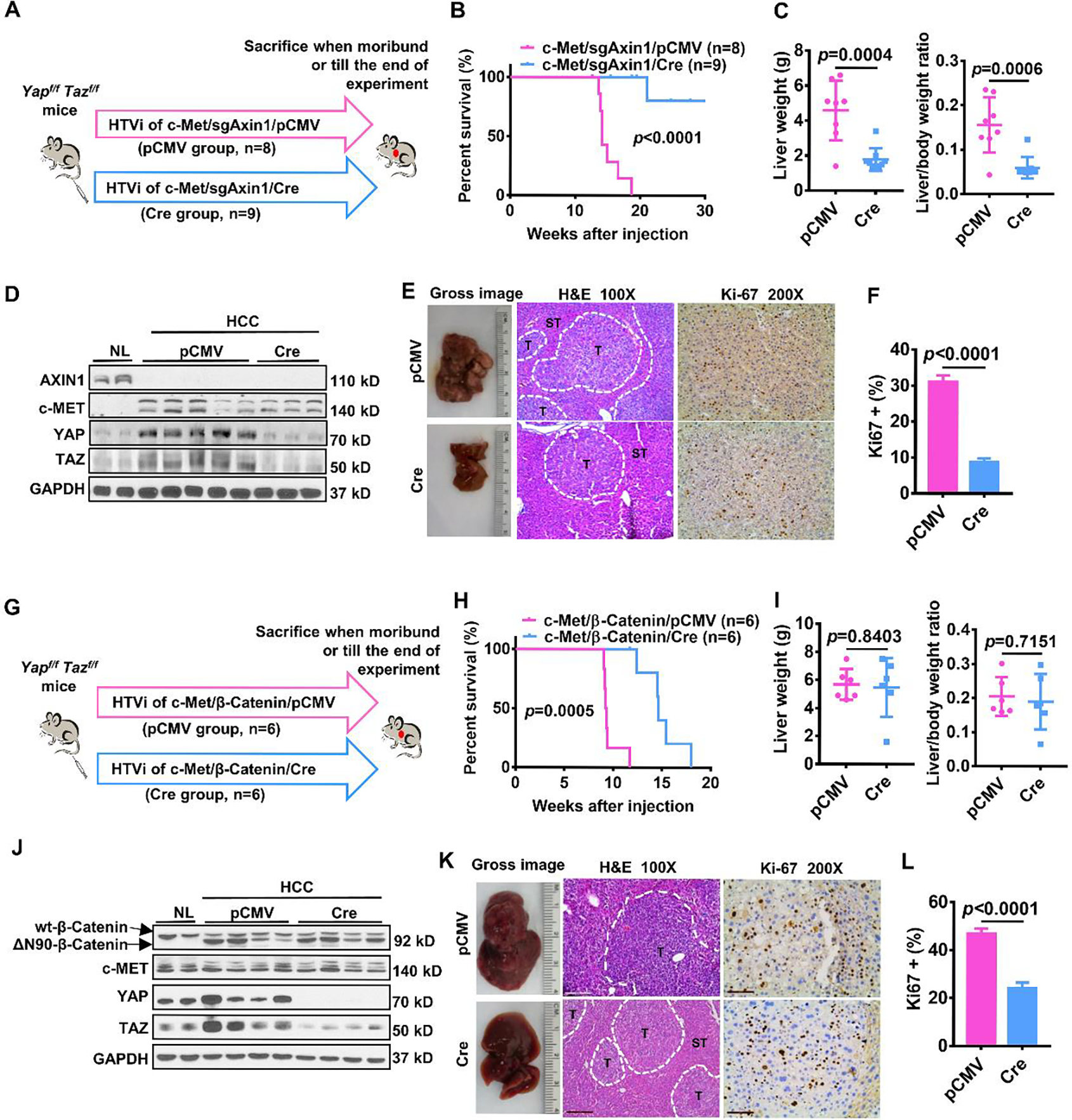
Figure 4. Depletion of Yap/Taz inhibits c-Met/sgAxin1 while slightly delaying c-Met/β-Catenin mouse HCC development.

(A) Study design. Yapflox/floxTazflox/flox mice were injected with c-Met/sgAxin1/pCMV (n=8) and c-Met/sgAxin1/Cre (n=9) plasmids using hydrodynamic tail vein injection (HTVi), respectively. (B) Survival curve of mice in both groups. Kaplan-Meier comparison was performed, p<0.0001. (C) Comparisons of liver weight and liver weight to body weight ratio in both groups. (D) Levels of AXIN1, c-MET, YAP, and TAZ determined by Western blot analysis. GAPDH was used as a loading control. (E) Representative images of macroscopic pictures, H&E, and Ki-67 staining of the tumor in both groups. Scale bars: 200μm for 100X, 100μm for 200X. (F) Quantification of Ki67 positive percentage in both groups. (G) Study design. Yapflox/floxTazflox/flox mice were injected with c-Met/ β-Catenin/pCMV (n=6) and c-Met/β-Catenin/Cre (n=6) plasmids using hydrodynamic tail vein injection (HTVi), respectively. (H) Survival curve of mice in both groups. Kaplan-Meier comparison was performed, p=0.0005. (I) Comparisons of liver weight and liver weight to body weight ratio in both groups. (J) Expression of β-Catenin, c-MET, YAP, and TAZ determined by Western blot analysis. GAPDH was used as a loading control. (K) Representative images of macroscopic pictures, H&E, and Ki-67 staining of the tumor in both groups. Scale bars: 200μm for 100X, 100μm for 200X. (L) Quantification of Ki67 positive percentage in both groups.
