ABSTRACT
Streptococcus parauberis is the dominant etiological agent of streptococcosis, the most devastating bacterial disease in the olive flounder farming industry in South Korea. In this study, the distribution of serotypes, antimicrobial susceptibility, and presence of antimicrobial resistance genes (ARGs) in S. parauberis isolates obtained between 1999 and 2021 was thoroughly investigated to gain insight into the dynamics of their presence and the relationship between serotypes and antimicrobial resistance. Disk diffusion testing of 103 isolates against 10 antimicrobial agents was performed, and epidemiological cut-off values generated through normalized resistance interpretation analysis were used to classify wild-type (WT) and non-wild-type (NWT) populations. Principal component analysis and hierarchical clustering were implemented to achieve an understanding on the relationship between serotypes and antimicrobial resistance patterns. PCR-based serotyping showed that serotype Ia (67.1%) was the most prevalent in South Korea, followed by serotypes Ib/Ic (25.2%) and II (7.7%). The highest proportion of isolates was assigned to NWT against amoxicillin (80.6%), followed by oxytetracycline (77.7%) and erythromycin (48.5%). The time-scale data showed that recently obtained serotypes Ib/Ic and II isolates tended to be categorized as NWT populations resistant to more antibiotics, possibly due to microbial adaptation to antibiotic pressure. ARGs responsible for resistance to oxytetracycline and erythromycin were found only in NWT populations in serotype Ia [tet(S) and erm(B), respectively], and serotype II [tet(M) and mef(J)-msr(I), respectively]. We also found that the mef-msr gene pair in S. parauberis serotype II might be involved in low-level resistance to erythromycin.
IMPORTANCE
This study presents serotype distribution and antimicrobial susceptibility data along with the antimicrobial resistance genes (ARGs) of Streptococcus parauberis, which is an important bacterial fish pathogen worldwide. In particular, almost all oxytetracycline and erythromycin non-wild-type (NWT) populations harbored tet(S) or tet(M), and erm(B) or mef(J)-msr(I), respectively. Interestingly, these ARGs were distributed in a highly serotype-dependent manner, resulting in a clear correlation between the antibiogram and serotype distribution. Moreover, recent isolates belonging to serotypes Ib/Ic and II tended to be more frequently categorized as NWT against antimicrobials, including amoxicillin and cefalexin compared to old isolates, while a dramatic decrease in erythromycin and clindamycin NWT frequencies was observed in recent serotype Ia isolates, which lacked erm(B). These variations might be attributed to shifts in the antibiotics employed in South Korean aquaculture over time. The overall findings would provide important background knowledge for understanding the epidemiology of S. parauberis infection in aquaculture.
KEYWORDS: Streptococcus parauberis, serotype, antimicrobial susceptibility, antimicrobial resistance gene
INTRODUCTION
Streptococcosis is one of the most important bacterial diseases in aquaculture worldwide, causing severe mortality in various fish species, including the olive flounder (Paralichthys olivaceus), starry flounder (Platichthys stellatus), turbot (Scophthalmus maximus), and striped bass (Morone saxatilis) (1 – 4). Since the early 2010s, Streptococcus parauberis has been a dominant causative agent of streptococcosis in farmed olive flounder in South Korea (5). Commercial formalin-killed cell vaccines and the use of antibiotics are options for the prevention and treatment of S. parauberis infections. However, S. parauberis infections still occur in the field (6). This might be due to the presence of different serotypes and the emergence of antibiotic-resistant strains.
Currently, several serotypes of S. parauberis (i.e., Ia, Ib, Ic, II, III, and IV) have been proposed, based on differences in the sequences of their capsular polysaccharide (CPS) genes (4, 7, 8). Tu et al. (9) developed a multiplex PCR assay to identify three serotypes (Ia, Ib/Ic, and II) of S. parauberis based on the sequence of the wzy gene encoding CPS polymerase, indicating that serotyping can be performed by PCR, as an alternative method to traditional serotyping using anti-sera. Previously, serotypes I and II were isolated from farmed olive flounder in South Korea and Japan (8, 10). Serotype III was isolated from turbot in Spain (4) and striped bass in the United States (2), and a single bovine isolate was assigned to serotype IV (11). In general, candidate vaccine strains are selected by taking into account the genotypes and serotypes currently circulating in the region (12). The selection of appropriate vaccine strains is an important element in implementing vaccination policies for the control of diseases (e.g., dengue and influenza) (13).
The emergence of drug-resistant bacteria under antibiotic selection pressure in aquaculture has become more evident and is a significant threat worldwide (14). Antimicrobial resistance can spread to microbial communities through the horizontal gene transfer of antimicrobial resistance genes (ARGs), which are often found in mobile genetic elements such as plasmids, prophages, and transposons (15). Previous studies (16 – 18) found that several S. parauberis isolates harbored tet(S), tet(M), or erm(B), exhibiting resistance to oxytetracycline or erythromycin. However, those studies applied clinical breakpoints as described in Clinical and Laboratory Standards Institute (CLSI) M31-A3 guidelines (19) developed for microbes derived from terrestrial animals, to discriminate resistance from susceptibility. For example, Woo et al. (18) reported that 18 erythromycin-susceptible isolates categorized by the breakpoint (19) harbored erm(B), indicating that incorrect interpretive criteria were used in the study and therefore not suitable for precise discrimination of antimicrobial resistance in S. parauberis. Instead, an epidemiological cut-off value (ECV) can be established based on arrays of antimicrobial susceptibility test results and applied as an interpretative criterion to categorize a bacterial population into two groups: those that are fully susceptible (wild type, WT), and those that exhibit reduced susceptibility by acquired mechanism of resistance (non-wild type, NWT) (20). Unlike the clinical breakpoint, the ECV is generated without consideration of pharmacokinetics/pharmacodynamics and results obtained from clinical trials. Therefore, the ECV should not be used as a predictor of clinical success but as epidemiological surveillance tool for bacterial antimicrobial resistance (20). Although we previously established ECVs for S. parauberis based on disk diffusion data using 75 isolates (21), serotyping and the detection of ARGs were not performed. This study used a total of 103 isolates, including recently isolated strains.
The relationship between serotype and antimicrobial resistance has been reported for S. parauberis (17, 22), as well as other streptococcal species (23 – 25). Studies (17, 22) using S. parauberis isolates from P. olivaceus between 2002 and 2008 showed that tet(M) was present in only 32 serotype II isolates and absent in 44 serotype I isolates. Meng et al. (17) found that S. parauberis serotype II isolates commonly exhibited intermediate resistance to erythromycin but were not able to specify any relevant ARGs. In Streptococcus agalactiae, macrolide-resistance genes were distributed with a clear correlation to serotypes: mef(A) and erm(A) were most common in serotypes Ia (55%) and Ib (75%), respectively, whereas erm(B) was most prevalent in both serotype III (73%) and V isolates (75%) (23). Streptococcus pneumoniae serotype 19A, a non-vaccine type, has become the most prevalent serotype worldwide since the implementation of the seven-valent pneumococcal conjugate vaccine (26). Previous studies have demonstrated that serotype 19A could frequently acquire and exhibit antimicrobial resistance to multiple drugs under antibiotic pressures, leading to its competitive survival and prevalence in multiple countries (24, 25). The overall findings from previous studies indicate that the surveillance of serotype distribution along with antimicrobial resistance could facilitate a better understanding of the epidemiology of S. parauberis infections in fish.
In this study, ECVs were estimated based on antibiotic disk diffusion data that were generated using 103 S. parauberis isolates collected from 1999 to 2021 and used to categorize WT and NWT populations of S. parauberis. Serotyping and ARG detection were also conducted to gain insight into the dynamics of serotype prevalence and their association with antimicrobial resistance patterns.
RESULTS
Serotyping of South Korean S. parauberis isolates
All bacterial isolates (n = 103) used in this study were identified as S. parauberis, and only three serotypes, Ia, Ib/Ic, and II, were found, as shown in Table S1. Of the 103 isolates, 75, 20, and 8 strains were isolated from flounder farms in Jeju Island, the Gyeongsang province, and the Jeolla province in South Korea, respectively (Table S1). Also, the majority of isolates were derived from olive flounder (98 out of 103 strains), while only five strains were from starry flounder. The most prevalent serotype in the South Korean S. parauberis isolates used in this study was Ia at ~67% (69 out of 103 strains), followed by Ib/Ic (~25%) and II (~8%) (Table 1). Serotype Ia was the most frequently isolated every year, except for 2005, when serotype Ib/Ic was the most dominant (93%). Serotype Ia was the predominant isolate in Jeju Island and the Gyeongsang province, while all isolates from the Jeolla province were serotype Ib/Ic.
TABLE 1.
Serotyping of 103 Streptococcus parauberis strains isolated in South Korea
| Year of isolation | Proportion of serotype | ||
|---|---|---|---|
| Ia | Ib/Ic | II | |
| 1999 | 100% (4/4) | 0% (0/4) | 0% (0/4) |
| 2003 | 50% (2/4) | 0% (0/4) | 50% (2/4) |
| 2004 | 50% (3/6) | 33% (2/6) | 17% (1/6) |
| 2005 | 0% (0/15) | 93% (14/15) | 7% (1/15) |
| 2007 | 100% (2/2) | 0% (0/2) | 0% (0/2) |
| 2013 | 100% (3/3) | 0% (0/3) | 0% (0/3) |
| 2014 | 83% (5/6) | 17% (1/6) | 0% (0/6) |
| 2015 | 100% (5/5) | 0% (0/5) | 0% (0/5) |
| 2018 | 73% (11/15) | 27% (4/15) | 0% (0/15) |
| 2019 | 75% (12/16) | 25% (4/16) | 0% (0/16) |
| 2021 | 81% (22/27) | 4% (1/27) | 15% (4/27) |
| Total | 67% (69/103) | 25% (26/103) | 8% (8/103) |
Analysis of zone diameters of S. parauberis using normalized resistance interpretation
The distribution of inhibitory zone sizes obtained from the disk diffusion testing of 103 S. parauberis isolates against 10 antimicrobial agents is shown in Table S2. The inhibitory zone diameters of the two reference strains were within the normal ranges in accordance with the CLSI guidelines (27). The histograms and ECVs of the respective antibiotics determined through normalized resistance interpretation (NRI) analysis are shown in Fig. S1 and Table 2. Isolates exhibiting inhibitory disk zone size ≥ECVs were designated the WT population since they fit the definition of the lower limit of the isolates lacking resistance mechanisms. Abbreviations for the antibiotic agents are summarized in Table 2. Overall, approximately 81% of the S. parauberis isolates used in this study were classified as NWT with resistance against AMX, followed by OTC (~77%) and ERY (~48%). In contrast, only ~6%, ~3%, 0%, and 0% of the S. parauberis isolates were categorized as NWT with resistance against ENR, FFC, CHL, and SXT, respectively.
TABLE 2.
Summary of results of normalized resistance interpretation analysis
| Antibiotics | ECV (mm) a | SD (mm) b | WT (%) c |
|---|---|---|---|
| Amoxicillin (AMX) | 34 | 2.3 | 19.4 |
| Cephalexin (CFL) | 23 | 4.6 | 83.8 |
| Ceftiofur (EFT) | 29 | 2.9 | 72.8 |
| Oxytetracycline (OTC) | 26 | 2.5 | 23.3 |
| Erythromycin (ERY) | 29 | 2.6 | 52.4 |
| Clindamycin (CLI) | 23 | 3.2 | 69.9 |
| Chloramphenicol (CHL) | 22 | 3.1 | 100 |
| Florfenicol (FFC) | 25 | 2.4 | 97.1 |
| Enrofloxacin (ENR) | 18 | 3.5 | 100 |
| Sulfamethoxazole/trimethoprim (SXT) | 20 | 2.2 | 94.2 |
ECV, epidemiological cut-off value.
SD, standard deviation.
WT, wild type.
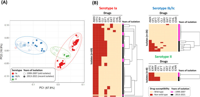
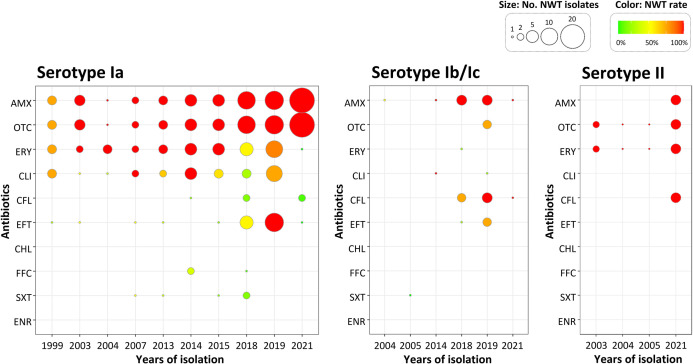
Based on principal component analysis (PCA) and hierarchical clustering analysis, the majority of isolates, with few exceptions, were significantly clustered depending on their serotype, and partially the year of isolation (Fig. 1). Based on the region of isolation, strains isolated from the Gyeongsang and Jeolla provinces could be grouped separately, while isolates from Jeju Island exhibited relatively more diverse antimicrobial susceptibility patterns (Fig. S2). Almost all isolates belonging to serotypes Ia and II were categorized as NWT with resistance against OTC and AMX, and OTC and ERY, respectively, indicating a possible decrease in the therapeutic effects of those antimicrobials (Fig. 1B). The median number of antibiotics to which the isolates exhibit NWT for serotype Ib/Ic was comparatively lower than that of the other two serotypes (Fig. S3). Interestingly, the recent isolates belonging to serotypes Ib/Ic and II were categorized as NWT with resistance against more antimicrobials (Fig. 2). Recent serotype Ib/Ic isolates (n = 10) were classified as NWT with resistance to seven tested antibiotics including AMX (100%) and CFL (80%), while old isolates (n = 16) were WT to most of the tested drugs (Fig. 1B and 2). However, in serotype Ia, a dramatic decrease in NWT frequency with resistance against ERY and CLI was observed from ~96% and ~71% in 1999–2015 to ~38% and ~27% in 2018–2021, respectively. In the case of EFT, the proportion of NWT in serotype Ia isolates was ~55% (6/11) in 2018 and 100% (12/12) in 2019, but dropped significantly to ~5% (1/21) in 2021 (Fig. S3). This might be linked to the recent introduction of injectable EFT in South Korean aquaculture, which has been released since 2018 (28).
Fig 1.
Principal component analysis (PCA; A) and hierarchical clustering analysis (B) based on the antibiogram of 103 Streptococcus parauberis isolates against 10 antibiotics. (A) The PCA plot captures the variance in the data set composed of normalized inhibitory zone diameters obtained from the disk diffusion tests of 103 S. parauberis strains. The X and Y-axes show principal components 1 and 2, which explain 67.8% and 16.9% of the total variance, respectively. The colors of the circles represent the serotype. Open and closed circles indicate the isolated year, categorized as 1999–2007 and 2013–2021, respectively. (B) Heatmaps show the wild-type (beige) and non-wild-type (red) distributions of S. parauberis isolates according to serotype. The black and pink ellipses located on the right side of the heatmap indicate the year of isolation, categorized as 1999–2007 and 2013–2021, respectively. Abbreviations for the antibiotic agents are shown in Table 2.
Fig 2.
Year-wide distribution of non-wild-type (NWT) populations (%) of three serotypes of Streptococcus parauberis isolates against 10 antibiotics. The color of the dots indicates the NWT proportion, while the size indicates the number of NWT populations in the respective isolation year. Abbreviations for the antibiotic agents are shown in Table 2.
Identification and detection of ARGs in S. parauberis
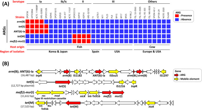
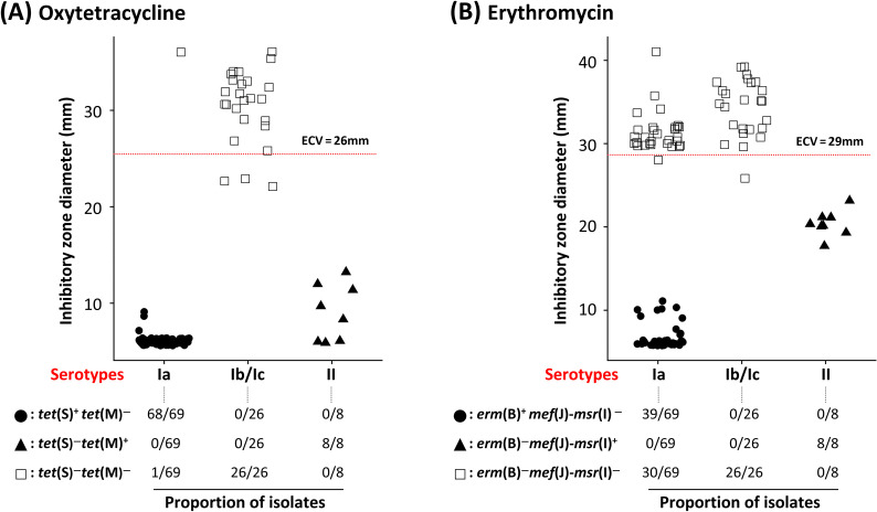
This study identified six ARGs from pan-genomic analysis using 31 S. parauberis genomes including four sequenced genomes in the current study (KSP45, SPOF19J15, SPOF21J2, and SPOF21J26), as shown in Fig. 3. Information on whole-genome sequences used in this study are available in the supplementary results and Table S4. Three ARGs, erm(B), tet(S), and ANT (6)-Ia, were found only in serotype Ia genomes, whereas tet(M) and mef(J)-msr(I) were present only in serotype II (Fig. 3A). tet(S) was present in a ~12 kb-sized plasmid, while two paralogous copies of erm(B) [with or without a leader peptide-encoding gene, erm(BL)] and ANT (6)-Ia were contained in the chromosome, as illustrated in Fig. 3B. Unlike other serotype Ia strains, the tet(S)-containing plasmid was exceptionally absent in the genome of KSP45, which exhibited WT to OTC (Fig. 3A). The transposon-mediated gene cluster carrying erm(B) could not be identified in genome sequence of KSP45 and SPOF21J26, which were WT to ERY (Fig. 3A). Consistent with this, among the serotype Ia isolates used in this study, NWT isolates with resistance to OTC and ERY harbored tet(S) (n = 68/69) and erm(B) (n = 39/69), respectively (Fig. 4A). All eight serotype II isolates used in this study were classified as NWT with resistance against OTC and ERY and harbored tet(M) and mef(J)-msr(I) (Fig. 4). Based on the inhibitory zones generated by serotype II isolates against ERY, the mef(J)-msr(I) genes might be responsible for the relatively low-level resistance to ERY (Fig. 4B). Although the ARGs mentioned above were not found in serotype Ib/Ic isolates in this study, three and one isolates were categorized as NWT with resistance against OTC and ERY, respectively. However, the inhibitory zone diameters of these isolates were distributed close to ECVs, unlike the other ARG-carrying isolates.
Fig 3.
Distribution of antibiotic resistance genes (ARGs) in 31 Streptococcus parauberis genome sequences. (A) The heatmap presents the presence and absence of ARGs in each isolate, as shown in the red and blue boxes, respectively. (B) Gene organization of four ARG-carrying mobile element structures in S. parauberis serotypes Ia and II. The direction of the arrow indicates the direction of transcription. ARGs and mobile element-related genes are shown in red and yellow arrows.
Fig 4.
Distribution of inhibitory zone diameters of 103 Streptococcus parauberis isolates to oxytetracycline (A) and erythromycin (B) shown with the proportion of isolates harboring antimicrobial-resistance genes. Inhibitory zone diameters of 103 S. parauberis isolates are shown in filled circles [tet(S)+ tet(M)− and erm(B)+ mef(J)-msr(I)− in panels A and B, respectively], filled triangles [tet(S) −tet(M)+ and erm(B)− mef(J)-msr(I)+ in panels A and B, respectively], and empty squares [tet(S)− tet(M)− and erm(B)− mef(J)-msr(I)− in panels A and B, respectively]. The horizontal red lines indicate the epidemiological cut-off values (ECVs). The proportion of respective genotypes in every serotype is shown below the scatter plots.
DISCUSSION
This study aimed to track the trends of the antibiotic-resistance patterns of S. parauberis isolated between 1999 and 2021, along with serotyping. In fact, we used 31 whole-genome sequencing of S. parauberis, including nine genomes sequenced in our laboratory, and compared our results with as relevant many references showing antimicrobial susceptibility and serotypes of the pathogen as possible (4, 7, 8, 10, 16 – 18, 21, 22, 29, 30) to overcome the limited numbers of strains (n = 103) used. We found profound diversity in terms of antibiotic resistance in S. parauberis through the establishment of ECVs for 10 antimicrobial agents based on disk diffusion test data using 103 isolates (Fig. 1). The number of WT isolates in the respective, tested antibiotics were diverse (n = 20–103), which satisfied the requirement of at least 20–30 observations from fully susceptible isolates in NRI analysis (31). In NRI analysis, standard deviations for normalized WT inhibitory zone sizes are calculated, thereby providing quantitative measure of the precision of the antimicrobial susceptibility data sets (31). The standard deviations of 9 out of the 10 tested antibiotics satisfied the criterion of ≤3.95 mm (31), except for CFL (4.3 mm) (Table 2). Based on the ECVs for OTC and ERY, with just a few exceptions, only NWT populations of S. parauberis harbored the respective ARGs, as shown in Fig. 4. This indicates that most ECVs obtained in this study were accurate and could be utilized as local criteria for the discrimination of WT from NWT S. parauberis isolates.
Based on similar but independent studies (10, 18), it can be concluded that serotype Ia has been the most dominant serotype in South Korea since the late 1990s, followed by serotypes Ib/Ic and II (Table 1). Since the appearance of approximately a dozen serotype Ib/Ic isolates collected in 2005, they have decreased, which is in partial agreement with the results of Kim et al. (10), showing that serotype Ib/Ic was the most prevalent serotype in the period of 2005–2009. Kim et al. (10) also pointed out that the isolation rate of serotype II was extremely low in the 2010s, but started to increase after 2019. In this study, it was found that serotypes Ia and Ib/Ic were predominant in the Gyeongsang (19 out of 20 isolates) and Jeolla provinces (8 out of 8 isolates), respectively (Table S1), which were also distinct in terms of their antibiotic susceptibility patterns (Fig. S2). However, as these isolates were clustered with the isolates of respective serotypes from other geographical sources based on the antibiogram (Fig. S2), it seems highly likely that the region-specific antimicrobial susceptibility patterns in the Gyeongsang and Jeolla provinces were substantially affected by the regional predominance of serotypes. Along with previous studies (17, 18), data from this study suggest that the existence of ARGs in S. parauberis has been significantly conserved in a serotype-dependent manner (Fig. 4). Similarly, Domelier et al. (23) found that among 119 ERY-resistant S. agalactiae isolates, 55% of serotype Ia, and 75% of serotype Ib populations, harbored mef(A) and erm(A), respectively, whereas erm(B) was present in both serotype III (73%) and V isolates (75%).
Besides antibiotic selective pressure, the dominance of certain populations within a microbial species (bio- and/or genogroup) might depend on host specificity and virulence (31, 32). In S. parauberis, only serotypes I and II have been isolated from fish in South Korea and Japan (9, 10, 30). Conversely, those serotypes have never been isolated from turbot in Spain, which are known to be significantly more susceptible to S. parauberis serotype III (4, 29). Yolanda et al. (29) demonstrated that the lethal dose of 50% (LD50) of S. parauberis serotype III isolates for turbot was at least 105 times lower than that of serotype I and II isolates, suggesting that S. parauberis might be a host-specific pathogen. Likewise, Han et al. (30) demonstrated that among serotypes of S. parauberis isolated from South Korea, serotype II was the least pathogenic to olive flounder, which might be associated with the very low frequency found in this study (~8%, 8/103).In this study, recent isolates belonging to serotype Ib/Ic and II tended to be classified as NWT with resistance against more antimicrobials, including AMX, OTC, and ERY, compared to the old isolates (Fig. 2 and Fig. S3). Those therapeutic agents are most commonly used in the field (33), and their frequent use might be associated with increased NWT populations with those serotypes. Buschmann et al. (34) showed a significant increase in the prevalence of ARGs in sediments from the vicinity of fish farms compared to those from control sites. Those results indicate that there is a clear correlation between the use of antibiotics and the prevalence of antibiotic resistance, as previously described for various bacterial species (35 – 37). While old serotype Ia isolates were commonly categorized as NWT with resistance to OTC and ERY, many recent serotype Ia isolates obtained from 2018 to 2021 (n = 28/45) were classified as NWT with resistance to only OTC, but not ERY, as they lacked erm(B) (Fig. 3 and 4B). Since erm(B) mediates resistance to both lincosamide and macrolide antibiotics (38), the rate of NWT populations with resistance to CLI was also significantly decreased (Fig. 2). Similarly, 19 serotype I isolates collected in the 2000s were commonly resistant to OTC and ERY (17), but 29 out of 56 serotype Ia isolates obtained in 2018–2019 were resistant only to OTC (18). Recent decreases in the number of serotype Ia isolates categorized as NWT with resistance to ERY might be due to reductions in ERY use by oral administration and increases in the use of amoxicillin by injection in the field, as shown in a study by Kim et al. (33).
Almost all serotype Ia isolates and about half of serotype Ib/Ic and II isolates were categorized as NWT populations with resistance to AMX, generally known to be effective for Gram-positive bacteria (39, 40). Resistance against AMX can be often introduced through point mutations in genes encoding for penicillin-binding proteins (PBPs) (41 – 43). As found by Woo et al. (18), it was presumed that the NWT populations of S. parauberis isolates used in this study were due to point mutations in pbp1A and pbp2x. Serotype Ia and II isolates classified as NWT with resistance to OTC and ERY harbored tet(S) and erm(B), and tet(M) and mef(J)-msr(I), respectively. These genes are responsible for resistance against OTC and ERY, respectively. Similarly, previous studies (17, 18) identified tet(S) and erm(B) in OTC and ERY-resistant populations of serotype Ia, respectively, and tet(M) in OTC-resistant serotype II. However, none of those studies identified the ARGs responsible for ERY resistance in serotype II. The genome-wide analysis in this study successfully identified the presence of an adjacent gene pair, mef(J)-msr(I) in S. parauberis serotype II genomes (Fig. 3 and 4B). Several variants of the mef-msr genes, which encode a dual-efflux system targeting macrolide antibiotics were previously reported (44, 45). In this study, mef(J) and msr(I) present in S. paruaberis serotype II showed ~98% and ~75% amino acid homology with mef(J)-msr(I) in Streptococcus pyogenes (Genbank accession number, QQA64075-6) and mef(D)-msr(F) in Macrococcus canis (Genbank accession number, QHW12307-8), respectively. Previous studies (46, 47) also showed that mef and erm mediated low- and high-level resistance to ERY, respectively, in both S. pneumoniae and S. pyogenes. It was also found that isolates harboring mef(J)-msr(I) showed low-level resistance against ERY, as shown in Fig. 4B.
BLAST search using the Comprehensive Antibiotic Resistance Database (48) indicated that all S. parauberis genomes used in this study harbored mre(A) and pat(A)-pat(B). Although mre(A) was previously identified as responsible for macrolide resistance in S. agalactiae (49), recent studies (50, 51) found that mre(A) was ubiquitous in S. agalactiae and was not directly related to macrolide resistance. The other genes, pat(A)-pat(B), are known to be ABC transporters that function as an efflux pump for various drugs, as well as toxic compounds in S. pneumoniae (52 – 54). However, pat(A)-pat(B) in S. parauberis shared only ~67% amino acid similarity with pat(A)-pat(B) in S. pneumoniae (Genbank accession numbers: AAK76136.1 and AAK76137.1) and were present in all S. parauberis genomes used in this study, indicating that these genes might be ubiquitous and not involved in drug resistance in S. parauberis. Hence, those two ARGs were excluded from the results since, as no evidence of antibiotic resistance could be found.
A study by Woo et al. (18), using 83 S. parauberis isolates derived from fish in South Korea, found that tet(S) and tet(M) were present in all OTC-resistant serotype Ia and II strains, respectively, which was in line with results of this study (Fig. 4A). However, Woo et al. (18) also reported that 5%, ~43%, and ~17% of the OTC-resistant serotype Ia isolates possessed tet(A), tet(B), and tet(Y), respectively, and ~67% of OTC resistant serotype II harbor tet(B). Some of the OTC-resistant isolates contained more than one tetracycline resistance genes, but we could not specify the exact proportion of those populations from the results presented by Woo et al. (18). We have not identified tet(A/B/Y) either by analysis of publicly available S. parauberis genome sequences and PCR tests using specific primer sets targeting those genes (data not shown). In general, tetracycline resistance in Streptococci is induced through an efflux pump encoded by the tet(K/L) gene and ribosomal protection mediated by tet(M/O/Q/S/T/W) (43). In fact, tet(A/B/Y) are frequently found in Gram-negative bacteria, whereas they are extremely rare (55, 56) or have never been reported (in case of tet(Y)) in other Streptococci.
MATERIALS AND METHODS
Bacterial isolation, identification, and serotyping
This study used a total of 103 strains, isolated from 1999 to 2021 from olive flounder and starry flounder and were identified as S. parauberis using the method previously described (57) (Table S1). Bacteria were isolated from swabs of the liver, kidney, and/or spleen of diseased fish by inoculating the swabs on brain heart infusion (BHI) agar plates supplemented with 1% NaCl (BN; Oxoid, UK). The plates were incubated at 28°C for 24–48 h. The bacterial isolates were pure-cultured on the same media and identified as S. parauberis using the PCR method developed by Mata et al. (58). For bacterial serotyping, a multiplex PCR assay developed by Tu et al. (9) was employed. Accordingly, the serotypes of S. parauberis isolates could be determined as Ia, Ib/Ic, and II types. Briefly, genomic DNA was extracted from the isolates using an AccuPrep Genomic DNA Extraction Kit (Bioneer, South Korea) and PCR was conducted using AccuPower PCR PreMix (Bioneer, South Korea) as described in previous studies (9, 58). The primer list is summarized in Table S3. The isolates were stored at −80°C in BHI broth supplemented with 10% glycerol.
Antimicrobial susceptibility testing
The drug susceptibility of S. parauberis isolates was determined by the disk diffusion test according to CLSI guideline VET03-A (27). Briefly, overnight cultured bacteria were adjusted to a concentration of 2 × 10⁸ CFU mL−1 and inoculated onto Mueller-Hinton agar containing 5% sheep blood using a sterile swab. The following commercial antibiotic disks (Oxoid) were placed on the lawn culture: amoxicillin 25 µg, oxytetracycline 30 µg, cephalexin 30 µg, ceftiofur 30 µg, enrofloxacin 5 µg, erythromycin 15 µg, sulfamethoxazole (23.75 µg)/trimethoprim (1.25 µg) 25 µg, clindamycin 10 µg, florfenicol 30 µg, and chloramphenicol 30 µg. Escherichia coli ATCC 25922 and Aeromonas salmonicida ATCC 33658 were used as the reference strains for quality control (27).
Data analysis
In this study, the ECV was determined through the NRI method developed by Kronvall and Smith (59). Mean values and standard deviations of WT normalized zone sizes were calculated using a plot of the probit values of the normalized accumulative frequencies of observations against zone diameters (59). The ECV was set at two and a half standard deviations below the mean (59). Principal component analysis and hierarchical clustering analysis were performed based on bacterial antibiotic susceptibility patterns using ClustVis (60) and Morpheus (https://software.broadinstitute.org/morpheus), respectively. Significant differences among groups were analyzed using the Kruskal–Wallis test, followed by the post hoc Dunn’s test with Bonferroni correction (significance at adjusted P values <0.05) using SPSS v23.0 (IBM, USA).
Identification of ARGs in whole-genomic sequences of S. parauberis
In addition to the four genome sequences obtained from this study (see supplementary results), 27 S. parauberis genomic sequences were obtained from the National Biotechnology Information Center (NCBI; https://www.ncbi.nlm.nih.gov/data-hub/genome/?taxon=1348) and used for the analysis (Table S4). The serotypes of a total of 31 strains were determined through in silico PCR (Primer-BLAST) using the primers listed in Table S3 (61). ARGs in the genomic sequences were identified using Resistance Gene Identifier in the Comprehensive Antibiotic Resistance Database (48) and illustrated using EasyFig v2.2.2 (62).
Detection of ARGs in S. parauberis isolates
Genomic DNA extracted from 103 S. parauberis strains was used for the identification of ARGs. PCR was conducted to evaluate the presence of three genes, erm(B), mef(J), and msr(I), involved in macrolide resistance, and two tetracycline resistance genes, tet(M) and tet(S). The primers used in this study are listed in Table S3. PCR was conducted as described in previous studies (63 – 65). In this study, primer sets for mef(J) and msr(I) were designed using Primer3Plus (66), based on coding DNA sequences retrieved from the whole-genomic sequence of S. paruaberis KSP10 (Table S4). The PCR conditions were as follows: pre-denaturation at 95°C for 5 min, 30 cycles of denaturation at 95°C for 30 s, annealing at 55°C for 30 s, extension at 72°C for 30 s, and a final extension at 72°C for 7 min. PCR products were identified by electrophoresis in 1.5% (wt/vol) agarose gel stained with ethidium bromide.
ACKNOWLEDGMENTS
This research was supported by a grant from the National Institute of Fisheries Science, South Korea (grant no. R2023051).
We are grateful to Korean Culture collection of Aquatic Microorganisms (KoCAM) of NIFS for providing us with bacterial strains (SPOF21J2, SPOF21J13, SPOF21J14, SPOF21J15, SPOF21J17, SPOF21J22, SPOF21J23, SPOF21J24, and SPOF21J25).
Contributor Information
Chan-Il Park, Email: vinus96@hanmail.net.
Do-Hyung Kim, Email: dhkim@pknu.ac.kr.
Luke R. Iwanowicz, USGS, Eastern Ecological Science Center, Kearneysville, West Virginia, USA
DATA AVAILABILITY
The complete genome sequences in this project have been deposited in BioProject accession number PRJNA940317. All data that support the findings of this study are available on reasonable request to the corresponding author.
SUPPLEMENTAL MATERIAL
The following material is available online at https://doi.org/10.1128/spectrum.04400-22.
This file includes supplementary materials and methods, supplementary results, Tables S1 to S4, Figures S1 to S3, and references.
ASM does not own the copyrights to Supplemental Material that may be linked to, or accessed through, an article. The authors have granted ASM a non-exclusive, world-wide license to publish the Supplemental Material files. Please contact the corresponding author directly for reuse.
REFERENCES
- 1. Baeck GW, Kim JH, Gomez DK, Park SC. 2006. Isolation and characterization of Streptococcus sp. from diseased flounder (Paralichthys olivaceus) in Jeju island. J Vet Sci 7:53–58. doi: 10.4142/jvs.2006.7.1.53 [DOI] [PMC free article] [PubMed] [Google Scholar]
- 2. Haines AN, Gauthier DT, Nebergall EE, Cole SD, Nguyen KM, Rhodes MW, Vogelbein WK. 2013. First report of Streptococcus parauberis in wild finfish from North America. Vet Microbiol 166:270–275. doi: 10.1016/j.vetmic.2013.05.002 [DOI] [PubMed] [Google Scholar]
- 3. Woo SH, Park SI. 2013. Streptococcous parauberis infection in starry flounder, Platichthys stellatus: characterization of innate immune responses following experimental infection. Fish Shellfish Immunol 35:413–420. doi: 10.1016/j.fsi.2013.04.047 [DOI] [PubMed] [Google Scholar]
- 4. Torres-Corral Y, Santos Y. 2020. Comparative genomics of Streptococcus parauberis: new target for molecular identification of serotype III. Appl Microbiol Biotechnol 104:6211–6222. doi: 10.1007/s00253-020-10683-z [DOI] [PMC free article] [PubMed] [Google Scholar]
- 5. Park SB, Nho SW, Jang HB, Cha IS, Kim MS, Lee W-J, Jung TS. 2016. Development of three-valent vaccine against streptococcal infections in olive flounder, Paralichthys olivaceus. Aquaculture 461:25–31. doi: 10.1016/j.aquaculture.2016.04.022 [DOI] [Google Scholar]
- 6. Shim J, Hwang S, Jang S, Kim T, Jeong J. 2019. Monitoring of the mortalities in oliver flounder (Paralichthys olivaceus) farms of Korea. Journal of fish pathology 32:29–35. doi: 10.7847/jfp.2019.32.1.029 [DOI] [Google Scholar]
- 7. Kanai Kinya, Yamada M, Meng F, Takahashi I, Nagano T, Kawakami H, Yamashita A, Matsuoka S, Fukuda Y, Miyoshi Y, Takami I, Nakano H, Hirae T, Shutou K, Honma T. 2009. Serological differentiation of Streptococcus parauberis strains isolated from cultured Japanese flounder in Japan. Fish Pathol 44:33–39. doi: 10.3147/jsfp.44.33 [DOI] [Google Scholar]
- 8. Kanai K, Tu C, Katayama N, Suga K. 2015. Existence of subserotypes in Streptococcus parauberis serotype I. 魚病研究 50:75–80. doi: 10.3147/jsfp.50.75 [DOI] [Google Scholar]
- 9. Tu C, Suga K, Kanai K. 2015. A multiplex PCR assay for different-Iation of Streptococcus parauberis serotypes. Fish Pathol 50:213–215. doi: 10.3147/jsfp.50.213 [DOI] [Google Scholar]
- 10. Kim KW, Yoo EH, Yang HY, Kang BJ. 2020. Isolation characteristics of causative agent of streptococcosis and serotype changes of Streptococcus parauberis from olive flounder (Paralichthys olivaceus) in jeju. Journal of fish pathology 33:119–125. doi: 10.7847/jfp.2020.33.2.119 [DOI] [Google Scholar]
- 11. Williams AM, Collins MD. 1990. Molecular taxonomic studies on Streptococcus uberis types I and II. description of Streptococcus parauberis sp. nov. J Appl Bacteriol 68:485–490. doi: 10.1111/j.1365-2672.1990.tb02900.x [DOI] [PubMed] [Google Scholar]
- 12. Bari FD, Parida S, Tekleghiorghis T, Dekker A, Sangula A, Reeve R, Haydon DT, Paton DJ, Mahapatra M. 2014. Genetic and antigenic characterisation of serotype A FMD viruses from East Africa to select new vaccine strains. Vaccine 32:5794–5800. doi: 10.1016/j.vaccine.2014.08.033 [DOI] [PMC free article] [PubMed] [Google Scholar]
- 13. Pollard AJ, Bijker EM. 2021. A guide to vaccinology: from basic principles to new developments. Nat Rev Immunol 21:83–100. doi: 10.1038/s41577-020-00497-5 [DOI] [PMC free article] [PubMed] [Google Scholar]
- 14. Preena PG, Swaminathan TR, Kumar VJR, Singh ISB. 2020. Antimicrobial resistance in aquaculture: a crisis for concern. Biologia 75:1497–1517. doi: 10.2478/s11756-020-00456-4 [DOI] [Google Scholar]
- 15. Partridge SR, Kwong SM, Firth N, Jensen SO. 2018. Mobile genetic elements associated with antimicrobial resistance. Clin Microbiol Rev 31:e00088–17. doi: 10.1128/CMR.00088-17 [DOI] [PMC free article] [PubMed] [Google Scholar]
- 16. Park Y-K, Nho S-W, Shin G-W, Park S-B, Jang H-B, Cha I-S, Ha M-A, Kim Y-R, Dalvi RS, Kang B-J, Jung T-S. 2009. Antibiotic susceptibility and resistance of Streptococcus iniae and Streptococcus parauberis isolated from olive flounder (Paralichthys olivaceus). Vet Microbiol 136:76–81. doi: 10.1016/j.vetmic.2008.10.002 [DOI] [PubMed] [Google Scholar]
- 17. Meng F, Kanai K, Yoshikoshi K. 2009. Characterization of drug resistance in Streptococcus parauberis isolated from Japanese flounder. Fish Pathol 44:40–46. doi: 10.3147/jsfp.44.40 [DOI] [PubMed] [Google Scholar]
- 18. Woo SJ, Do MY, Jeong MG, Kim NY, Kim MS. 2021. Prevalence, antibiotic susceptibility and Serotyping of Streptococcus parauberis isolates from diseased marine fish. Aquacult Res 52:6525–6536. doi: 10.1111/are.15523 [DOI] [Google Scholar]
- 19. CLSI . 2008. Performance standards for antimicrobial disk and dilution susceptibility tests for bacteria isolated from animals. Approved standard M31–A3, 3rd ed. CLSI, Wayne, PA. [Google Scholar]
- 20. Michael A, Kelman T, Pitesky M. 2020. Overview of quantitative methodologies to understand antimicrobial resistance via minimum inhibitory concentration. Animals (Basel) 10:1405. doi: 10.3390/ani10081405 [DOI] [PMC free article] [PubMed] [Google Scholar]
- 21. Chun W, Lee Y, Kim Y, Roh HJ, Kim A, Kim N, Seo J, Kwon M, Lee JH, Kim D. 2019. Epidemiological cut-off values generated for disc diffusion data from Streptococcus parauberis. Korean Journal of Fisheries and Aquatic Sciences 52:382–388. doi: 10.5657/KFAS.2019.0382 [DOI] [Google Scholar]
- 22. Meng F, Kanai K, Yoshikoshi K. 2009. Structural characterization of Tn916‐Like element in Streptococcus parauberis serotype II strains isolated from diseased Japanese flounder. Lett Appl Microbiol 48:770–776. doi: 10.1111/j.1472-765X.2009.02609.x [DOI] [PubMed] [Google Scholar]
- 23. Domelier A-S, van der Mee-Marquet N, Arnault L, Mereghetti L, Lanotte P, Rosenau A, Lartigue M-F, Quentin R. 2008. Molecular characterization of erythromycin-resistant Streptococcus agalactiae strains. J Antimicrob Chemother 62:1227–1233. doi: 10.1093/jac/dkn388 [DOI] [PubMed] [Google Scholar]
- 24. Pillai DR, Shahinas D, Buzina A, Pollock RA, Lau R, Khairnar K, Wong A, Farrell DJ, Green K, McGeer A, Low DE. 2009. Genome-wide dissection of globally emergent multi-drug resistant serotype 19A Streptococcus pneumoniae. BMC Genomics 10:1–13. doi: 10.1186/1471-2164-10-642 [DOI] [PMC free article] [PubMed] [Google Scholar]
- 25. Ruiz García Y, Nieto Guevara J, Izurieta P, Vojtek I, Ortega-Barría E, Guzman-Holst A. 2021. Circulating clonal complexes and sequence types of Streptococcus pneumoniae serotype 19A worldwide: the importance of multidrug resistance: a systematic literature review. Expert Rev Vaccines 20:45–57. doi: 10.1080/14760584.2021.1873136 [DOI] [PubMed] [Google Scholar]
- 26. Isturiz R, Sings HL, Hilton B, Arguedas A, Reinert R-R, Jodar L. 2017. Streptococcus pneumoniae serotype 19A: worldwide epidemiology. Expert Rev Vaccines 16:1007–1027. doi: 10.1080/14760584.2017.1362339 [DOI] [PubMed] [Google Scholar]
- 27. CLSI . 2006. Methods for antimicrobial disk susceptibility testing of bacteria isolated from aquatic animals; approved guideline M42-A. CLSI, Wayne, PA. [Google Scholar]
- 28. Lee J-H, Seo JS, Kim GW, Kwon M-G, Kim D-H, Park C-I, Kim KT, Park J. 2022. Effect of lincomycin, an injectable lincosamide antibiotic, against streptococcosis in cultured olive flounder Paralichthys olivaceus and its pharmacokinetic-pharmacodynamic profile. Aquaculture 548:737667. doi: 10.1016/j.aquaculture.2021.737667 [DOI] [Google Scholar]
- 29. Yolanda T-C, Clara F-Á, Ysabel S. 2019. Proteomic and molecular fingerprinting for identification and tracking of fish pathogenic Streptococcus. Aquaculture 498:322–334. doi: 10.1016/j.aquaculture.2018.08.041 [DOI] [Google Scholar]
- 30. Han SY, Kang BK, Kang BJ, Shin SP, Soen BH, Kim JM, Kim JH, Choresca CH, Han JE, Jun JW, Park SC. 2011. Prevalence and different characteristics of two serotypes of Streptococcus parauberis isolated from the farmed olive flounder, Paralichthys olivaceus (Temminck and Schlegel), in Korea. J Fish Dis 34:731–739. doi: 10.1111/j.1365-2761.2011.01289.x [DOI] [PubMed] [Google Scholar]
- 31. Smith P. 2020. Eight rules for improving the quality of papers on the antimicrobial susceptibility of bacteria isolated from aquatic animals. Dis Aquat Organ 139:87–92. doi: 10.3354/dao03476 [DOI] [PubMed] [Google Scholar]
- 32. Moreno E, Andreu A, Pigrau C, Kuskowski MA, Johnson JR, Prats G. 2008. Relationship between Escherichia coli strains causing acute cystitis in women and the fecal E. J Clin Microbiol 46:2529–2534. doi: 10.1128/JCM.00813-08 [DOI] [PMC free article] [PubMed] [Google Scholar]
- 33. Kim Y, Seo J, Park J, Jeong AR, Lee J. 2019. Monitoring of aquatic medicine managements in South Korea. J Fish Pathol 32:37–43. doi: 10.7847/jfp.2019.32.1.037 [DOI] [Google Scholar]
- 34. Buschmann AH, Tomova A, López A, Maldonado MA, Henríquez LA, Ivanova L, Moy F, Godfrey HP, Cabello FC. 2012. Salmon aquaculture and antimicrobial resistance in the marine environment. PLoS One 7:e42724. doi: 10.1371/journal.pone.0042724 [DOI] [PMC free article] [PubMed] [Google Scholar]
- 35. Albrich WC, Monnet DL, Harbarth S. 2004. Antibiotic selection pressure and resistance in Streptococcus pneumoniae and Streptococcus pyogenes. Emerg Infect Dis 10:514–517. doi: 10.3201/eid1003.030252 [DOI] [PMC free article] [PubMed] [Google Scholar]
- 36. Hsueh P-R, Shyr J-M, Wu J-J. 2006. Changes in macrolide resistance among respiratory pathogens after decreased erythromycin consumption in Taiwan. Clin Microbiol Infect 12:296–298. doi: 10.1111/j.1469-0691.2005.01348.x [DOI] [PubMed] [Google Scholar]
- 37. Skalet AH, Cevallos V, Ayele B, Gebre T, Zhou Z, Jorgensen JH, Zerihun M, Habte D, Assefa Y, Emerson PM, Gaynor BD, Porco TC, Lietman TM, Keenan JD. 2010. Antibiotic selection pressure and macrolide resistance in nasopharyngeal Streptococcus pneumoniae: a cluster-randomized clinical trial. PLoS Med 7:e1000377. doi: 10.1371/journal.pmed.1000377 [DOI] [PMC free article] [PubMed] [Google Scholar]
- 38. Roberts MC, Sutcliffe J, Courvalin P, Jensen LB, Rood J, Seppala H. 1999. Nomenclature for macrolide and macrolide-lincosamide-streptogramin B resistance determinants. Antimicrob Agents Chemother 43:2823–2830. doi: 10.1128/AAC.43.12.2823 [DOI] [PMC free article] [PubMed] [Google Scholar]
- 39. Kaur SP, Rao R, Nanda S. 2011. Amoxicillin: a broad spectrum antibiotic. Int J Pharm Pharm Sci 3:30–37. [Google Scholar]
- 40. Lulijwa R, Rupia EJ, Alfaro AC. 2020. Antibiotic use in aquaculture, policies and regulation, health and environmental risks: a review of the top 15 major producers. Rev Aquacult 12:640–663. doi: 10.1111/raq.12344 [DOI] [Google Scholar]
- 41. Zapun A, Contreras-Martel C, Vernet T. 2008. Penicillin-binding proteins and β-lactam resistance. FEMS Microbiol Rev 32:361–385. doi: 10.1111/j.1574-6976.2007.00095.x [DOI] [PubMed] [Google Scholar]
- 42. Hakenbeck R, Brückner R, Denapaite D, Maurer P. 2012. Molecular mechanisms of β-lactam resistance in Streptococcus pneumoniae. Future Microbiol 7:395–410. doi: 10.2217/fmb.12.2 [DOI] [PubMed] [Google Scholar]
- 43. Haenni M, Lupo A, Madec J. 2018. Antimicrobial resistance in Streptococcus spp. Microbiol Spectr 6:6–2. doi: 10.1128/microbiolspec.ARBA-0008-2017 [DOI] [PMC free article] [PubMed] [Google Scholar]
- 44. Varaldo PE, Montanari MP, Giovanetti E. 2009. Genetic elements responsible for erythromycin resistance in streptococci. Antimicrob Agents Chemother 53:343–353. doi: 10.1128/AAC.00781-08 [DOI] [PMC free article] [PubMed] [Google Scholar]
- 45. Schwendener S, Donà V, Perreten V. 2020. The novel macrolide resistance genes mef (D), mSR (F), and mSR (H) are present on resistance islands in Macrococcus canis, Macrococcus caseolyticus, and Staphylococcus aureus. Antimicrob Agents Chemother 64:e00160-20. doi: 10.1128/AAC.00160-20 [DOI] [PMC free article] [PubMed] [Google Scholar]
- 46. Morosini M-I, Cantón R, Loza E, del Campo R, Almaraz F, Baquero F. 2003. Streptococcus pyogenes isolates with characterized macrolide resistance mechanisms in Spain: in vitro activities of telithromycin and cethromycin. J Antimicrob Chemother 52:50–55. doi: 10.1093/jac/dkg303 [DOI] [PubMed] [Google Scholar]
- 47. Isozumi R, Ito Y, Ishida T, Osawa M, Hirai T, Ito I, Maniwa K, Hayashi M, Kagioka H, Hirabayashi M, Onari K, Tomioka H, Tomii K, Gohma I, Imai S, Takakura S, Iinuma Y, Ichiyama S, Mishima M, Kansai Community Acquired Pneumococcal Pneumonia Study Groupe . 2007. Genotypes and related factors reflecting macrolide resistance in Pneumococcal pneumonia infections in Japan. J Clin Microbiol 45:1440–1446. doi: 10.1128/JCM.01430-06 [DOI] [PMC free article] [PubMed] [Google Scholar]
- 48. Alcock BP, Raphenya AR, Lau TTY, Tsang KK, Bouchard M, Edalatmand A, Huynh W, Nguyen A-LV, Cheng AA, Liu S, Min SY, Miroshnichenko A, Tran H-K, Werfalli RE, Nasir JA, Oloni M, Speicher DJ, Florescu A, Singh B, Faltyn M, Hernandez-Koutoucheva A, Sharma AN, Bordeleau E, Pawlowski AC, Zubyk HL, Dooley D, Griffiths E, Maguire F, Winsor GL, Beiko RG, Brinkman FSL, Hsiao WWL, Domselaar GV, McArthur AG. 2020. CARD 2020: antibiotic resistome surveillance with the comprehensive antibiotic resistance database. Nucleic Acids Res 48:D517–D525. doi: 10.1093/nar/gkz935 [DOI] [PMC free article] [PubMed] [Google Scholar]
- 49. Clancy J, Dib-Hajj F, Petitpas JW, Yuan W. 1997. Cloning and characterization of a novel macrolide efflux gene, mreA, from Streptococcus agalactiae. Antimicrob Agents Chemother 41:2719–2723. doi: 10.1128/AAC.41.12.2719 [DOI] [PMC free article] [PubMed] [Google Scholar]
- 50. Koide S, Nagano Y, Takizawa S, Sakaguchi K, Soga E, Hayashi W, Tanabe M, Denda T, Kimura K, Arakawa Y, Nagano N. 2022. Genomic traits associated with virulence and antimicrobial resistance of invasive group B Streptococcus isolates with reduced penicillin susceptibility from elderly adults. Microbiol Spectr 10:e0056822. doi: 10.1128/spectrum.00568-22 [DOI] [PMC free article] [PubMed] [Google Scholar]
- 51. Rebelo AR, Bortolaia V, Leekitcharoenphon P, Hansen DS, Nielsen HL, Ellermann-Eriksen S, Kemp M, Røder BL, Frimodt-Møller N, Søndergaard TS, Coia JE, Østergaard C, Westh H, Aarestrup FM. 2022. One day in Denmark: comparison of phenotypic and genotypic antimicrobial susceptibility testing in bacterial isolates from clinical settings. Front Microbiol 13:804627. doi: 10.3389/fmicb.2022.804627 [DOI] [PMC free article] [PubMed] [Google Scholar]
- 52. Feng J, Lupien A, Gingras H, Wasserscheid J, Dewar K, Légaré D, Ouellette M. 2009. Genome sequencing of linezolid-resistant Streptococcus pneumoniae mutants reveals novel mechanisms of resistance. Genome Res 19:1214–1223. doi: 10.1101/gr.089342.108 [DOI] [PMC free article] [PubMed] [Google Scholar]
- 53. Boncoeur E, Durmort C, Bernay B, Ebel C, Di Guilmi AM, Croizé J, Vernet T, Jault J-M. 2012. PatA and PatB form a functional heterodimeric ABC multidrug efflux transporter responsible for the resistance of Streptococcus pneumoniae to fluoroquinolones. Biochemistry 51:7755–7765. doi: 10.1021/bi300762p [DOI] [PubMed] [Google Scholar]
- 54. Lupien A, Gingras H, Bergeron MG, Leprohon P, Ouellette M. 2015. Multiple mutations and increased RNA expression in tetracycline-resistant Streptococcus pneumoniae as determined by genome-wide DNA and mRNA sequencing. J Antimicrob Chemother 70:1946–1959. doi: 10.1093/jac/dkv060 [DOI] [PMC free article] [PubMed] [Google Scholar]
- 55. Arioli S, Guglielmetti S, Amalfitano S, Viti C, Marchi E, Decorosi F, Giovannetti L, Mora D. 2014. Characterization of tetA-like gene encoding for a major facilitator superfamily efflux pump in Streptococcus thermophilus. FEMS Microbiol Lett 355:61–70. doi: 10.1111/1574-6968.12449 [DOI] [PubMed] [Google Scholar]
- 56. Arredondo A, Àlvarez G, Nart J, Mor C, Blanc V, León R. 2019. Detection and expression analysis of tet (B) in Streptococcus oralis. J Oral Microbiol 11:1643204. doi: 10.1080/20002297.2019.1643204 [DOI] [PMC free article] [PubMed] [Google Scholar]
- 57. Lee Y, Nguyen TL, Kim A, Kim N, Roh HJ, Han H-J, Jung S-H, Cho M-Y, Kang HY, Kim D-H. 2018. Complete genome sequence of multiple-antibiotic-resistant Streptococcus parauberis strain SPOF3K, isolated from diseased olive flounder (Paralichthys olivaceus). Genome Announc 6:e00248-18. doi: 10.1128/genomeA.00248-18 [DOI] [PMC free article] [PubMed] [Google Scholar]
- 58. Mata AI, Gibello A, Casamayor A, Blanco MM, Domínguez L, Fernández-Garayzábal JF. 2004. Multiplex PCR assay for detection of bacterial pathogens associated with warm-water streptococcosis in fish. Appl Environ Microbiol 70:3183–3187. doi: 10.1128/AEM.70.5.3183-3187.2004 [DOI] [PMC free article] [PubMed] [Google Scholar]
- 59. Kronvall G, Smith P. 2016. Normalized resistance interpretation, the NRI method: review of NRI disc test applications and guide to calculations. APMIS 124:1023–1030. doi: 10.1111/apm.12624 [DOI] [PubMed] [Google Scholar]
- 60. Metsalu T, Vilo J. 2015. ClustVis: a web tool for visualizing clustering of multivariate data using principal component analysis and heatmap. Nucleic Acids Res 43:W566–70. doi: 10.1093/nar/gkv468 [DOI] [PMC free article] [PubMed] [Google Scholar]
- 61. Ye J, Coulouris G, Zaretskaya I, Cutcutache I, Rozen S, Madden TL. 2012. Primer-BLAST: a tool to design target-specific primers for polymerase chain reaction. BMC Bioinformatics 13:1–11. doi: 10.1186/1471-2105-13-134 [DOI] [PMC free article] [PubMed] [Google Scholar]
- 62. Sullivan MJ, Petty NK, Beatson SA. 2011. Easyfig: a genome comparison visualizer. Bioinformatics 27:1009–1010. doi: 10.1093/bioinformatics/btr039 [DOI] [PMC free article] [PubMed] [Google Scholar]
- 63. Aarestrup FM, Agerso Y, Gerner-Smidt P, Madsen M, Jensen LB. 2000. Comparison of antimicrobial resistance phenotypes and resistance genes in Enterococcus faecalis and Enterococcus faecium from humans in the community, broilers, and pigs in Denmark. Diagn Microbiol Infect Dis 37:127–137. doi: 10.1016/s0732-8893(00)00130-9 [DOI] [PubMed] [Google Scholar]
- 64. Ng LK, Martin I, Alfa M, Mulvey M. 2001. Multiplex PCR for the detection of tetracycline resistant genes. Mol Cell Probes 15:209–215. doi: 10.1006/mcpr.2001.0363 [DOI] [PubMed] [Google Scholar]
- 65. Nagai K, Shibasaki Y, Hasegawa K, Davies TA, Jacobs MR, Ubukata K, Appelbaum PC. 2001. Evaluation of PCR primers to screen for Streptococcus pneumoniae isolates and β-lactam resistance, and to detect common macrolide resistance determinants. J Antimicrob Chemother 48:915–918. doi: 10.1093/jac/48.6.915 [DOI] [PubMed] [Google Scholar]
- 66. Untergasser A, Nijveen H, Rao X, Bisseling T, Geurts R, Leunissen JAM. 2007. Primer3Plus, an enhanced web interface to Primer3. Nucleic Acids Res 35:W71–4. doi: 10.1093/nar/gkm306 [DOI] [PMC free article] [PubMed] [Google Scholar]
Associated Data
This section collects any data citations, data availability statements, or supplementary materials included in this article.
Supplementary Materials
This file includes supplementary materials and methods, supplementary results, Tables S1 to S4, Figures S1 to S3, and references.
Data Availability Statement
The complete genome sequences in this project have been deposited in BioProject accession number PRJNA940317. All data that support the findings of this study are available on reasonable request to the corresponding author.


