

Abstract
Thirteen adult healthy captive skinks, six Gidgee spiny-tailed skinks (Egernia stokesii), three Hosmer’s spiny-tailed skinks (Egernia hosmeri) and four tree crevice-skinks (Egernia striolata), were submitted to the study. The weight of the animals ranged from 28 g to 146 g. All skinks were explored during their putative mating season, which is December. Lizards were restrained manually and positioned in ventral recumbence. Cloacoscopy was performed with the rigid endoscope, protecting channel, endocamera and recording system Telepack-Pal (Karl Storz Endoskope, Tuttlingen, Germany). While the endoscope was gently introduced into the cloaca and advanced into the coprodeum sterile saline was flushed into the cloaca through the protecting channel. The endoscope was then slowly withdrawn to allow visualization of the main structures of the urodeum and proctodeum. Male skinks are characterized by the presence of urethral papillae and only one horizontal septum which divides the chamber of the urodeum into two subchambers. Female skinks are characterised by the presence of two septa. The central-dorsal fold that divides the urodeum into two pouches in female skinks is absent in males. Cloacoscopy proved an effective method of sex identification and can be considered a valuable method for breeding and conservation in monomorphic skink lizards.
Keywords: lizard cloaca, monomorphic reptiles, urodeum, vaginal pouch
Sex identification in monomorphic lizard species is challenging. Detailed knowledge of reptile cloacal anatomy is necessary for sex determination and breeding programs ex-situ for endangered reptile species (Fox 1977; Trauth et al. 1987; O’Malley 2005; Oliveri et al. 2016; Oliveri et al. 2018; Spadola et al. 2021; Spadola et al. 2022). Hemipenal pouches probing or hemipenes eversion have been described in snakes and lizards as feasible methods for sexing (Stahl and DeNardo 2019), however, misinterpretations are common in varanids, helodermatids, and skinks. Ultrasound and endoscopy proved to be valuable methods for clinical sex identification in monomorphic lizard species (Schildger and Wicker 1989; Hochleithner and Sharma 2019). Sexual dimorphism of the cloaca has been described in the broad-headed skink, Eumeces laticeps, using physical examination and light microscopy, with the most striking differences occurring in the urodeum (Trauth et al. 1987). Males have more prominent urogenital papillae than females, whereas females exhibit well-developed urodeal chambers that are reduced in males. Our previous studies described the cloacoscopy as a feasible method for sex identification in horned vipers and tegu lizards (Oliveri et al. 2016; Morici et al. 2017; Spadola et al. 2022).
Opening in the dorsal urodeum, vaginal pouches have been described in female vipers and tegus (Oliveri et al. 2016). Recently, cloacal anatomy and sex identification with the use of cloacoscopy have been published for another monomorphic lizard, Tiliqua sp. (Spadola et al. 2021).
Lizards of the genus Egernia are diurnal, viviparous species, endemic to Australia. They are monogamous and form stable, long-term social aggregations. They are omnivorous, eating insects, leaves, plants, and berries. The primary diet of E. striolata consists of hard-bodied insects. Potential predators include cats, foxes, dingos, snakes, and birds of prey. Members of genus Egernia have moderately-sized heads. Tails are thick and tapering and the tongue is unpigmented. Egernia stokesii ranges in colour from olive to reddish brown, with white to yellow ventral scales. Egernia hosmeri is reddish-brown on top, with both scattered darker and paler spots along the back, legs, and tail. It has a brown head and neck, a white abdomen, and a few dark brown blotches under the chin. Egernia striolata is colored dark-black to grey-brown with a pale stripe going from the head to the tail. The snout-to-vent length (SVL) of a mature E. stokessi and E. hosmeri is 155–190 mm and 180 mm, respectively. The SVL of a mature E. striolata is 100–118 mm. E. striolata is highly territorial and tends to defaecate in or by the home site, creating scat piles to mark the territory.
The aim of this study was to validate a cloacoscopic sex identification technique (CSI) in Egernia stokesii, E. hosmeri and E. striolata.
MATERIAL AND METHODS
Thirteen adult captive skinks of the Egernia species were examined: six one-to-three-year-old E. stokesii (Figure 1), three eight-year-old E. hosmeri (Figure 2), and four one-to-two-year-old E. striolata (Table 1).
Figure 1. Female Gidgee spiny-tailed skink (Egernia stokesii).

Figure 2. Male Hosmer’s spiny-tailed skink (Egernia hosmeri).
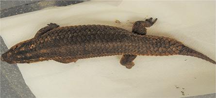
Table 1. Skinks used for cloacoscopy sex identification.
| Skinks | Species | Body weight (g) | Sex (M/F)* | Age (years) |
| 1 | Egernia stokesii | 146 | F | 3 |
| 2 | Egernia hosmeri | 133 | F | 8 |
| 3 | Egernia stokesii | 126 | M | 3 |
| 4 | Egernia stokesii | 116 | M | 3 |
| 5 | Egernia hosmeri | 128 | M | 8 |
| 6 | Egernia hosmeri | 125 | M | 8 |
| 7 | Egernia stokesii | 70 | F | 2 |
| 8 | Egernia stokesii | 72 | M | 2 |
| 9 | Egernia stokesii | 62 | M | 1 |
| 10 | Egernia striolata | 33 | M | 2 |
| 11 | Egernia striolata | 34 | M | 2 |
| 12 | Egernia striolata | 28 | M | 1 |
| 13 | Egernia striolata | 33 | F | 2 |
*Identified by cloacoscopy
The cloacoscopy procedure was performed in compliance with directive 2010/63/EU of the European Parliament and of the Council of the European Union and after ethical approval; with high standards of veterinary care followed. Moreover, in addition, informed consent was signed by the breeders of the skinks. The skinks originated from a professional breeding collection. A complete clinical examination was performed prior to the procedure, with all animals were found to be in good health. The weight of the animals ranged from 28 g to 146 g (85.08 ± 45 g). All skinks were examined during the putative mating season, which is December. A cloacoscopy of the lizards was carried out without sedation or anaesthesia. Animals were gently restrained by using a towel and positioned on an electric heating pad. Cloacoscopy was performed with the rigid endoscope (Hopkins Telescope 2.7 mm diameter, 18 cm length, angle of 30°; Karl Storz Endoskope, Tuttlingen, Germany) and a protecting channel. Images were recorded using an endoscopic camera Telecam (Karl Storz Endoskope, Tuttlingen, Germany) connected to the recording system Telepack-Pal (Karl Storz Endoskope, Tuttlingen, Germany). The endoscope was gently introduced into the cloaca through the vent and advanced 1 cm to 2 cm into the coprodeum. Simultaneously, sterile saline (0.9% NaCl; B. Braun, Melsungen, Germany) was flushed through the protecting channel to clean the optic and dilate the cloacal mucosa. The endoscope was then slowly withdrawn to allow visualization of the main structures of the urodeum and proctodeum.
RESULTS
Cloacoscopy offers excellent imaging of the coprodeum. Retraction of the endoscope in a caudal direction clearly shows the two septa in a female skink, or in the case of a male, one septum. The sphincter of the coprodeum (SC) divides the urodeum from the coprodeum, and it is visible ventrally. Dorsally, two vaginal pouches (VPs) are visible in the urodeum. Compared to other squamata, the VPs are small structures and it is rather difficult to identify them. The urodeum is divided by two septa, one separating the sphincter of the coprodeum from the vaginal pouches (A), and the other (B) separating the two vaginal pouches (Figure 3).
Figure 3. Female tree crevice-skink (E. striolata) urodeum.

All structures of the urodeum are divided by two septa: One separating the sphincter of the coprodeum from the vaginal pouches (A), and the other (B) separating the two vaginal pouches
The urodeum of female skinks has a central-dorsal fold dividing it into two pouches. In males this is absent. The male urodeum is characterised by the presence of one horizontal septum. Ventrally, the SC is visible. Dorsally, two urethral papillae (UP) are visible (Figure 4). Finally, the optic is gently withdrawn from the vent.
Figure 4. Male tree crevice-skink (E. striolata) urodeum.

Dorsally, the sphincter of the coprodeum is visible (A) The urethral papillae (black asterisks) are visible on the dorsal urodeum
DISCUSSION
A number of methods of sex identification in monomorphic lizard species have been investigated (Morris and Alberts 1996; Di Ianni et al. 2015; Stahl and DeNardo 2019), but many have limitations due to a variety of factors (Philips et al. 2016). Some methods are species-specific, such as the use of radiography for identifying mineralized hemibacula in monitor lizards (Varanus spp.; Shea and Reddacliff 1986; Phillips et al. 2016) or ultrasonography for identifying the presence of ovarian follicles in adult female viviparous lizards (Gartrell et al. 2002). In the eastern blue-tongued lizard careful manual eversion of the hemipenes would identify a male, but a negative result cannot exclude a male (Phillips et al. 2016). The study by Phillips et al. (2016) is a non-invasive morphometric technique for sex identification in a population of eastern blue-tongued lizards. However, the degree of differences was subtle, requiring the use of ratios. Di Ianni et al. (2015) used ultrasonography, computed tomography, and radiography as methods for sexing eastern blue-tongued lizards. The presence of osteoderms and skin appendages, however, impedes the visualization of the gonads and hemipenes with ultrasonography in Egernia spp. (Oliveri, personal experience). Other proposed imaging methods such as computed tomography (Morris and Alberts 1996) require sedation and entail prohibitive costs for owners and research institutions. Classical endoscopic methods (coelioscopy) or surgical visualization of gonads are invasive and require anaesthesia and analgesia. Cloacoscopy is a minimally invasive and accurate method of sex identification due to the distinct morphological differences between sexes. Trauth et al. (1987) described in detail the subdivisions within the cloacal complex of the broad-headed skink and the differences between the sexes – the most striking structural modifications occurring in the urodeum. Males had more prominent urogenital papillae than did females, whereas females exhibited well-developed urodeal chambers which were reduced in males.
The existence of similar structures has been described in tegu lizards (Morici et al. 2017; Spadola et al. 2022), male Tiliqua sp. (Spadola et al. 2021) and has been recently confirmed also in another monomorphic genus, the helodermatid lizards (our unpublished results).
Female skinks of the Egernia genus (Egernia hosmeri, E. stokesi, E. striolata) are characterised by the presence of a central-dorsal fold dividing urodeum in two pouches, which is not found in males. Male skinks are characterised by the presence of one septum which divides the chamber of the urodeum into two sub-chambers. Urethral papillae are visible in the dorsal subchamber. These structures and the division of the cloaca into two sub-chambers are major characteristics of the male urodeum in the skinks’ genus Egernia.
In conclusion, it could be stated that this study demonstrates that cloacoscopy is a useful technique for sex identification in a monomorphic skink genus, Egernia, even in small species (E. striolata). Furthermore, cloacoscopy can be considered to be used in the breeding and conservation of endangered skink species.
Funding Statement
Supported by the Internal Creative Agency of the University of Veterinary Sciences Brno (Project No. 2023ITA15).
Conflict of interest
The authors declare no conflict of interest.
REFERENCES
- Di Ianni F, Volta A, Pelizzone I, Manfredi S, Gnudi G, Parmigiani E. Diagnostic sensitivity of ultrasound, radiography and computed tomography for gender determination in four species of lizards. Vet Radiol Ultrasound. 2015 Jan-Feb;56(1):40-5. [DOI] [PubMed] [Google Scholar]
- Fox H. The urinogenital system of reptiles. In: Gans C, editor. Biology of the peptilia. New York: Academic Press; 1977. p. 1-157. [Google Scholar]
- Gartrell BD, Girling JE, Edwards A, Jones SM. Comparison of noninvasive methods for the evaluation of female reproductive condition in a large viviparous lizard, Tiliqua nigrolutea. Zoo Biol. 2002;21(3):253-68. [Google Scholar]
- Hochleithner C, Sharma A. Ultrasonography. In: Divers SJ, Stahl SJ, editors. Mader’s reptile and amphibian medicine and surgery. 3rd ed. St. Louis, USA: Saunders/Elsevier; 2019. p. 543-59. [Google Scholar]
- Morici M, Di Giuseppe M, Oliveri M, Knotek Z, Spadola F. Cloacoscopy sex determination in tegu lizards. In: ICARE, 3rd International Conference on Avian, Herpetological and Exotic Mammal Medicine; 25-29 Mar 2017; Venice, Italy; 2017. p. 544-5. [Google Scholar]
- Morris PJ, Alberts AC. Determination of sex in white-throated monitors (Varanus albigularis), Gila Monsters (Heloderma suspectum) and bearded lizards (H. horridum) using two-dimensional ultrasound imaging. J Zoo Wildl Med. 1996 Sep;27(3):371-7. [Google Scholar]
- O’Malley B. Snakes. In: O’Malley B, editor. Clinical anatomy and physiology of exotic species. Philadelphia: Elsevier Saunders; 2005. p. 77-93. [Google Scholar]
- Oliveri M, Morici M, Novotny R, Bartoskova A, Knotek Z. Cloacoscopy in the horned viper (Vipera ammodytes). Acta Vet Brno. 2016;85(3):251-3. [Google Scholar]
- Oliveri M, Bartoskova A, Spadola F, Morici M, Di Giuseppe M, Knotek Z. Method of semen collection and artificial insemination in snakes. J Exot Pet Med. 2018;27(2):75-80. [Google Scholar]
- Phillips CA, Roffey JB, Hall E, Johnson R. Sex identification in the eastern blue-tongued lizard (Tiliqua scincoides White, ex Shaw, 1790) using morphometrics. Aust Vet J. 2016 Jul;94(7):256-9. [DOI] [PubMed] [Google Scholar]
- Schildger BJ, Wicker R. Sex determination and clinical examination in reptiles using endoscopy. Herp Rev. 1989;20(1):9-10. [Google Scholar]
- Shea GM, Reddacliff GL. Ossifications in the hemipenes of Varanids. J Herpetol. 1986;20(4):566-8. [Google Scholar]
- Spadola F, Sgroi P, Lubian E, Morici M. Cloacal anatomy and sex determination in Tiliqua sp. Herpetol Bull. 2021;156:11-3. [Google Scholar]
- Spadola F, Morici M, Di Giuseppe M, Lubian E, Oliveri M, Bartosovska A, Knotek Z. Evaluation of cloacoscopy for sex determination in the Argentine black and white tegu (Salvator merianae). Herpetol Bull. 2022 Sep;161:1-4. [Google Scholar]
- Stahl SJ, DeNardo DF. Theriogenology. In: Divers SJ, Stahl SJ, editors. Mader’s reptile and amphibian medicine and surgery. 3rd ed. St. Louis, USA: Saunders/Elsevier; 2019. p. 849-93. [Google Scholar]
- Trauth SE, Cooper WE, Vitt LJ, Perrill SA. Cloacal anatomy of the broad-headed skink, Eumeces laticeps, with a description of a female pheromonal gland. Herpetologica. 1987 Dec;43(4):458-66. [Google Scholar]