Abstract
Background
The cyclin-dependent kinase subunit 2 (CKS2) is recognized to have a substantial impact on the pathogenesis and advancement of several malignant neoplasms. Nevertheless, its biological function and prognostic significance in oral squamous cell carcinoma (OSCC) have yet to be thoroughly investigated. Our primary objective was to clarify the contribution of CKS2 in the progression and prognosis of OSCC.
Methods
We first conducted a thorough examination of online databases to investigate the expression of CKS2, and subsequently corroborated our discoveries by analyzing clinical specimens that we collected. According to the clinicopathological data, we then explored the prognostic significance of CKS2. Furthermore, we predicted the role of CKS2 in OSCC progression by employing weighted gene co-expression network analysis (WGCNA) in conjunction with functional enrichment analysis. We conducted functional experiments in vitro to confirm our speculations. Additionally, we explored other potential functions of CKS2 in immune infiltration, tumor mutation burden (TMB), and drug sensitivity. Finally, we established and validated a nomogram that effectively integrated CKS2-related genes and other relevant clinical factors.
Results
Our findings indicated a significant upregulation of CKS2 expression in OSCC tissues compared to normal groups, which was positively associated with poor clinical outcomes. We also predicted and validated the role of CKS2 in promoting proliferation by regulating the cell cycle. Additionally, its upregulation was significantly correlated to enhanced immune cell infiltration, high TMB, and increased sensitivity of anti-tumor agents. Following verification, the nomogram was conducted to quantify an individual’s survival probability.
Conclusions
In general, our study indicates that CKS2 is a novel prognostic biomarker and potential therapeutic target in OSCC.
Keywords: Cyclin-dependent kinase subunit 2 (CKS2), oral squamous cell carcinoma (OSCC), prognosis, immunity, cell proliferation
Highlight box.
Key findings
• Cyclin-dependent kinase subunit 2 (CKS2) is a novel prognostic biomarker and potential therapeutic target in oral squamous cell carcinoma (OSCC).
What is known and what is new?
• CKS2 is significantly upregulated and is known to exert a substantial impact on the pathogenesis and progression of several malignant neoplasms.
• CKS2 is recognized as a novel prognostic biomarker and potential therapeutic target in OSCC, as it promotes cell proliferation through the regulation of the cell cycle, correlates with immune infiltration and immunotherapy, enhances drug sensitivity, and offers a prognostic prediction value.
What is the implication, and what should change now?
• It is imperative to give clinical consideration to the CKS2 expression in patients with OSCC, as it can aid in the prognostication of their condition and optimize the treatment strategy.
Introduction
Oral squamous cell carcinoma (OSCC) is the most prevalent type of oral malignant neoplasms, representing over 90% of all cases. Despite the progress made in diagnostic and therapeutic approaches, the prognosis of OSCC has exhibited minimal advancement in recent decades (1,2). The 5-year survival rate of OSCC is still around 50% because of the high rate of metastasis and recurrence (3). Therefore, it is imperative to unravel the underlying molecular mechanisms and find innovative biomarkers to enhance the prognostic potential of OSCC.
Cyclin-dependent kinase subunit 2 (CKS2), a member of the human CKS family, is located on chromosome 9q22 (4). CKS1, a critical paralog of CKS2, shares 81% identical amino acids with CKS2 but performs distinct functions. CKS2 is a protein that interacts with cyclin-dependent kinases (CDK) and plays vital, conserved roles in the cell cycle (5). Previous research found that CKS2 acts as a crucial nexus between the proteins necessary for meiotic chromosome recombination and those required for homologous chromosome segregation at meiotic anaphase (6). Further studies revealed that CKS2 opposes CKS1 in controlling cyclin A and CDK2 activity, thereby preserving replication reliability (7). Recent evidence showed that CKS2 overlaps the DNA damage response barrier activated by oncoproteins and inhibits programmed cell death (8,9). Over the years, multiple studies have reported high expression levels of CKS2 in various malignant tumors, such as gastric cancer (10), hepatocellular carcinoma (11), pediatric retinoblastoma (12), colorectal cancer (13), prostatic cancer (9), lymphatic cancer (14), bladder cancer (15) and cervical cancer (16). These findings indicate a pivotal role of CKS2 in malignant tumor progression. However, the precise role of CKS2 in OSCC remains to be fully elucidated.
The primary aim of this study was to thoroughly investigate the role of CKS2 in OSCC progression and prognosis. Specifically, we analyzed CKS2 expression in OSCC samples as well as in paired adjacent non-cancerous tissues (ANCT). Meanwhile, we found that CKS2 expression was correlated with the prognosis and some clinical characteristics of OSCC patients. Weighted gene co-expression network analysis (WGCNA) was performed to identify key modules associated with CKS2 expression in OSCC. After that, we conducted Gene Ontology (GO), Kyoto Encyclopedia of Genes and Genomes (KEGG), and Gene Set Enrichment Analysis (GSEA) to deepen our comprehension of the biological functions of CKS2. To confirm the effect of CKS2 on cell proliferation, we carried out functional experiments in OSCC cell lines. Apart from the role in proliferation, we continued to explore other potential functions of CKS2 in immune infiltration, tumor mutation burden (TMB), and drug sensitivity. Basing on the CKS2-related risk score and other clinical factors, we finally constructed and validated a risk model to predict the prognosis of OSCC patients. We present this article in accordance with the REMARK (17) and the TRIPOD reporting checklists (available at https://tcr.amegroups.com/article/view/10.21037/tcr-23-511/rc).
Methods
Clinical samples and OSCC cell lines
Totally 39 OSCC specimens and 25 ANCT specimens were obtained from the Hospital of Stomatology, Sun Yat-sen University. Additionally, we included 46 OSCC samples and five ANCT samples from the tissue chip (HOraC060PG01, Shanghai Outdo Biotech Co., China) in our analysis. The study was conducted in accordance with the Declaration of Helsinki (as revised in 2013). The study was approved by the ethics committee of the Hospital of Stomatology, Sun Yat-sen University (IRB AF/SC-07/v3.0; No. KQEC-2022-15-01) and informed consent was granted by each patient involved in this study.
The human OSCC cell lines CAL33, SCC1, SCC15, and SCC25 cells were utilized in the present study. Specifically, SCC15 and SCC25 cells were procured from America Type Culture Collection. SCC1 cells were obtained from the University of Michigan, and CAL33 cells were provided by the Deutsche Sammlung von Mikroorganismen und Zellkulturen (DSMZ, Germany). The Dulbecco’s modified Eagle’s medium (DMEM, Gibco, USA), which included 10% fetal bovine serum (FBS, WISENT, Montreal, QC, Canada), was used to cultivate CAL33 and SCC1 cells. The SCC25 and SCC15 cell lines were cultured in a medium composed of DMEM/Hams F12 (DMEM/F12, Gibco, USA), supplemented with 400 ng/mL hydrocortisone (MACKLIN, Shanghai, China) and 10% FBS. All the cells were cultured at a temperature of 37 ℃ in a humidified incubator with 5% CO2.
Data acquisition and pre-processing
Patients who met the following criteria were included in the study: (I) histologically verified primary OSCC; (II) sample size in the dataset was more than 70; (III) patients with complete RNA-seq data and survival data. We downloaded RNA-seq, clinical, and TMB data of OSCC patients (306 tumor samples and 30 normal controls) from the The Cancer Genome Atlas (TCGA) database (https://www.cancer.gov/tcga) (18). The RNA-seq data and associated clinical data were obtained from the GEO database, which included data on 45 normal samples and 167 OSCC samples (GSE30784), 29 normal samples and 74 OSCC samples (GSE42743), and 97 OSCC samples (GSE46163). The RNA-seq data was filtered, missing and duplicated data were removed before it was converted to log2(TPM +1). The OSCC samples with complete clinical data in TCGA-OSCC and GSE4743 datasets were included in the development of the Nomogram.
CKS2 differential expression and prognosis correlation
The Gene Expression Profiling Interactive Analysis (GEPIA2) web server (http://gepia2.cancer-pku.cn/) was used to analyse gene expression in tumor and normal samples obtained from the TCGA and Genotype-Tissue Expression (GTEx) databases (19). We performed differential expression analysis and overall survival (OS) analysis of CKS2 by the GEPIA2 on the 33 cancer subtypes in the TCGA pan-cancer datasets. After that, we utilized the R package “limma” to calculate CKS2 mRNA expression between normal specimens and OSCC specimens in GSE30784, GSE42743, and TCGA datasets. The relationships between the mRNA level of CKS2 and several clinical features of the TCGA-OSCC and GSE41613 cohorts were also examined. The OSCC patients were divided into CKS2-high expression group and CKS2-low expression group based on the median value of CKS2 expression. Kaplan-Meier survival analysis was then conducted to compare the OS and PFS in these two groups using the “Survival” package.
WGCNA
The R package “WGCNA” (20) was utilized for the analysis of the top 25% of the most variant genes in the TCGA dataset and GSE30784 dataset. To ensure data integrity, the “goodSamplesGenes” function was implemented. Following the confirmation of the optimal soft threshold value (β), the expression matrix was transformed into multiple modules via the “blockwiseModules” function. Subsequently, we determined the correlation coefficients for CKS2 expression level and eigengene in the module. In conclusion, the pivotal module was identified for further analysis.
Functional enrichment analysis
The GO and KEGG analysis of the hub module gene were performed using the R package “clusterProfiler” (21,22).
We applied the GSEA to analyze the enriched biological functions and pathways between the groups with low and high CKS2 expression (23). Gene sets with FDR and P value less than 0.05 were considered highly enriched.
Immunohistochemistry (IHC) staining
Expression of the CKS2 protein was investigated by immunostaining in OSCC and paired ANCT. After dewaxing, dehydrating, and antigen repairing, we blocked the tissue slices with goat serum (AR0009, BosterBio, China). Next, we incubated the tissue slices overnight at 4 ℃ with a primary antibody against CKS2 (1:100; ab155078, Abcam). The sections were subsequently treated with secondary antibodies for 30 minutes at 25 ℃ and stained with diaminobenzidine (DAB, GK600510, Gene Tech, China). The percentage of positively stained cells (0–100%) and the intensity of tissue staining (0: no staining, 1: weak staining, 2: moderate staining, and 3: strong staining) were estimated, and the staining index (ranging from 0 to 300) was used to calculate the immunoscore (percentage of positive cells multiplied by staining intensity).
Small interfering RNA (siRNA) and lentiviral transfection
CAL33 and SCC25 cells were seeded and incubated for approximately 24 hours until they reached the logarithmic growth phase. Following the product instructions, we applied the Pepmute Transfection Reagent (Signagen, Rockville, MD, USA) to transfect 30 nM CKS2-specific si-RNAs or negative controls (si-NC) in each well. The two independent siRNA sequences are: si-CKS2#1: 5'-GAGTCTAGGCTGGGTTCATTA-3', si-CKS2#2: 5'-CTTGGTGTCCAACAGAGTCTA-3'.
The lentiviral vector pCDH-CMV-MCS carrying the CKS2 coding sequence was employed to achieve stable overexpression of CKS2. According to the product instructions, cotransfection of the 293T cells were achieved using the lentivirus packaging plasmids psPAX2, pMD2G (GenePharma, China), and CKS2 plasmids. An empty vector was used as the control group. After 48 hours of transfection, the supernatant containing virus was collected, filtered, concentrated, and then transfected into SCC15 and SCC1 cells with 3 µg/mL Polybrene (40804ES76, Yeasen, China). After infection, puromycin (P8230, Solarbio, China) was used to screen the cells for 14 days (SCC1: 2 µg/mL, SCC15: 2.5 µg/mL).
Western blot
The cells were lysed using the RIPA buffer (CW2333S, CWBIO, China). The proteins were measured and distinguished through the process of electrophoresis on a 15% SDS-PAGE gel. Following this, the proteins were transferred onto PVDF membranes (ISEQ00010, Millipore, USA). After blocking them with 5% bovine serum albumin (0332, Amresco) at room temperature for one hour, the membranes were incubated with primary antibodies against CKS2 (1:2,000; ab155078, Abcam) and β-actin (1:1,500; 30102ES60, Yeasen, China) overnight at 4 ℃, followed by incubation with horseradish peroxidase-conjugated secondary antibodies (GK600510, Gene Tech, China) for one hour at room temperature. Finally, the bands were quantified using Image-J (https://imagej.nih.gov/ij/download.html) after exposure to a chemiluminescent HRP substrate.
RNA extraction and quantitative real-time PCR (qRT-PCR)
According to the guidelines provided by the manufacturer, we extracted the RNA using the RNAzol reagent (RN190, Molecular Research Center, USA) and then reverse transcribed it to cDNA through utilization of the the HifairTM III 1st Strand cDNA Synthesis SuperMix (11141ES60, Yeasen, China). In order to conduct the qPCR reactions, we utilized the SYBR Green Master Mix (11201ES08; Yeasen, China) in the LightCycler 96 System (Roche, Basil, Switzerland). The 2−ΔΔCt method was used to determine the relative RNA expression of CKS2 compared to the control. The following are the primer sequences we utilized: β-ACTIN forward: CTACCTCATGAAGATCCTCACCGA, reverse: TTCTCCTTAATGTCACGCACGATT; CKS2 forward: TTCGACGAACACTACGAGTACC; reverse: GGACACCAAGTCTCCTCCAC.
Cell counting kit-8 (CCK-8) assay
CAL33, SCC25, SCC1, and SCC15 cells were seeded into 96-well plates at a density of 2,000 cells per well. The CCK-8 (40203ES80, Yeasen, China) was utilized to quantify the proliferation of cells at 0, 24, 48, 72, and 96 hours. The growth curve was eventually produced using the absorbance values obtained from a microplate reader (Bio-Rad, USA) operating at 450 nm.
Colony formation assay
Following the corresponding treatment, 800 cells of CAL33, SCC25, SCC1, and SCC15 were seeded in each well of 6-well plates. Following a 10–14-day culture period to facilitate colony formation, the cells were immobilized with methanol and subsequently stained with crystal violet. Upon examination under a microscope, the colonies were quantified.
Immune infiltration and TMB analysis
The CIBERSORT algorithm was employed to calculate the abundances of 22 immune cells in TCGA-OSCC cohort. Subsequently, the association between the abundances of these 22 immune cells and CKS2 expression was examined. To investigate the correlation between CKS2 expression and TMB in OSCC, the Spearman’s correlation was computed in the TMB analysis.
Drug sensitivity analysis
To estimate the IC50 value of frequently used anti-tumor agents in the TCGA-OSCC cohort, tumor drug response data were collected from the Genomics of Drug Sensitivity in Cancer (GDSC) (https://www.cancerRxgene.org) (24). The R package “oncopredict” was applied to explore the relationship between CKS2 expression and drug sensitivity (25). P value <0.05 was considered statistically significant.
Establishment of a risk model based on prognostic genes associated with CKS2
By conducting differential analysis on CKS2 expression and univariate Cox regression analysis, the prognostic genes related to CKS2 were identified in the TCGA-OSCC cohort. Moreover, we seek out those genes with optimal performance and calculate their coefficients using least absolute shrinkage and selection operator (LASSO) Cox regression algorithm. Following this, the risk score for each sample was calculated by multiplying the expression of each gene by its respective regression coefficient (β), and adding them together. Patients with OSCC were divided into high- and low-risk groups according to the median value of the risk score.
Development and verification of the nomogram
To estimate the predictive accuracy of the risk score in the TCGA-OSCC cohort, we calculated the area under the ROC curve (AUC) at 1, 3, and 5 years. Additionally, the prognostic value of the risk score and other clinical features (including age, gender, stage, T, and N) was evaluated using univariate and multivariate COX regression analyses. Based on the risk score and these clinical factors, we formulated a nomogram utilizing the R package “rms” nomogram to predict the 1-, 3-, and 5-year survival probabilities of OSCC patients. The calibration curves and ROC were utilized to verified it. Furthermore, we validated the nomogram in the external validation dataset GSE42743 using the same methodology.
Statistical analysis
R software (version 4.2.2, https://www.Rproject.org) and GraphPad Prism 9.0 software were used for statistical analysis in this study. Wilcoxon test was employed to compare the expression of CKS2 among the two groups. Correlation assessment was conducted by calculating Pearson’s r value. Mann-Whitney U, Kruskal-Wallis, and Chi-square tests were used to access the association between CKS2 expression and clinicopathological parameter. One way ANOVA and student’s t-test were utilized to compare the control group and the experimental group. A P value of 0.05 or less was considered statistically significant.
Results
CKS2 is overexpressed in OSCC and correlated with poor prognosis
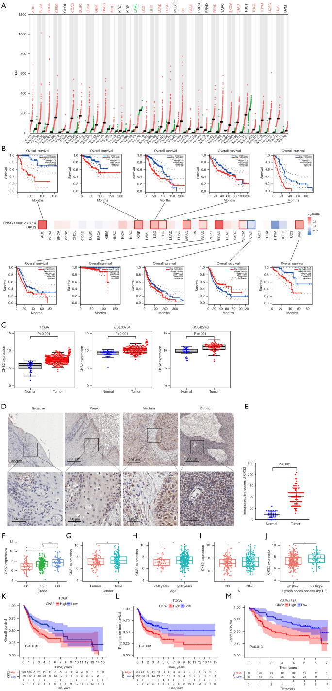
Firstly, we used the GEPIA web server to explore the expression level and the prognostic significance of CKS2 in a comprehensive pan-cancer assessment. Our analysis encompassed RNA-seq and clinical data of 33 cancer subtypes gathered from the GTEx and TCGA databases. Notably, in comparison to normal tissue, we observed a substantially elevated expression level of CKS2 in tumor tissue across the most subtypes (Figure 1A). Additionally, through our OS analysis, we concluded that high CKS2 expression was a predictor of a poor prognosis in adrenocortical cancer (ACC), kidney papillary cell carcinoma (KIRP), brain lower grade glioma (LGG), liver hepatocellular carcinoma (LIHC), mesothelioma (MESO), pancreatic adenocarcinoma (PAAD), prostate adenocarcinoma (PRAD), skin cutaneous melanoma (SKCM), and uveal melanoma (UVM) (Figure 1B). Based on our pan-cancer analysis, we proceeded to study CKS2 expression specifically in OSCC. We compared data obtained from TCGA, GSE30784, and GSE42743 datasets, which demonstrated a significant upregulation of CKS2 in OSCC samples when compared with normal tissue samples (Figure 1C, P<0.001). In addition, the IHC staining analysis of the clinical samples corroborated our findings (Figure 1D,1E, P<0.001).
Figure 1.
CKS2 was overexpressed in OSCC tissues and associated with poor prognosis. (A) Differential expression of CKS2 in 33 different tumor tissues and paired normal tissues from TCGA and GTEx databases. Each dot represents the expression of samples. (B) The prognostic impact of CKS2 expression level based on the survival heatmap, showing significance in ACC, KIRP, LGG, LIHC, MESO, PAAD, PRAD, SKCM, STAD and UVM. (C) Differential analysis of CKS2 expression between OSCC and normal samples in OSCC-TCGA, GSE30784, and GSE42743 datasets. (D) Representative images of immunohistochemistry staining for CKS2 in ANCT and OSCC tissues. Magnification at 50× (up) and 200× (down). (E) Immunohistochemical staining score of CKS2 in ANCT (n=30) and OSCC (n=85) tissues. (F-J) The correlation between CKS2 expression and clinical features in TAGA-OSCC cohort, including the grade, gender, age, N, and lymph nodes positive (by hematoxylin-eosin staining). (K-L) Kaplan-Meier survival curves of overall survival (K) and progression free survival (L) based on TCGA-OSCC patients with high- and low-expression CKS2. (M) Kaplan-Meier survival curves of overall survival based on GSE41613 dataset. Differences between the two groups were compared using a log-rank test. *, P<0.05; **, P<0.01; ***, P<0.001. CKS2, cyclin-dependent kinase subunit 2; TCGA, The Cancer Genome Atlas; N, node; HE, hematoxylin and eosin; OSCC, oral squamous cell carcinoma; GTEx, Genotype-Tissue Expression; ACC, adrenocortical cancer; KIRP, kidney papillary cell carcinoma; LGG, brain lower grade glioma; LIHC, liver hepatocellular carcinoma; MESO, mesothelioma; PAAD, pancreatic adenocarcinoma; PRAD, prostate adenocarcinoma; SKCM, skin cutaneous melanoma; STAD, stomach cancer; UVM, uveal melanoma; ANCT, adjacent non-cancerous tissues.
We separated the 306 OSCC samples chosen from the TCGA dataset into groups with high and low CKS2 mRNA expression levels, using the median expression level of CKS2 as the dividing point. Our findings indicate that elevated CKS2 expression is notably correlated with higher grade (Figure 1F, P<0.01), male gender (Figure 1G, P<0.05), older age (Figure 1H, P<0.05), higher N stage (Figure 1I, P<0.05), and greater positivity of lymph nodes (Figure 1J, P<0.05).
Considering that CKS2 expression was found to be high in OSCC and this high expression was associated with unfavorable clinical characteristics, we proceeded to investigate the prognostic significance of CKS2 in both TCGA-OSCC and GSE41613 datasets. Through Kaplan-Meier survival analysis of OS and PFS, we observed that elevated CKS2 expression levels were significantly linked to a poorer prognosis (Figure 1K, P=0.0018; Figure 1L, P=0.001; Figure 1M, P=0.013).
Construction of a weighted co‑expression network and the detection of the key module associated with CKS2
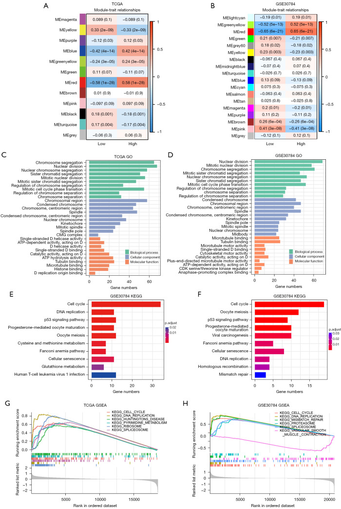
Using the WGCNA method, we aimed to uncover gene modules that cooperatively express and investigate their relationship with CKS2 expression. Initially, we produced clustering dendrograms and eliminated outliers before selecting a power of β=5 to attain the desired scale-free topology (Figure S1A,S1B). As the clustering dendrogram presented, we merged modules with an eigenvalue similarity >0.75 for further examination (Figure S1C,S1D). The WGCNA algorithm recognized 18 modules and 12 modules in GSE30784 and TCGA-OSCC cohorts, respectively. In both datasets, the red module with the largest correlation coefficient with CKS2 (r=0.58, P=1×10⁻²⁸; r=0.65, P=6×10⁻²¹) was recognized as the key module for further analysis (Figure 2A,2B and website: https://cdn.amegroups.cn/static/public/tcr-23-511-1.xlsx, website: https://cdn.amegroups.cn/static/public/tcr-23-511-2.xlsx).
Figure 2.
Identification of the key module relayed to CKS2 and functional enrichment analysis in TCGA-OSCC and GSE30784 datasets. (A,B) The correlation coefficients and P values of module-trait relationships Each row corresponds to a module eigengene. (C,D) GO analysis for CKS2-realated key module. (E,F) KEGG enrichment analysis for CKS2-realated key module. (G,H) GSEA results show the significant enriched pathway based on CKS2 expression. CKS2, cyclin-dependent kinase subunit 2; TCGA, The Cancer Genome Atlas; OSCC, oral squamous cell carcinoma; GO, Gene Ontology; KEGG, Kyoto Encyclopedia of Genes and Genomes; GSEA, The Gene Set Enrichment Analysis.
Functional enrichment analysis
We conducted KEGG enrichment and GO annotation analyses on the red module to explore the pathways associated with CKS2 in OSCC. The GO analysis results demonstrated that genes in the red module were significantly correlated with cell division (Figure 2C,2D and website: https://cdn.amegroups.cn/static/public/tcr-23-511-3.xlsx, website: https://cdn.amegroups.cn/static/public/tcr-23-511-4.xlsx). The KEGG analysis results indicated that genes in the red module were predominantly enriched in the cell cycle (Figure 2E,2F and website: https://cdn.amegroups.cn/static/public/tcr-23-511-5.xlsx, website: https://cdn.amegroups.cn/static/public/tcr-23-511-6.xlsx).
Furthermore, we conducted GSEA between groups with high and low CKS2 expression using the R package “clusterProfiler”. A similar outcome was obtained from the analysis, which revealed that group with high CKS2 expression was primarily enriched with “Cell Cycle” and “DNA Replication” (Figure 2G,2H and website: https://cdn.amegroups.cn/static/public/tcr-23-511-7.xlsx, website: https://cdn.amegroups.cn/static/public/tcr-23-511-8.xlsx).
CKS2 promotes the proliferation of OSCC cells
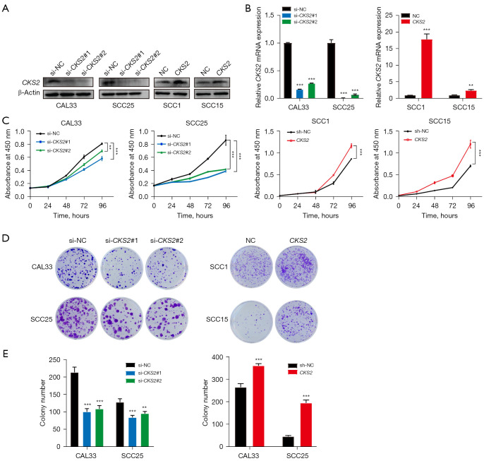
Based on the aforementioned analysis, our hypothesis suggests that the regulation of cell cycle by CKS2 promotes the proliferation of OSCC cells. To validate our conjecture, we developed cellular models for CKS2 knockdown and overexpression. We transfected siRNA, which was specifically designed to target CKS2, into CAL33 and SCC25 cell lines to suppress CKS2 expression. Additionally, we constructed a lentiviral vector that carries the CKS2 coding sequence to stably overexpress CKS2 in SCC1 and SCC15. The knockdown and overexpression effects of CKS2 were determined using PCR and Western blotting (Figure 3A,3B). Following this, we conducted colony formation and CCK-8 assays to assess the cell growth. The results indicate that downregulation of CKS2 considerably reduced cell proliferation and clonogenicity in CAL33 and SCC25 cells. Conversely, upregulation of CKS2 significantly amplified cell proliferation and clonogenicity in SCC1 and SCC15 (Figure 3C-3E).
Figure 3.
CKS2 promotes proliferation of OSCC by regulating cell cycle. (A,B) Western blot and RT-qPCR were used to detect the CKS2 expression after transfection in OSCC cell lines. (C) CCK-8 assay showed that down regulation of CKS2 expression inhibited the proliferation of CAL33 and SCC25 cells, up-regulation of CKS2 expression promoted the proliferation of SCC1 and SCC15 cells. (D,E) Clone formation assay showed that down regulation of CKS2 expression inhibited the proliferation of CAL33 and SCC25 cells, up-regulation of CKS2 expression promoted the proliferation of SCC1 and SCC15 cells. Cells were stained with crystal violet. Magnification at 1×. **, P<0.01; ***, P<0.001. OSCC, oral squamous cell carcinoma; CKS2, cyclin-dependent kinase subunit 2; TCGA, The Cancer Genome Atlas; RT-qPCR, quantitative reverse transcription PCR; CCK-8, cell counting kit-8; NC, negative control.
Immunological significance of CKS2 in OSCC
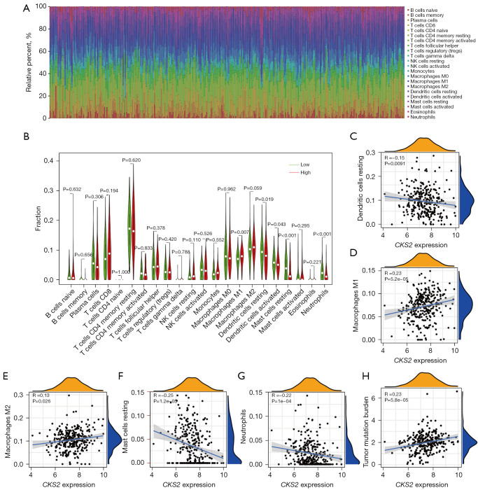
Apart from its role in proliferation, we further investigated other potential functions of CKS2 in the regulation of OSCC. Initially, we employed the CIBERSORT algorithm to illustrate the presence of 22 immune cells infiltrating the TCGA-OSCC cohort, wherein significant differences were observed between the high and low CKS2 expression groups (Figure 4A,4B). Correlation analyses were also conducted between CKS2 expression and the 22 immune cells (Figure 4C-4G). Our findings suggest that CKS2 has a positive correlation with the infiltration of macrophage M1 (R=0.23, P=5.2×10⁻⁵) and macrophage M2 (R=0.13, P=0.026), while exhibiting a negative correlation with immune infiltration of dendritic cells resting (R=−0.15, P=0.0091), mast cells resting (R=−0.25, P=1.2×10⁻⁵), and neutrophils (R=−0.22, P=1×10⁻⁴).
Figure 4.
Immunological significance of CKS2 in OSCC. (A) The distribution of infiltrating immune cells in 306 TCGA-OSCC samples based on CKS2 expression. (B) The differences in the infiltrating levels of 22 immune cells between the high- and low-CKS2 expression groups. (C-G) The correlations between the CKS2 expression and the enrichment of five core immune cells. (H) The correlations between the CKS2 expression and the tumor mutation burden. CKS2, cyclin-dependent kinase subunit 2; OSCC, oral squamous cell carcinoma; TCGA, The Cancer Genome Atlas.
Emerging evidence shows that TMB is a novel prognostic biomarker for immunotherapy in various cancers. We collected TMB data from the TCGA-OSCC cohort and included it in the analysis along with CKS2 expression. As a result, high expression of CKS2 was positively related to TMB (Figure 4H, R=0.23, P=5.8×10⁻⁵).
Drug sensitivity analysis
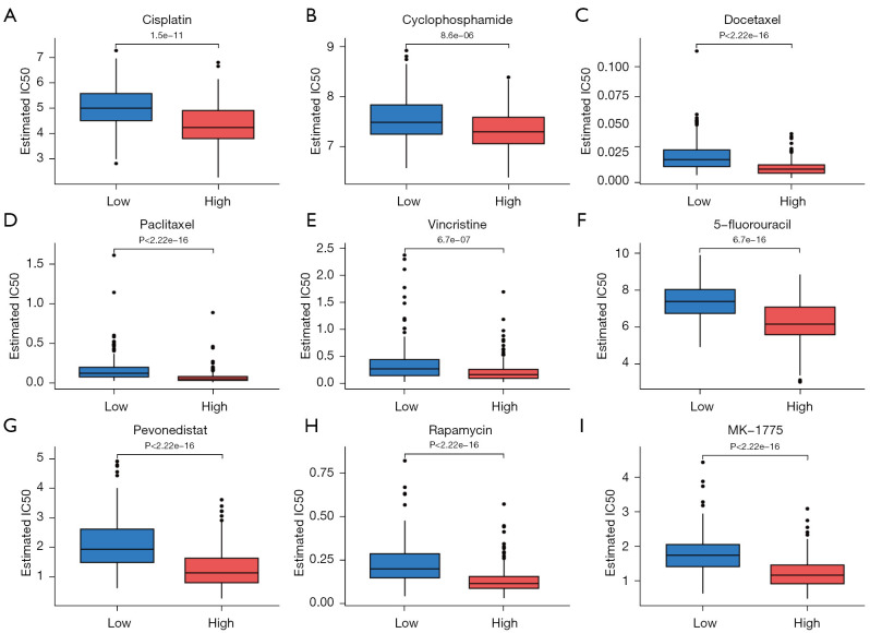
Antitumor drugs play a vital role in OSCC therapy. Hence, we explored the potential association between CKS2 expression level and anti-tumor drug sensitivity. Following the NCCN guidelines, we focused on the recommended therapeutic drugs, which included cisplatin, carboplatin, 5-FU, paclitaxel, docetaxel, hydroxyurea, cetuximab, vincristine, cyclophosphamide, and methotrexate (26). Our findings indicated that a high level of CKS2 expression was significantly correlated with a low IC50 of cisplatin, 5-FU, paclitaxel, vincristine, cyclophosphamide, and docetaxel (Figure 5A-5F). Furthermore, a high level of CKS2 expression was also significantly correlated with a low IC50 of pevonedistat, rapamycin, and MK-1775, suggesting that they could be potentially effective treatments (Figure 5G-5I).
Figure 5.
Drug prediction of IC50 differences in CKS2 high and low group based on the GDSC database. (A-F) High level of CKS2 expression was significantly correlated with low IC50 of recommended therapeutic drugs in OSCC. (G-I) Other commonly used anti-tumor drugs most associated with the expression level of CKS2. IC50, half maximal inhibitory concentration; CKS2, cyclin-dependent kinase subunit 2; GDSC, Genomics of Drug Sensitivity in Cancer; OSCC, oral squamous cell carcinoma.
A nomogram based on CKS2-related genes
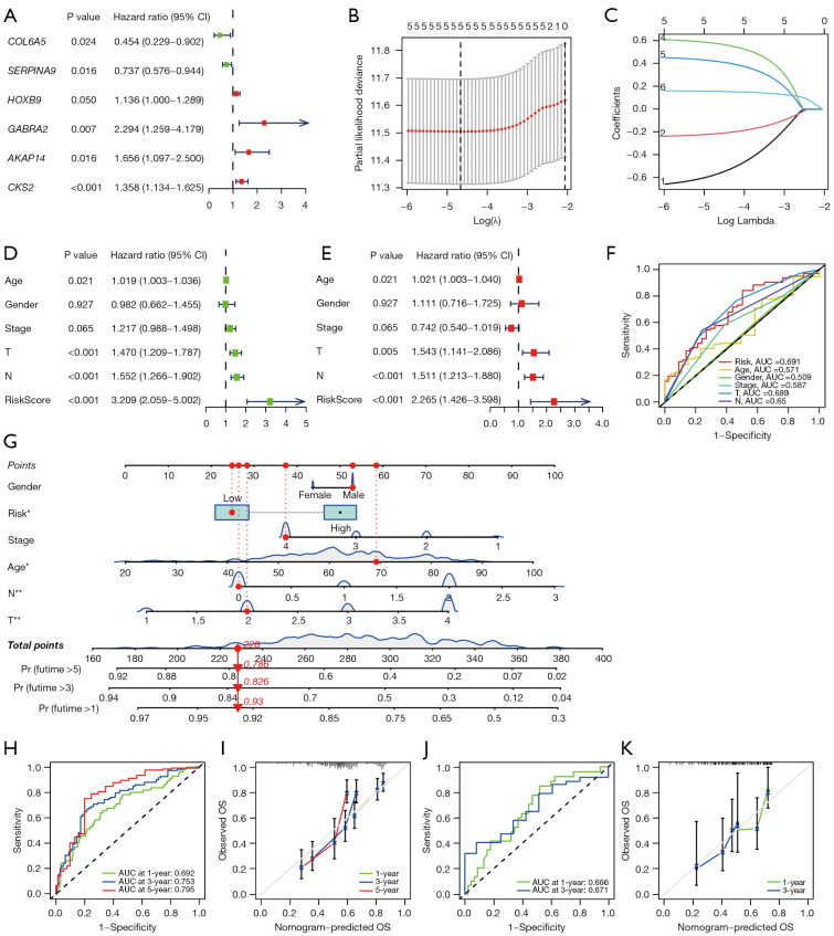
The study above showed that CKS2 has significant functions in OSCC, leading us to investigate its clinical predictive value. By utilizing a cutoff of logFC >1 and an adjusted P value of <0.05, we detected 110 genes related to CKS2 expression level in the TCGA-OSCC cohort (website: https://cdn.amegroups.cn/static/public/tcr-23-511-9.xlsx). Univariate Cox regression models indicated that six genes, namely COL6A5 (HR =0.454, 95% CI: 0.229–0.902, P=0.024), SERPINA9 (HR =0.737, 95% CI: 0.576–0.944, P=0.016), HOXB9 (HR =1.136, 95% CI: 1.000–1.289, P=0.050), GABRA (HR =2.294, 95% CI: 1.259–4.179, P=0.007), AKAP14 (HR =1.656, 95% CI: 1.097–2.500, P=0.016), and CKS2 (HR =1.358, 95% CI: 1.134–1.625, P<0.001), were linked to OSCC patients’ prognosis (Figure 6A). LASSO regression analysis determined five CKS2-related genes to create a prognostic gene model (Figure 6B,6C). The CKS2-derived risk score = (−0.5676* COL6A5 expression) + (−0.2141* SERPINA9 expression) + (0.5742* GABRA expression) + (0.4183* AKAP14 expression) + (0.1560* CKS2 expression). 253 samples in TCGA-OSCC and 74 samples in GSE42743 were selected to construct the nomogram. Our research in univariate Cox regression analysis further revealed that the risk score (HR =3.209, 95% CI: 2.059–5.002, P<0.001), T (HR =1.470, 95% CI: 1.209–1.787, P<0.001), N (HR =1.552, 95% CI: 1.266–1.902, P<0.001), and age (HR =1.019, 95% CI: 1.003–1.036, P=0.021) were significantly correlated with OSCC prognosis (Figure 6D). A multivariate Cox regression analysis was conducted, which indicated that the risk score (HR =2.265, 95% CI: 1.426–3.598, P<0.001), T (HR =1.543, 95% CI: 1.141–2.086, P=0.005), N (HR =1.511, 95% CI: 1.213–1.880, P<0.001), and age (HR =1.021, 95% CI: 1.003–1.040, P=0.026) were independent prognostic indicators of OSCC (Figure 6E). Notably, the risk score based on CKS2 exhibited greater predictive potential for OSCC compared to conventional clinical indicators such as age, gender, T, N, and stage (Figure 6F). Finally, we developed a nomogram that effectively integrates the risk score and related clinical factors, within the training set, to predict the OS of OSCC patients (Figure 6G). The points of each factor add up to the total points, which indicate 1-, 3-, and 5-year survival probabilities. The ROC curves confirmed the prediction accuracy of the nomogram in predicting OSCC patients’ survival outcomes (Figure 6H). And the calibration curves revealed that the predicted OS results were highly consistent with actual results for the 1-, 3-, and 5-year periods (Figure 6I). In addition, data from GSE42743 was utilized to validate the nomogram, which indicated the nomogram possessed the potential to function as a quantitative tool for predicting OS in patients with OSCC (Figure 6J,6K).
Figure 6.
Construction and validation of a nomogram based on CKS2-related genes. (A) Forest plot of univariate Cox regression analysis of 6 hub genes associated with CKS2 expression and OS in TCGA-OSCC cohort. (B,C) LASSO regression analysis screened 5 CKS2-related genes. (D,E) Uni- and multivariate Cox regression models are conducted to uncover the association of clinical features and CKS2-derived risk score with OSCC survival outcome. (F) The ROC curves show the predictive power of CKS2-derived risk score and clinical features on the prognosis of OSCC patients. (G) Construction of prognostic nomogram for predicting OS of OSCC patients based on TCGA dataset. (H,I) The ROC curves and calibration curves for 1-, 3-, and 5-year in the TAGA-OSCC training set. (J,K) The ROC curves and calibration curves for 1- and 3-year in the GSE42743 testing set. *, P<0.05; **, P<0.01. T, tumor; N, node; AUC, area under the curve; CI, confidence interval; OS, overall survival; CKS2, cyclin-dependent kinase subunit 2; TCGA, The Cancer Genome Atlas; OSCC, oral squamous cell carcinoma; LASSO, least absolute shrinkage and selection operator; ROC, receiver operating characteristic.
Discussion
As one of the most common malignant tumors globally, OSCC is typically treated with surgery, chemotherapy, radiotherapy, or a combination of these modalities (26). Unfortunately, despite these efforts, around 50% of OSCC patients eventually succumb to recurrence or metastasis (3). Given the high heterogeneity of OSCC, identifying new biomarkers could lead to significant clinical benefits for OSCC patients, as evidenced by recent studies (27). CKS2, a protein that has been shown to be highly expressed and correlated with unfavourable prognosis in various cancers such as colorectal cancer, epithelial ovarian cancer, and osteosarcoma (28-30), is among the promising candidates. However, the precise roles of CKS2 in OSCC remain unclear, which prompted us to investigate it. Our study unveiled that CKS2 was overexpressed in OSCC samples and associated with unfavorable prognosis. Through WGCNA and functional enrichment analysis, we discovered that CKS2 may promote OSCC cell proliferation by modulating the cell cycle, a finding that was validated through functional assays conducted in OSCC cell lines. Additionally, we examined the roles of CKS2 in immune infiltration, TMB, and drug sensitivity. Finally, an established and validated risk model based on prognostic CKS2-related genes was developed in light of the abundant and significant functions played by CKS2.
The pan-cancer analysis findings indicated that CKS2 was highly expressed in most subtypes, and negatively correlated with OS, consistent with studies on adrenocortical carcinoma (31), lower-grade glioma (32), hepatocellular carcinoma (11), pancreatic adenocarcinoma (33), and skin cutaneous melanoma (34). Both several independent datasets and the IHC assay data confirmed upregulation of CKS2 in OSCC samples. Notably, the IHC assay showed that CKS2 was prominently stained in the region close to the stratum basale of normal epithelial tissue. It can be hypothesized that this result could be attributed to the actively proliferating cells in the stratum basale. The Kaplan-Meier survival analysis demonstrated that increased levels of CKS2 expression were linked to unfavorable OS and PFS. Despite the lack of statistical significance between CKS2 expression and the OS rate of head and neck squamous cell carcinoma (HNSC), it was postulated that this outcome might have resulted from the limited sample size and non-OSCC HNSC. Additional analysis revealed a significant association between high CKS2 expression level and older age, male gender, higher grade, and greater lymph node positivity—all of which are commonly observed adverse clinical features in carcinoma.
To investigate the role of CKS2 in OSCC, we conducted WGCNA and functional enrichment analyses in GSE30784 and TCGA-OSCC cohorts. The results of GSEA and KEGG analyses showed that CKS2 mainly activates cell cycle and DNA replication pathways, which was consistent with the findings of GO analysis that showed enrichment in chromosome segregation and nuclear division of hub module genes. Recently, CKS2 was found to induce cell cycle progression and cell proliferation through downregulating p21, p53, and PTEN in non-small cell lung cancer (35). A study has reported that the interaction between CDK2 and CKS1 or CKS2 provides cells with partial resistance to inhibitory tyrosine phosphorylation mediated by the intra-S-phase checkpoint, enabling them to continue DNA replication even in the presence of replicative stress (8). Several studies have shown that CKS2 is essential for cell proliferation (12,33,36). These findings indicated that upregulation of CKS2 might promote OSCC growth and proliferation by promoting the cell cycle. Our in vitro experiments confirmed that downregulation of CKS2 reduced the proliferation and clonogenicity of OSCC cells, while overexpression of CKS2 significantly enhanced them. To summarize, CKS2 potentially expedites the proliferation of OSCC cells through its promotion of the cell cycle.
Substantial evidence suggests that the tumor microenvironment (TME) is widely recognized as a critical contributor to cancer initiation and promotion of tumor growth (37). To gain further insight into the potential effects of CKS2 on OSCC, we conducted an immune infiltration analysis on the TCGA-OSCC cohort. The results revealed that CKS2 expression was positively correlated with tumor-associated macrophages (TAMs), including Macrophage M1 and M2, and negatively associated with the infiltration of resting dendritic cells, resting mast cells, and neutrophils. It is worth noting that TAMs are known to contribute to tumor initiation, progression, angiogenesis, and metastasis (38,39). Recent studies have shown that targeting TAMs enhances immunotherapy (40-42). In light of these findings, CKS2 is probably involved in the formation of the immune-tolerant microenvironment. Besides, CKS2 may activate dendritic cells and mast cells by regulating the cell cycle.
Moreover, our investigation revealed that elevated CKS2 expression in the TCGA-OSCC cohort was positively correlated with TMB, which has been shown to be a valuable biomarker for immunotherapy in certain cancer types (43). While additional verification is required, it is plausible that CKS2 has multifaceted roles within the OSCC TME and is linked to the effectiveness of the antitumor immune response.
Due to the use of genomics to predict medication responses and the development of precise gene editing technologies, new evidence points to intriguing potential in the search for improved possible treatments for OSCC patients (27). The analysis of drug sensitivity revealed that TCGA-OSCC patients with high CKS2 expression were sensitive to commonly used OSCC therapeutic agents, including cisplatin, 5-FU, paclitaxel, vincristine, cyclophosphamide, and docetaxel. From this perspective, CKS2 may serve as a biomarker to identify the most sensitive therapeutic medications, thus minimizing adverse effects of anti-tumor treatments and improving OS in OSCC patients.
Clinical prognostic models are already widely used for many human diseases (44-46). However, there were methodological differences in model development and validation (47). In accordance with the REMARK reporting checklist and TRIPOD checklist, we finally established a nomogram based on CKS2-related genes and other clinical factors from the TCGA-OSCC cohort. In 1-, 3-, and 5-year AUC and calibration analyses, the nomogram presents customized and precise survival prediction results. Furthermore, we applied the GSE42743 dataset for further validation. Due to the limitation of sample size and follow-up time, we only performed 1- and 3-year analyses, which showed the same results.
However, there are still some limitations in the study. Considering the nomogram was constructed and verified based on retrospective data from public databases, it is necessary to collect additional prospective clinical data for validation. Additionally, the role of CKS2 in cell cycle regulation, TMB, and drug sensitivity necessitates further validation through a comprehensive set of cellular experiments.
Conclusions
In summary, our research demonstrated that CKS2 was upregulated and associated with poor prognosis in OSCC. Moreover, CKS2 influences the malignant progression and treatment of OSCC through various mechanisms, including cell cycle, proliferation, immune infiltration, TMB, and drug sensitivity. All these findings indicated that CKS2 was a novel prognostic biomarker and potential therapeutic target in OSCC.
Supplementary
The article’s supplementary files as
Acknowledgments
Funding: This research was funded by the National Natural Science Foundation of China (Nos. 81874128, and 82072994), Sun Yat-sen University Clinical Research 5010 Program (No. 2015018).
Ethical Statement: The authors are accountable for all aspects of the work in ensuring that questions related to the accuracy or integrity of any part of the work are appropriately investigated and resolved. The study was conducted in accordance with the Declaration of Helsinki (as revised in 2013). The ethics committee at the Hospital of Stomatology, Sun Yat-sen University granted approval for this study (No. KQEC-2022-15-01). Informed consent was granted by each patient involved in this study.
Footnotes
Reporting Checklist: The authors have completed the REMARK and TRIPOD reporting checklists. Available at https://tcr.amegroups.com/article/view/10.21037/tcr-23-511/rc
Data Sharing Statement: Available at https://tcr.amegroups.com/article/view/10.21037/tcr-23-511/dss
Peer Review File: Available at https://tcr.amegroups.com/article/view/10.21037/tcr-23-511/prf
Conflicts of Interest: All authors have completed the ICMJE uniform disclosure form (available at https://tcr.amegroups.com/article/view/10.21037/tcr-23-511/coif). The authors have no conflicts of interest to declare.
References
- 1.Bray F, Ferlay J, Soerjomataram I, et al. Global cancer statistics 2018: GLOBOCAN estimates of incidence and mortality worldwide for 36 cancers in 185 countries. CA Cancer J Clin 2018;68:394-424. 10.3322/caac.21492 [DOI] [PubMed] [Google Scholar]
- 2.Warnakulasuriya S. Global epidemiology of oral and oropharyngeal cancer. Oral Oncol 2009;45:309-16. 10.1016/j.oraloncology.2008.06.002 [DOI] [PubMed] [Google Scholar]
- 3.Zeng H, Chen W, Zheng R, et al. Changing cancer survival in China during 2003-15: a pooled analysis of 17 population-based cancer registries. Lancet Glob Health 2018;6:e555-67. 10.1016/S2214-109X(18)30127-X [DOI] [PubMed] [Google Scholar]
- 4.Demetrick DJ, Zhang H, Beach DH. Chromosomal mapping of the human genes CKS1 to 8q21 and CKS2 to 9q22. Cytogenet Cell Genet 1996;73:250-4. 10.1159/000134349 [DOI] [PubMed] [Google Scholar]
- 5.Pines J. Cell cycle: reaching for a role for the Cks proteins. Curr Biol 1996;6:1399-402. 10.1016/S0960-9822(96)00741-5 [DOI] [PubMed] [Google Scholar]
- 6.Spruck CH, de Miguel MP, Smith AP, et al. Requirement of Cks2 for the first metaphase/anaphase transition of mammalian meiosis. Science 2003;300:647-50. 10.1126/science.1084149 [DOI] [PubMed] [Google Scholar]
- 7.Frontini M, Kukalev A, Leo E, et al. The CDK subunit CKS2 counteracts CKS1 to control cyclin A/CDK2 activity in maintaining replicative fidelity and neurodevelopment. Dev Cell 2012;23:356-70. 10.1016/j.devcel.2012.06.018 [DOI] [PMC free article] [PubMed] [Google Scholar]
- 8.Liberal V, Martinsson-Ahlzén HS, Liberal J, et al. Cyclin-dependent kinase subunit (Cks) 1 or Cks2 overexpression overrides the DNA damage response barrier triggered by activated oncoproteins. Proc Natl Acad Sci U S A 2012;109:2754-9. 10.1073/pnas.1102434108 [DOI] [PMC free article] [PubMed] [Google Scholar]
- 9.Lan Y, Zhang Y, Wang J, et al. Aberrant expression of Cks1 and Cks2 contributes to prostate tumorigenesis by promoting proliferation and inhibiting programmed cell death. Int J Cancer 2008;123:543-51. 10.1002/ijc.23548 [DOI] [PMC free article] [PubMed] [Google Scholar]
- 10.Tanaka F, Matsuzaki S, Mimori K, et al. Clinicopathological and biological significance of CDC28 protein kinase regulatory subunit 2 overexpression in human gastric cancer. Int J Oncol 2011;39:361-72. [DOI] [PubMed] [Google Scholar]
- 11.Shen DY, Fang ZX, You P, et al. Clinical significance and expression of cyclin kinase subunits 1 and 2 in hepatocellular carcinoma. Liver Int 2010;30:119-25. 10.1111/j.1478-3231.2009.02106.x [DOI] [PubMed] [Google Scholar]
- 12.Chen M, Zhao Z, Wu L, et al. E2F1/CKS2/PTEN signaling axis regulates malignant phenotypes in pediatric retinoblastoma. Cell Death Dis 2022;13:784. 10.1038/s41419-022-05222-9 [DOI] [PMC free article] [PubMed] [Google Scholar]
- 13.Jung Y, Lee S, Choi HS, et al. Clinical validation of colorectal cancer biomarkers identified from bioinformatics analysis of public expression data. Clin Cancer Res 2011;17:700-9. 10.1158/1078-0432.CCR-10-1300 [DOI] [PubMed] [Google Scholar]
- 14.Urbanowicz-Kachnowicz I, Baghdassarian N, Nakache C, et al. ckshs expression is linked to cell proliferation in normal and malignant human lymphoid cells. Int J Cancer 1999;82:98-104. [DOI] [PubMed] [Google Scholar]
- 15.Kawakami K, Enokida H, Tachiwada T, et al. Identification of differentially expressed genes in human bladder cancer through genome-wide gene expression profiling. Oncol Rep 2006;16:521-31. 10.3892/or.16.3.521 [DOI] [PubMed] [Google Scholar]
- 16.Jonsson M, Fjeldbo CS, Holm R, et al. Mitochondrial Function of CKS2 Oncoprotein Links Oxidative Phosphorylation with Cell Division in Chemoradioresistant Cervical Cancer. Neoplasia 2019;21:353-62. 10.1016/j.neo.2019.01.002 [DOI] [PMC free article] [PubMed] [Google Scholar]
- 17.Altman DG, McShane LM, Sauerbrei W, et al. Reporting Recommendations for Tumor Marker Prognostic Studies (REMARK): explanation and elaboration. PLoS Med 2012;9:e1001216. 10.1371/journal.pmed.1001216 [DOI] [PMC free article] [PubMed] [Google Scholar]
- 18.Goldman MJ, Craft B, Hastie M, et al. Visualizing and interpreting cancer genomics data via the Xena platform. Nat Biotechnol 2020;38:675-8. 10.1038/s41587-020-0546-8 [DOI] [PMC free article] [PubMed] [Google Scholar]
- 19.Tang Z, Kang B, Li C, et al. GEPIA2: an enhanced web server for large-scale expression profiling and interactive analysis. Nucleic Acids Res 2019;47:W556-60. 10.1093/nar/gkz430 [DOI] [PMC free article] [PubMed] [Google Scholar]
- 20.Langfelder P, Horvath S. WGCNA: an R package for weighted correlation network analysis. BMC Bioinformatics 2008;9:559. 10.1186/1471-2105-9-559 [DOI] [PMC free article] [PubMed] [Google Scholar]
- 21.The Gene Ontology Resource : 20 years and still GOing strong. Nucleic Acids Res 2019;47:D330-8. 10.1093/nar/gky1055 [DOI] [PMC free article] [PubMed] [Google Scholar]
- 22.Kanehisa M, Goto S. KEGG: kyoto encyclopedia of genes and genomes. Nucleic Acids Res 2000;28:27-30. 10.1093/nar/28.1.27 [DOI] [PMC free article] [PubMed] [Google Scholar]
- 23.Subramanian A, Tamayo P, Mootha VK, et al. Gene set enrichment analysis: a knowledge-based approach for interpreting genome-wide expression profiles. Proc Natl Acad Sci U S A 2005;102:15545-50. 10.1073/pnas.0506580102 [DOI] [PMC free article] [PubMed] [Google Scholar]
- 24.Yang W, Soares J, Greninger P, et al. Genomics of Drug Sensitivity in Cancer (GDSC): a resource for therapeutic biomarker discovery in cancer cells. Nucleic Acids Res 2013;41:D955-61. 10.1093/nar/gks1111 [DOI] [PMC free article] [PubMed] [Google Scholar]
- 25.Maeser D, Gruener RF, Huang RS. oncoPredict: an R package for predicting in vivo or cancer patient drug response and biomarkers from cell line screening data. Brief Bioinform 2021;22:bbab260. 10.1093/bib/bbab260 [DOI] [PMC free article] [PubMed] [Google Scholar]
- 26.Caudell JJ, Gillison ML, Maghami E, et al. NCCN Guidelines® Insights: Head and Neck Cancers, Version 1.2022. J Natl Compr Canc Netw 2022;20:224-34. 10.6004/jnccn.2022.0016 [DOI] [PubMed] [Google Scholar]
- 27.Chai AWY, Lim KP, Cheong SC. Translational genomics and recent advances in oral squamous cell carcinoma. Semin Cancer Biol 2020;61:71-83. 10.1016/j.semcancer.2019.09.011 [DOI] [PubMed] [Google Scholar]
- 28.Xu JH, Wang Y, Xu D. CKS2 promotes tumor progression and metastasis and is an independent predictor of poor prognosis in epithelial ovarian cancer. Eur Rev Med Pharmacol Sci 2019;23:3225-34. [DOI] [PubMed] [Google Scholar]
- 29.Mo C, Wu Y, Ma J, et al. Clinicopathological value of the upregulation of cyclin-dependent kinases regulatory subunit 2 in osteosarcoma. BMC Med Genomics 2022;15:81. 10.1186/s12920-022-01234-8 [DOI] [PMC free article] [PubMed] [Google Scholar]
- 30.Yu MH, Luo Y, Qin SL, et al. Up-regulated CKS2 promotes tumor progression and predicts a poor prognosis in human colorectal cancer. Am J Cancer Res 2015;5:2708-18. [PMC free article] [PubMed] [Google Scholar]
- 31.Yang Z, Cheng H, Zhang Y, et al. Identification of NDRG Family Member 4 (NDRG4) and CDC28 Protein Kinase Regulatory Subunit 2 (CKS2) as Key Prognostic Genes in Adrenocortical Carcinoma by Transcriptomic Analysis. Med Sci Monit 2021;27:e928523. 10.12659/MSM.928523 [DOI] [PMC free article] [PubMed] [Google Scholar]
- 32.Hu M, Li Z, Qiu J, et al. CKS2 (CDC28 protein kinase regulatory subunit 2) is a prognostic biomarker in lower grade glioma: a study based on bioinformatic analysis and immunohistochemistry. Bioengineered 2021;12:5996-6009. 10.1080/21655979.2021.1972197 [DOI] [PMC free article] [PubMed] [Google Scholar]
- 33.Li J, Tan W, Peng L, et al. Integrative analysis of gene expression profiles reveals specific signaling pathways associated with pancreatic duct adenocarcinoma. Cancer Commun (Lond) 2018;38:13. 10.1186/s40880-018-0289-9 [DOI] [PMC free article] [PubMed] [Google Scholar]
- 34.Huang CH, Han W, Wu YZ, et al. Identification of aberrantly methylated differentially expressed genes and pro-tumorigenic role of KIF2C in melanoma. Front Genet 2022;13:817656. 10.3389/fgene.2022.817656 [DOI] [PMC free article] [PubMed] [Google Scholar]
- 35.Wan Z, Wang L, Yang D, et al. CKS2 Promotes the Growth in Non-Small-Cell Lung Cancer by Downregulating Cyclin-Dependent Kinase Inhibitor. Pathobiology 2022;89:13-22. 10.1159/000517755 [DOI] [PubMed] [Google Scholar]
- 36.Shen DY, Zhan YH, Wang QM, et al. Oncogenic potential of cyclin kinase subunit-2 in cholangiocarcinoma. Liver Int 2013;33:137-48. 10.1111/liv.12014 [DOI] [PubMed] [Google Scholar]
- 37.Bissell MJ, Hines WC. Why don't we get more cancer? A proposed role of the microenvironment in restraining cancer progression. Nat Med 2011;17:320-9. 10.1038/nm.2328 [DOI] [PMC free article] [PubMed] [Google Scholar]
- 38.Cassetta L, Pollard JW. Tumor-associated macrophages. Curr Biol 2020;30:R246-8. 10.1016/j.cub.2020.01.031 [DOI] [PubMed] [Google Scholar]
- 39.Chen Y, Song Y, Du W, et al. Tumor-associated macrophages: an accomplice in solid tumor progression. J Biomed Sci 2019;26:78. 10.1186/s12929-019-0568-z [DOI] [PMC free article] [PubMed] [Google Scholar]
- 40.Xiang X, Wang J, Lu D, et al. Targeting tumor-associated macrophages to synergize tumor immunotherapy. Signal Transduct Target Ther 2021;6:75. 10.1038/s41392-021-00484-9 [DOI] [PMC free article] [PubMed] [Google Scholar]
- 41.Mantovani A, Marchesi F, Malesci A, et al. Tumour-associated macrophages as treatment targets in oncology. Nat Rev Clin Oncol 2017;14:399-416. 10.1038/nrclinonc.2016.217 [DOI] [PMC free article] [PubMed] [Google Scholar]
- 42.Binnewies M, Pollack JL, Rudolph J, et al. Targeting TREM2 on tumor-associated macrophages enhances immunotherapy. Cell Rep 2021;37:109844. 10.1016/j.celrep.2021.109844 [DOI] [PubMed] [Google Scholar]
- 43.Chan TA, Yarchoan M, Jaffee E, et al. Development of tumor mutation burden as an immunotherapy biomarker: utility for the oncology clinic. Ann Oncol 2019;30:44-56. 10.1093/annonc/mdy495 [DOI] [PMC free article] [PubMed] [Google Scholar]
- 44.Schiborn C, Schulze MB. Precision prognostics for the development of complications in diabetes. Diabetologia 2022;65:1867-82. 10.1007/s00125-022-05731-4 [DOI] [PMC free article] [PubMed] [Google Scholar]
- 45.Schinke H, Shi E, Lin Z, et al. A transcriptomic map of EGFR-induced epithelial-to-mesenchymal transition identifies prognostic and therapeutic targets for head and neck cancer. Mol Cancer 2022;21:178. 10.1186/s12943-022-01646-1 [DOI] [PMC free article] [PubMed] [Google Scholar]
- 46.Zhao Z, Li W, Zhu L, et al. Construction and Verification of a Fibroblast-Related Prognostic Signature Model for Colon Cancer. Front Genet 2022;13:908957. 10.3389/fgene.2022.908957 [DOI] [PMC free article] [PubMed] [Google Scholar]
- 47.Russo D, Mariani P, Caponio VCA, et al. Development and Validation of Prognostic Models for Oral Squamous Cell Carcinoma: A Systematic Review and Appraisal of the Literature. Cancers (Basel) 2021;13:5755. 10.3390/cancers13225755 [DOI] [PMC free article] [PubMed] [Google Scholar]
Associated Data
This section collects any data citations, data availability statements, or supplementary materials included in this article.
Supplementary Materials
The article’s supplementary files as


