Summary:
Venomous snakebites can cause severe injury. The loss of tendon and skin of the hand is incredibly challenging for the surgeon. A single-staged reconstruction with the free composite anterolateral thigh flap is an acceptable option for a complex thumb injury. In this case, reconstruction for a 23-year-old patient with a complex cobra-induced thumb injury had failed to cover the defect with a skin graft. There was a limitation in choice, and the patient was treated with the free composite anterolateral thigh (ALT) flap and fascia lata flap in one stage to reconstruct both the extensor tendon and the soft tissue coverage. The flap was well-vascularized, and no complications were reported. A single-stage reconstruction with a composite ALT flap with vascularized fascia was chosen as a suitable alternative. The result is satisfying both aesthetically and functionally. This technique can help shorten treatment time and restore function quickly, allowing patients to return to work in less time. The disadvantages of this technique are flap thickness, which can affect finger movement and aesthetics. The composite ALT flap with vascularized fascia lata shows that it is a reliable procedure for single-staged reconstruction, especially combined with the tendon preparation in the hand.
Cobra bites can cause severe complex injury in the hand and finger, such as necrosis; infection; and a range of clinical manifestations involving skin, soft tissue, tendon, and bone defects.1 Reconstructive surgical treatment for cobra bite injuries helps restore function and aesthetics of the hand. Traditionally, tendon and skin injury reconstruction would be performed through multiple stages, and this method requires a long treatment time.2,3 Microsurgical flaps with a single-stage composite design are a great step in reconstructing complex hand injuries. This technique can help shorten treatment time and restore function quickly, allowing patients to return to work in less time.4,5 Free anterolateral thigh flaps (ALTs), especially composite ALT flaps with fascial lata (FL), have been successfully used to reconstruct complex foot and hand defects. We present a clinical case of a patient with a complex cobra-induced thumb injury treated with the free composite ALT and FL flap in one stage.
CASE REPORT
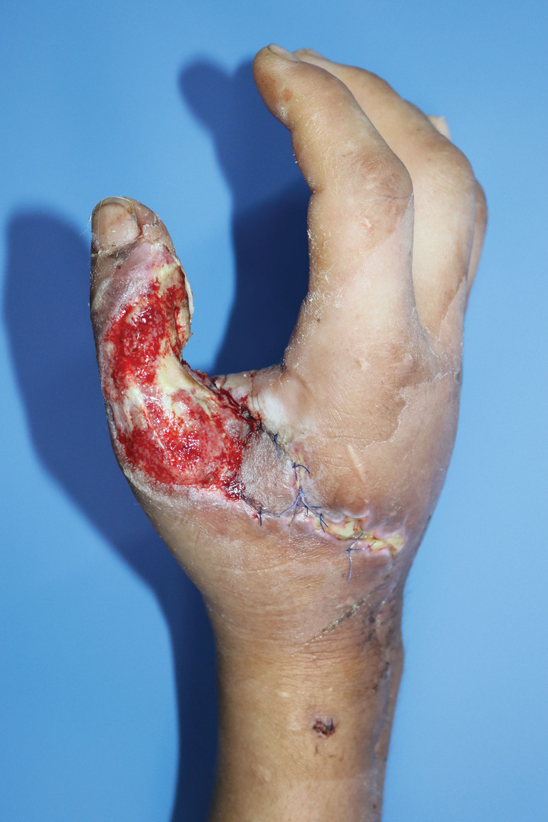
A 23-year-old male patient with no previous medical history was admitted to our hospital because of a cobra bite on his right thumb. The patient was immediately treated with antivenom in the intensive care unit (ICU). After 5 days of hospitalization, the patient developed swelling, bullae formation, and dark discoloration of the thumb, with no systemic findings. The ICU then referred the patient to our department with the diagnosis of thumb tissue necrosis and severe inflammation spreading to one-third of the forearm. We debrided necrotic tissue and covered it with a full-thickness skin graft. After 1 week, the patient showed signs of an infectious process with areas of full-thickness skin loss. Culture sensitivity showed Pseudomonas aeruginosa sensitive to Ciprofloxacin. After debridement, the wound presented with complex defects, including a 4 × 12 cm dorsal soft tissue defect of the thumb and a 6-cm extensor pollicis longus (EPL) tendon loss in zones II–IV (Fig. 1). There was no damage to the bone structure. A composite ALT and FL flap was designed to reconstruct the extensor tendons and soft tissue defects. The perforator was located using a handheld Doppler. The descending branch of the lateral circumflex femoral artery and its perforators were identified and isolated. A 5 × 13 cm skin flap and a 4 × 10 cm fascia flap were harvested, and the pedicle length was measured to be 11 cm. The FL flap is attached to the skin paddle by a 3 × 5 cm island and a perforator in the center of the flap (Fig. 2). The skin paddle was thinned with blunt scissors, and the thickness was reduced from 15 to 5 mm. The flap was elevated entirely from the thigh. The FL flap was used for tendon reconstruction and subsequently attached to healthy tendon remnants with 2-0 Prolene and simple interrupted sutures. An end-to-end anastomosis was used to connect the flap pedicle and the dorsal branch of the radial vessel. After confirmation of blood perfusion, the thinned skin paddle covered the defect entirely. The donor sites were primarily closed. Postoperatively, the flap was well-vascularized, and no complication was reported.
Fig. 1.
After debridement of skin graft failure in the snakebite wound, EPL tendons, and dorsal overlying skin defects.
Fig. 2.
Composite-free ALT and FL flap with one perforator were harvested. Tendon defect reconstruction was done by FL flap, and soft tissue defect was covered by thinned ALT flap. “A” indicates the flap pedicle; B, the FL flap; C, the skin paddle.
The patient could easily do all his daily activities after the 4-month follow-up. Twenty months later, the patient could perform thumb abduction, adduction, flexion, extension, and opposition (Figs. 3 and 4). The range of movement of the interphalangeal joint was from 10 degrees of extension to 60 degrees of flexion. The range of motion of the metacarpophalangeal joint was 60 degrees of flexion and 0 degrees of extension.
Fig. 3.
Twenty months follow-up result. The patient can fully adduct the hand.
Fig. 4.
Twenty months follow-up result. There was some limitation of the IP joint of the thumb when flexed.
DISCUSSION
Snake bites can cause severe problems, and are quite common in Vietnam, with an estimated 30,000 cases annually.6 Cobra bites can provoke severe local complications, followed by tissue necrosis, and without acute life-threatening systemic neurotoxicity. The degree of tissue damage can be cutaneous, necrotizing fasciitis, and tendon necrosis. The condition shares the same symptoms of swelling, redness, pain, and the spread of ulcers.1 The hand is one of the most injured areas. The thumb is a unique structure with a delicate function, and any damage to these units may lead to impairment of the whole finger. The approach to the complex injuries of the thumb requires reconstruction for skin coverage and the affected functional components. This process can be a multi-staged or single procedure. The reconstructive stages typically take two or more procedures to cover and repair the functional structures such as bones and tendons.3 Our patient had a specific presence of necrotic tissue and local infection from a snakebite. Subsequently, the defects after debridement were loss of the dorsal skin and the EPL tendon. There was a limited choice of material for this case, including the local and regional flap. The multi-staged procedure is more likely to succeed but increases the risk of infection, wound dehiscence, contracture, and prolonged rehabilitation. A single-staged operation for coverage and tendon reconstruction is performed by free flaps.4 Free flap selection should meet the requirements of being thin, flexible, capable of withstanding strong tendon forces, and can provide a clear gliding surface for tendon movement. A single-stage reconstruction with a composite ALT flap with vascularized fascia was chosen as a suitable alternative.7
The anterolateral thigh flap (ALT) has evolved into the workhorse flap for soft-tissue reconstruction. An ALT flap can have many components (skin, fascia, adipose, muscle). The composite ALT and FL flap is harvested as a fasciocutaneous type, including skin paddle with FL.8 More data about this flap choice need to be collected for reconstructing complex hand injuries.8,9 The fascia is used for tendon reconstruction and shares the same blood perfusion source with the skin paddle. Research about the perfusion of the fascia lata has shown that it has a reliable vascular supply, and the vascularized FL by a perforator can be used as a free flap. The FL has the advantages of providing excellent protection, accelerating the healing process, and significantly improving tendon mobility. With the composite flap, the FL attaches to the middle skin paddle, through which the perforator passes. The fascia is dissected from the skin paddle at two ends of the flap, and each head is sutured to the remaining tendon segment. Before that, the fascia is folded two to three times into the tendon-like structure. The skin paddle covering the defect should also be adjusted in width and height dimensions. The rehabilitation process begins at week 3, with a maximal range of motion at weeks 4–6, and removal of all splints and strength training at weeks 7–12.
This technique still has some disadvantages, such as pedicle instability and flap thickness that can affect finger movement and aesthetics, as well as the need for secondary thinning procedures to improve final finger contour and extensor function. We also recommend harvesting this flap in the patient with body mass index less than 22 because its thickness did not compromise the finger function.
CONCLUSIONS
Composite ALT with vascularized FL has proven to be a reliable material for soft tissue reconstruction, especially in combination with thumb EPL tendon reconstruction. These techniques have the following advantages: reducing operation times and length of hospital stay and allowing patients to return to normal life activities sooner.
DISCLOSURE
The authors have no financial interest to declare in relation to the content of this article.
Footnotes
Published online 18 October 2023.
Disclosure statements are at the end of this article, following the correspondence information.
REFERENCES
- 1.Edgerton MT, Koepplinger ME. Management of snakebites in the upper extremity. J Hand Surg. 2019;44:137–142. [DOI] [PubMed] [Google Scholar]
- 2.Schubert CD, Giunta RE. Extensor tendon repair and reconstruction. Clin Plast Surg. 2014;41:525–531. [DOI] [PubMed] [Google Scholar]
- 3.Sundine M, Scheker LR. A comparison of immediate and staged reconstruction of the dorsum of the hand. J Hand Surg Edinb Scotl. 1996;21:216–221. [DOI] [PubMed] [Google Scholar]
- 4.Scheker LR, Langley SJ, Martin DL, et al. Primary extensor tendon reconstruction in dorsal hand defects requiring free flaps. J Hand Surg Edinb Scotl. 1993;18:568–575. [DOI] [PubMed] [Google Scholar]
- 5.Adani R, Marcoccio I, Tarallo L. Flap coverage of dorsum of hand associated with extensor tendons injuries: a completely vascularized single-stage reconstruction. Microsurgery. 2003;23:32–39. [DOI] [PubMed] [Google Scholar]
- 6.Patra A, Mukherjee AK. Assessment of snakebite burdens, clinical features of envenomation, and strategies to improve snakebite management in Vietnam. Acta Trop. 2021;216:105833. [DOI] [PubMed] [Google Scholar]
- 7.Cui MY, Shen H. Anterolateral thigh free flap for simultaneous reconstruction of digital extensor tendon and defect of the dorsal hand: a case report. Chin J Traumatol Zhonghua Chuang Shang Za Zhi. 2016;19:309–310. [DOI] [PMC free article] [PubMed] [Google Scholar]
- 8.Meky M, Safoury Y. Composite anterolateral thigh perforator flaps in the management of complex hand injuries. J Hand Surg Eur Vol. 2013;38:366–370. [DOI] [PubMed] [Google Scholar]
- 9.Yazar S, Gideroglu K, Kilic B, et al. Use of composite anterolateral thigh flap as double-vascularised layers for reconstruction of complex hand dorsum defect. J Plast Reconstr Aesthetic Surg JPRAS. 2008;61:1549–1550. [DOI] [PubMed] [Google Scholar]


