Abstract
Objectives
Anti-IgLON5 disease (IgLON5-D) may present with a bulbar-onset motor neuron disease-like phenotype, mimicking bulbar-onset amyotrophic lateral sclerosis. Recognition of their distinctive clinical and paraclinical features may help for differential diagnosis. We report 2 cases of atypical trigeminal neuropathy in bulbar-onset IgLON5-D.
Methods
Trigeminal nerve involvement was assessed using comprehensive clinical, laboratory, electrophysiologic, and MRI workup.
Results
Both patients were referred for progressive dysphagia, sialorrhea, and hoarseness. They were treated with bilevel positive airway pressure for nocturnal hypoventilation. Patient 1 complained of continuous facial burning pain with allodynia, exacerbated by mastication and prolonged speech. Patient 2 reported no facial pain. Anti-IgLON5 autoantibodies (IgLON5-Abs) were positive in serum for both patients and CSF for patient 1. Cerebral MRI revealed bilateral T2 fluid-attenuated inversion recovery (FLAIR) hyperintensity and enlargement of trigeminal nerves without gadolinium enhancement in both patients. Needle myography showed fasciculations in masseter muscles. Blink-reflex study confirmed bilateral trigeminal neuropathy only in patient 2. Cortical laser-evoked potentials showed a bilateral small-fiber dysfunction in the trigeminal nerve ophthalmic branch in patient 1.
Discussion
In case of progressive atypical bulbar symptoms, the presence of a trigeminal neuropathy or trigeminal nerve abnormalities on MRI should encourage the testing of IgLON5-Abs in serum and CSF.
Introduction
Anti-IgLON5 disease (IgLON5-D) is a progressive-onset autoimmune neurologic disorder characterized by the variable association of parasomnia, sleep apnea, bulbar symptoms, gait abnormalities, cognitive decline, and chorea, combined with serum and/or CSF anti-IgLON5 autoantibodies (IgLON5-Abs).1,2 Although brain autopsy may not reveal inflammatory infiltrates, in vitro and in vivo animal experiments suggest a potentially direct pathogenicity of IgLON5-Abs,3-5 and the strong association with the human leukocyte antigen (HLA) system (DRB1*1001 and DQB1*0501) suggests an immune-mediated process.1,2 Recently, several cohorts and case series have enlarged the spectrum of IgLON5-D, helping to better recognize their various phenotypes.5-7 Among them, a bulbar-onset motor neuron disease (MND)–like phenotype, mimicking MND, has been described.8 The differential diagnosis with diseases, such as bulbar-onset amyotrophic lateral sclerosis (ALS), is crucial, as IgLON5-D may require prompt immunotherapy and may rely on the identification of other signs of brainstem involvement.1,3 When bulbar symptoms are prominent, clinical and paraclinical clues should thus be carefully assessed. In this article, we describe 2 cases of bulbar-onset IgLON5-D with clinical, electrophysiologic, and MRI evidence of trigeminal nerve involvement.
Methods
The 2 cases were initially referred to our ALS center for suspicion of bulbar-onset ALS. They had clinical and paraclinical assessment, including IgLON5-Abs testing by immunofluorescence on rat brain slices and cell-based assay (HEK293 cells) in serum and CSF as previously described.9 3-T cerebral MRI scan, nerve conduction study, blink-reflex testing,10 needle EMG, and laser-evoked potentials (LEPs) performed with a pulse laser (Nd:YAP laser) stimulating skin A-delta sensory nerve fibers. Written informed consent was obtained from both patients.
Results
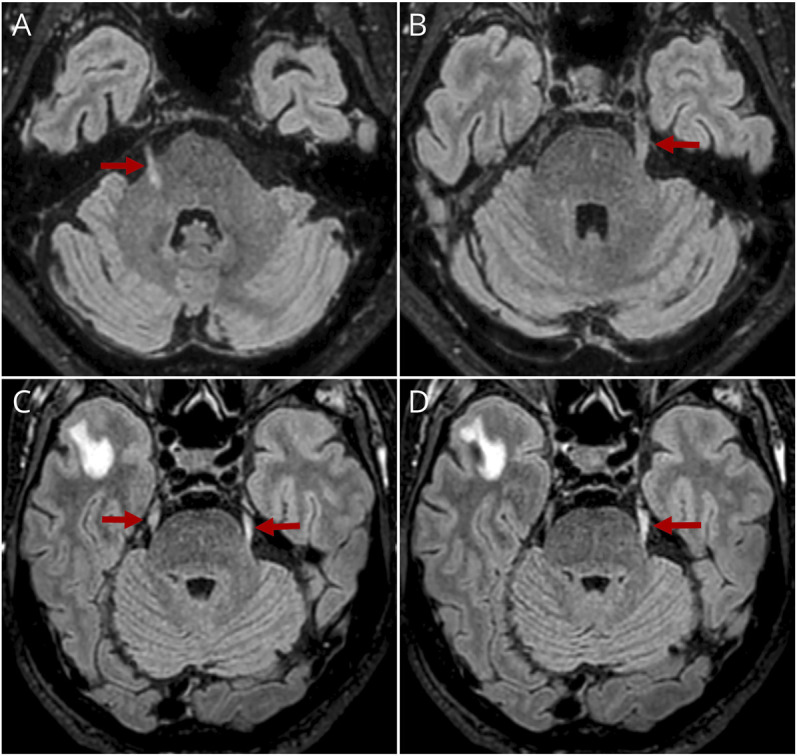
Patient 1, 65-year-old, had a 5-year history of severe dysphagia, sialorrhea, and hoarseness, complicated 6 months before admission by a cardiac arrest due to choking during meal. He also presented with severe obstructive sleep apnea (OSA) requiring nocturnal bilevel positive airway pressure (BiPAP) since 5 years. The patient and his wife reported episodic insomnia, agitation and vocalization during sleep, daytime sudden sleep attacks, and mild gait instability. In addition, his main complaint was a continuous bilateral facial burning pain for the past 4 years, predominant on the right hemiface and in the jaw region, sometimes exacerbated by mastication, jaw opening, or prolonged speech. Neurologic examination found no dysarthria despite severe dysphagia, no tongue paresis or atrophy, face allodynia, and diffuse limb fasciculations without muscle weakness or wasting. Deep tendon reflexes were abolished in lower limbs, and no upper motor neuron signs were noted. Odontologic examination ruled out temporomandibular disorder. Cerebral MRI showed bilateral T2 fluid-attenuated inversion recovery (FLAIR) hyperintensity involving the cisternal part of both trigeminal nerves and the intra-axial part on the right side, without enlargement, atrophy, nor contrast enhancement (Figure 1). Cell count and protein level in CSF were normal. EMG showed many fasciculation potentials and a few fibrillation potentials with chronic neurogenic changes in some limb muscles, possibly explained by degenerative changes in the cervical and lumbar spine with multiple root impingement observed on MRI. Fasciculations were also recorded in masseter muscles. The blink-reflex study was normal. LEPs were absent after stimulation of the right supraorbital region and showed low amplitude of N2/P2 complex (8 μV) on the left side (normal value: 26.2 ± 6),11 suggesting a bilateral small-fiber dysfunction in the ophthalmic branch (V1) of the trigeminal nerves, more severe on the right side. Video-polysomnography confirmed severe sleep architecture abnormalities, simple and unpurposeful abnormal movements, and severe OSA. Finally, given the atypical features presented by the patient, the ALS diagnostic criteria were considered unfulfilled,12 and after a general screening for neuronal autoantibodies, IgLON5-Abs were detected in serum and CSF. HLA typing was DRB1*1001 and DQB1*0501 positive. Immunotherapy was promptly engaged, comprising 3-day IV corticosteroids followed by cyclophosphamide and rituximab association for 12 months. At 12 months, the facial burning pain and MRI trigeminal nerve signal abnormalities remained stable, but the patient reported an improvement of his bulbar symptoms: Despite the persistent dysphagia, he was able to continue a normal diet and return from sparkling to still water and his mealtime decreased from 60 to 35 minutes. He suffered no more choking episodes, and the sialorrhea improved dramatically.
Figure 1. Trigeminal Nerve Abnormalities on MRI of the Brain.

Axial T2 FLAIR sequences showing hyperintensity and slight nerve enlargement of both trigeminal nerves in patients 1 (A, B) and 2 (C, D) (red arrows), including the intra-axial portion of the right trigeminal nerve in patient 1 (A). In patient 2, the right temporal pole abnormalities were attributed to coincidental opercular enlargement of perivascular space with peripheral gliosis.
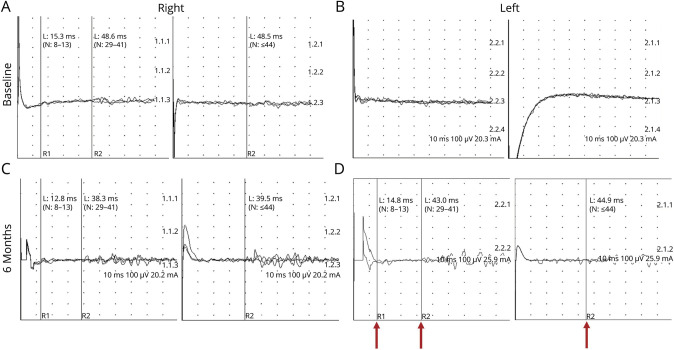
Patient 2, 77-year-old, also presented with a 6-year history of progressive dysphagia, sialorrhea, and hoarseness. Over the past 6 months, the swallowing difficulties were complicated by repetitive aspiration pneumonia, leading to percutaneous endoscopic gastrostomy. Hypercapnic respiratory insufficiency was diagnosed in this context, prompting nocturnal BiPAP, although he had no sleep complaint. Most prominently, the patient experienced several daytime episodes of stridor, requiring tracheostomy shortly after his admission. Clinically, he had no dysarthria, no tongue paresis or atrophy, a few limb fasciculations without muscle weakness or wasting, increased deep tendon reflexes, bilateral Hoffman sign, no gait instability, and no facial pain nor sensitive symptoms. Cerebral MRI revealed bilateral T2 FLAIR hyperintensity of the cisternal part of both trigeminal nerves, without gadolinium enhancement or size anomaly (Figure 1), and subtle T2 FLAIR hyperintensity of the lateral and medial pterygoid muscles suggesting denervation edema.13 Cell count and protein level in CSF were normal. EMG showed fasciculation potentials in trigeminal-innervated temporal and masseter muscles, bulbar muscles, and all spinal regions. However, no evidence of ongoing denervation or chronic neurogenic changes was recorded; ALS diagnostic criteria were considered unfulfilled.12 The blink-reflex study favored a bilateral trigeminal neuropathy, predominant on the left side (Figure 2). After our experience with patient 1, IgLON5-Abs were specifically tested and returned positive in serum and negative in CSF. HLA typing was DRB1*1001 and DQB1*0501 positive. The same therapeutic regimen as patient 1 was started. At the 6-month follow-up, the patient remained clinically stable, still had tracheostomy (no ablation was attempted), and underwent no other emergency hospitalization. He remained painless, and the MRI trigeminal nerve signal abnormalities were stable. However, the blink-reflex study showed a marked improvement of the bilateral trigeminal neuropathy (Figure 2).
Figure 2. Bilateral Trigeminal Neuropathy on Blink-Reflex Study in Patient 2: Baseline and 6-Month Follow-up Evaluations.
At baseline evaluation (A, B), after stimulation of the right supraorbital nerve (A), R1 and ipsilateral and contralateral R2 latencies were all prolonged (R1 latency: 15.3 ms, normal value: 8–13 ms; ipsilateral R2 latency: 48.6 ms, normal value 29–41 ms; contralateral R2 latency: 48.5 ms, normal value ≤44 ms), suggesting right trigeminal neuropathy according to previously described normative values.10 After stimulation of the left supraorbital nerve (B), R1 and ipsilateral and contralateral R2 responses were all absent, indicating a severe left trigeminal neuropathy. After 6 months of immunotherapy (C, D), normal latency responses were recorded after right side stimulation (R1 latency: 12.8 ms, ipsilateral R2 latency: 38.3 ms, contralateral R2 latency: 39.5 ms) (C). After left side stimulation (D), R1 and ipsilateral and bilateral R2 responses were obtained but with slightly prolonged latencies (R1 latency: 14.8 ms, ipsilateral R2 latency: 43.0 ms, contralateral R2 latency: 44.9 ms) (red arrows), indicating a clear improvement of both left and right trigeminal neuropathy. L = latency; N = normal value.
Discussion
In this article, we describe clinical, electrophysiologic, and radiologic features indicating various degrees of trigeminal nerve injury in 2 cases of bulbar-onset IgLON5-D. Patient 1 had a major complaint of facial burning pain evocative of trigeminal neuropathy. The blink-reflex study was unremarkable, but LEPs brought electrophysiologic evidence of a selective small-fiber dysfunction in both trigeminal nerves, which correlated well with the clinical features. Indeed, although LEPs specifically study A-delta fibers, the blink-reflex loop involves large myelinated fibers10 and may be preserved in case of a selective small-fiber disease. Electrophysiology and neuroimaging were concordant as both LEPs and MRI abnormalities were more marked on the right side. Patient 2 had no sensitive nor neuralgic symptoms but displayed radiologic and electrophysiologic signs of trigeminal nerve involvement, both were more severe on the left side. The blink-reflex study was abnormal, indicating a large-fiber trigeminal neuropathy. The R1 and R2 responses showed prolonged latencies, suggesting a demyelinating mechanism. In addition, a brainstem dysfunction might also have contributed to the prolonged R2 latencies because these responses rely on polysynaptic pathways running through the dorsolateral pons and medulla.10 LEPs were not performed because he had no symptom evocative of small-fiber involvement.
Bilateral trigeminal nerve abnormalities on MRI are not specific of IgLON5-D as they may be found in other settings,14 including connective tissue diseases such as Sjögren syndrome, sarcoidosis, neoplasms, MS, vasculitis, infections, or amyloidosis. However, symmetric T2 FLAIR hyperintensity of the cisternal part of the trigeminal nerves without tumoral enlargement, involvement of other cranial nerves, leptomeningeal enhancement, or focal cerebral lesions is an uncommon finding and should point toward IgLON5-D in case of evocative symptoms.
Cranial nerve involvement is infrequent in IgLON5-D: Vocal cord paralysis is classical but may be of central origin, rare cases of peripheral facial palsy have been reported,15 but to the best of our knowledge, trigeminal neuropathy has not been reported yet. The underlying mechanisms of trigeminal involvement remain to be elucidated. Neuronal accumulation of hyperphosphorylated tau has been described at autopsy in the tegmental nuclei of the brainstem, including the trigeminal nuclei.3 Nevertheless, primary inflammation of the nerve may also participate, as suggested by the reversibility of the blink-reflex abnormalities under immunotherapy in patient 2 and by the slightly swollen and T2 FLAIR hyperintense appearance of the nerves in both patients. Altogether, these findings support the need for early initiation of immunotherapy to address the inflammatory part of the disease.
In conclusion, in case of progressive atypical bulbar symptoms unfulfilling the ALS diagnostic criteria, the presence of a trigeminal neuropathy or trigeminal nerve T2 FLAIR abnormalities on MRI should lead to IgLON5-Abs testing in serum and CSF. IgLON5-D should also be suspected in patients with trigeminal neuropathy of unknown underlying cause, especially in the presence of bulbar symptoms or sleep disorders, as nontraumatic and noniatrogenic trigeminal neuropathies have no identified etiology in approximately 60% of cases.14
Acknowledgment
The authors thank Véréna Landel (DRS, Hospices Civils de Lyon) for her help in language editing and word processing during manuscript preparation.
Appendix. Authors

Study Funding
This work is supported by a public grant overseen by the Agence Nationale de la Recherche (ANR) as part of the “Investissements d'Avenir” program (ANR-18-RHUS-0012).
Disclosure
The authors report no relevant disclosures. Go to Neurology.org/NN for full disclosures.
References
- 1.Sabater L, Gaig C, Gelpi Eet al. A novel non-rapid-eye movement and rapid-eye-movement parasomnia with sleep breathing disorder associated with antibodies to IgLON5: a case series, characterisation of the antigen, and post-mortem study. Lancet Neurol. 2014;13(6):575-586. doi. 10.1016/S1474-4422(14)70051-1. Erratum in: Lancet Neurol. 2015;14(1):28. [DOI] [PMC free article] [PubMed] [Google Scholar]
- 2.Gaig C, Graus F, Compta Y, et al. Clinical manifestations of the anti-IgLON5 disease. Neurology. 2017;88(18):1736-1743. doi. 10.1212/WNL.0000000000003887 [DOI] [PMC free article] [PubMed] [Google Scholar]
- 3.Gelpi E, Höftberger R, Graus F, et al. Neuropathological criteria of anti-IgLON5-related tauopathy. Acta Neuropathol. 2016;132(4):531-543. [DOI] [PMC free article] [PubMed] [Google Scholar]
- 4.Sabater L, Planagumà J, Dalmau J, Graus F. Cellular investigations with human antibodies associated with the anti-IgLON5 syndrome. J Neuroinflammation. 2016;13(1):226. doi. 10.1186/s12974-016-0689-1 [DOI] [PMC free article] [PubMed] [Google Scholar]
- 5.Ni Y, Shen D, Zhang Y, et al. Expanding the clinical spectrum of anti-IgLON5 disease: a multicenter retrospective study. Eur J Neurol. 2022;29(1):267-276. doi. 10.1111/ene.15117 [DOI] [PubMed] [Google Scholar]
- 6.Aghelan Z, Karima S, Ghadami MR, et al. IgLON5 autoimmunity tested positive in patients with isolated chronic insomnia disease. Clin Exp Immunol. 2022;207(2):237-240. doi. 10.1093/cei/uxab017 [DOI] [PMC free article] [PubMed] [Google Scholar]
- 7.Grüter T, Möllers FE, Tietz A, et al. , German Network for Research on Autoimmune Encephalitis (GENERATE). Clinical, serological and genetic predictors of response to immunotherapy in anti-IgLON5 disease. Brain. 2022;146(2):600-611. doi. 10.1093/brain/awac090 [DOI] [PubMed] [Google Scholar]
- 8.Werner J, Jelcic I, Schwarz EI, et al. Anti-IgLON5 disease: a new bulbar-onset motor neuron mimic syndrome. Neurol Neuroimmunol Neuroinflamm. 2021;8(2):e962. doi. 10.1212/NXI.0000000000000962 [DOI] [PMC free article] [PubMed] [Google Scholar]
- 9.Shambrook P, Hesters A, Marois C, et al. Delayed benefit from aggressive immunotherapy in waxing and waning anti-IgLON5 disease. Neurol Neuroimmunol Neuroinflamm. 2021;8(4):e1009. doi. 10.1212/NXI.0000000000001009 [DOI] [PMC free article] [PubMed] [Google Scholar]
- 10.Muzyka IM, Estephan B. Electrophysiology of cranial nerve testing: trigeminal and facial nerves. J Clin Neurophysiol. 2018;35(1):16-24. doi. 10.1097/WNP.0000000000000445 [DOI] [PubMed] [Google Scholar]
- 11.Verdugo RJ, Matamala JM, Inui K, et al. Review of techniques useful for the assessment of sensory small fiber neuropathies: report from an IFCN expert group. Clin Neurophysiol. 2022;136:13-38. doi. 10.1016/j.clinph.2022.01.002 [DOI] [PubMed] [Google Scholar]
- 12.Shefner JM, Al-Chalabi A, Baker MR, et al. A proposal for new diagnostic criteria for ALS. Clin Neurophysiol. 2020;131(8):1975-1978. doi. 10.1016/j.clinph.2020.04.005 [DOI] [PubMed] [Google Scholar]
- 13.Russo CP, Smoker WR,Weissman JL. MR appearance of trigeminal and hypoglossal motor denervation. AJNR Am J Neuroradiol. 1997;18(7):1375-1383. [PMC free article] [PubMed] [Google Scholar]
- 14.Ghislain B, Rabinstein AA,Braksick SA. Etiologies and utility of diagnostic tests in trigeminal neuropathy. Mayo Clin Proc. 2022;97(7):1318-1325. doi. 10.1016/j.mayocp.2022.01.006 [DOI] [PubMed] [Google Scholar]
- 15.Gaig C, Compta Y. Neurological profiles beyond the sleep disorder in patients with anti-IgLON5 disease. Curr Opin Neurol. 2019;32(3):493-499. doi. 10.1097/WCO.0000000000000677 [DOI] [PubMed] [Google Scholar]


