Abstract
Background
Magnetic resonance sialography (MRS) can be used to clearly examine the main duct of the parotid and is widely applied in the diagnosis of chronic obstructive parotitis (COP). However, there are few studies on the classification, treatment options and prognosis of COP using MRS.
Methods
Clinical and imaging data were retrospectively collected from 41 patients with COP between January 2010 and December 2020 at the Ninth People’s Hospital affiliated with Shanghai Jiao Tong University School of Medicine. All patients underwent MRS and were treated with intraductal irrigation. The patients were divided into 2 groups according to the presence or absence of symptomatic relapse during the 6-month follow-up period. The imaging features of parotid MRS included three parts: gland volume, stenosis classification and dilatation classification. The location/length of dilatation, the widest diameter of the dilated duct, and the condition of the branch ducts were also recorded and compared between the groups.
Results
A mean of 14.8±12.3 irrigations were performed. There were 15 patients with recurrence and 26 without recurrence. There was no significant difference in the parotid volume (P=0.460), stenosis grade (P=0.738) or maximum diameter of dilatation of the branch duct (P=0.723) between the recurrence and non-recurrence groups. Statistically significant differences were found in dilatation classification (P=0.009), length of dilatation (P=0.043), condition of the branch ducts (P=0.017) and dexamethasone use (P=0.031).
Conclusions
MRS is an available diagnostic and grading modality for COP. The imaging features and classification of the parotid main duct in MRS could be helpful for treatment selection. Patients who accept irrigation could be less likely to experience recurrence with a low dilatation grade and no branch duct dilatation.
Keywords: Magnetic resonance sialography (MRS), chronic obstructive parotiditis (COP), dilatation, stenosis, intraductal irrigation
Introduction
Chronic obstructive parotiditis (COP) is a common disorder characterized by recurrent swelling and pain in the parotids, sometimes with bacterial infection and inflammation, and it accounts for approximately half of benign parotid diseases. COP is caused by ductal diseases (lithiasis, stenosis), as well as infections (bacterial and viral), inflammatory and autoimmune diseases. Recurrent infections may lead to the destruction of alveoli and progressive loss of glandular function (1). Its inciting factors include stones, strictures, mucus plugs and debris (2).
Currently, there is a consensus to use sialoendoscopy for the basic management of COP, while imaging examination can be a helpful guide for the final diagnosis (3). Different imaging modalities are used in the clinical setting, including ultrasound, sialography, computed tomography (CT), magnetic resonance (MR) and radionuclide imaging using 99m Tc-pertechnetate scintigraphy (4). The method selected depends on the suspected cause of parotiditis (5). Ultrasound (US) is an inexpensive, noninvasive but operator-dependent examination for early suspicion and monitoring during follow-up (6). Sialography and CT are also usually used for diagnosis, but they both have the disadvantage of involving ionizing radiation. Bertin et al. described the procedure and unique advantages of three-dimensional cone beam computed tomographic sialography (3D-CBCT sialography). It is believed that 3D-CBCT has high accuracy in mapping the parotid ducts and salivary stones (7). The application of MR in salivary gland diseases was first proposed in 1996 by Lomas et al. (8). MR sialography (MRS) utilizes the characteristics of water with a long T2-weighted sequence relaxation time and heavy T2-weighted imaging (T2WI) combined with fat suppression technology to highlight the high signal of static water in the duct to obtain an image of the duct. MRS depicts the main ductal system and even the first- and second-order branches (5). Patients undergoing MRS do not have to accept ionizing radiation, contrast medium and ductal catheterization. MRS is considered the best method for COP diagnosis, with high overall sensitivity and specificity.
Conservative treatment can be a choice for COP, including sialagogues and gland massage. The efficacy of medical therapy is unclear, and the duration is long, so some patients may choose other treatments, such as intraductal irrigation, injection of botulinum toxin, sialoendoscopy-assisted salivary duct surgery or even parotid removal (9). Intraductal irrigation using different agents has been reported to be a simple and effective treatment in some studies (10-12).
Currently, MR is applied in the diagnosis of strictures, grading of Sjogren’s syndrome and follow-up after radiotherapy (5). However, far too little attention has been given to the MR classification of COP and the relationship between MRS images and clinical prognosis in COP. This retrospective study investigated the use of MRS images and a classification system to assess the prognosis in patients treated with ductal irrigation for COP. We present this article in accordance with the STROBE reporting checklist (available at https://qims.amegroups.com/article/view/10.21037/qims-22-1452/rc).
Methods
Patients
Between January 2010 and December 2020, the data of 41 patients who were diagnosed with COP at the Ninth People’s Hospital affiliated with Shanghai Jiao Tong University School of Medicine were retrospectively collected in the study. The inclusion criteria were as follows: (I) COP diagnosed according to clinical symptoms (swelling, pain, itch in the parotid region or dry mouth) and MR examination results; and (II) prior parotid duct irrigation treatments and follow-up for half a year. Patients diagnosed with sialolithiasis, tumors or other systemic diseases such as Sjogren syndrome were excluded.
Patients underwent MRS at the first visit and subsequently agreed to parotid duct irrigation treatments without anesthesia. Every parotid duct received 5–10 mL of irrigation agent (saline, chloramphenicol, chymotrypsin, gentamicin and dexamethasone) dissolved or diluted in saline. The irrigation agents could be decided by both doctors and patients; the doctors took the MRS classifications and severity of symptoms into account, while for patients with higher grades of dilation/stenosis and more frequency, chymotrypsin with dexamethasone was recommended. Additionally, the patients were informed of the effects and price of the irrigation agents. After treatments, the patients were followed up every 3 months for half a year. When patients experienced swelling and pain of the parotid again, it was considered recurrence. During the follow-up period, patients were divided into two groups based on the presence or absence of recurrence.
The study was conducted in accordance with the Declaration of Helsinki (as revised in 2013). This study was approved by the ethics committee of Shanghai Ninth People’s Hospital in China, and individual consent for this retrospective analysis was waived.
MRS
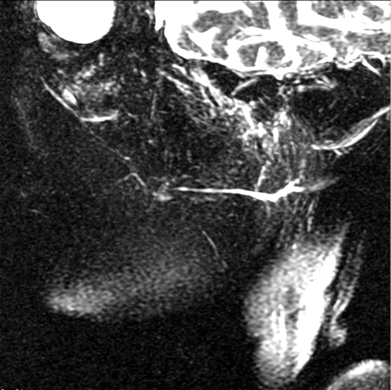
All MRS examinations were performed using a 3.0T MRI scanner (Siemens MagnetomVerio, Germany) equipped with a carotid surface coil. The parameters of MRS in the axial plane were as follows: T1WI, repetition time (TR) 479 ms, echo time (TE) 12 ms, slice thickness 5 mm; T2WI, TR 3,800 ms, TE 70 ms, slice thickness 5 mm. Some images were 3D reconstructed to visualize the oblique sagittal plane (Figure 1). Before examination, patients were required to take vitamin C to stimulate salivary secretion to obtain better images of the ducts.
Figure 1.

Images in a 55-year-old woman with chronic obstructive parotiditis in the right parotid. 3DMRSi shows dilatation of grade 1 in the main duct without dilatation of branch ducts. There are no findings of stenosis in the main duct. 3DMRSi, three-dimensional magnetic resonance sialography.
Grade assessment and feature measurement
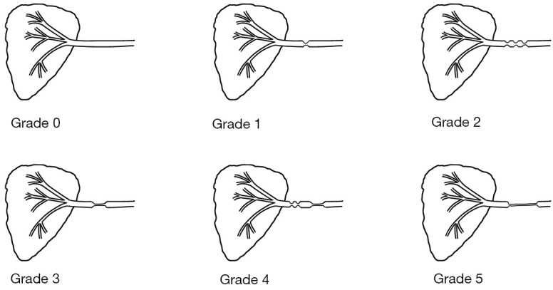
All imaging data were downloaded from the medical image system after the follow-up period. Grade assessments of the parotid and Stensen’s ducts based on MRS images were completed by two radiologists with 3 years of experience. If there were disagreements, a third experienced radiologist checked the results and determined the final grades through discussion. Initially, sialectasis and dilatation of the main duct were classified. Stenosis was classified as follows: grade 0= normal; grade 1= single focal stenosis; grade 2= multifocal stenosis; grade 3= segmental stenosis (stenotic portion less than one-third of total ductal length); grade 4= the composition of grades 2–3; and grade 5= diffuse stenosis (13) (Figure 2). The classification of dilatation was as follows: grade 0= normal; grade 1= slight irregular dilatation of the main duct (length shorter than 2/3 of the main duct) with no disease within the parotid; grade 2= more ductal changes (length longer than 2/3 of the main duct) or branch duct dilatation; and grade 3= diffused dilatation with complete sialectasis and formation of cavities (14) (Figure 3). Moreover, the gland volume, dilatation length/location/width and condition of branch ducts were measured on axial T2WI and recorded. The gland volume was evaluated and categorized according to three states: normal, swelling and atrophy. The parotid main duct was divided into 3 segments according to MRS landmarks: the distal part of the duct (from the papilla to the anterior edge of the masseter); the middle of the duct (up to the hilum and parenchymal duct system); and the proximal duct system (the duct inside the parenchyma). The location depended on the first dilating segment. If there was a dilated branch duct, the most severe branch duct with the widest diameter was measured.
Figure 2.

Stenosis classification scheme.
Figure 3.
Dilatation classification scheme.
Statistical analysis
All statistical analyses were performed by SPSS 16.0 (IBM Corp., Armonk, NY, USA) and the R software “effectsize” package (15). Measurement data are presented as the means with standard deviations. Differences were assessed by the t-test and Mann-Whitney U test depending on whether the data conformed to a normal distribution. Enumeration data are expressed as absolute frequencies and were compared with the chi-squared test (unordered variables) or Mann-Whitney U test (graded variables). The effect size of the difference between groups was measured by the phi or rank-biserial (rrb) correlation coefficient. The weighted Cohen’s kappa and intraclass correlation coefficient were used for enumeration data and measurement data, respectively. A P value of <0.05 was considered to indicate statistical significance.
Results
Patient characteristics
A total of 41 patients who were diagnosed with COP on the basis of clinical examination and MRI findings were included in the present study. The patients’ age (mean ± SD) was 49.8±14.5 years. Among 41 patients, 24 were women and 17 were men. Table 1 shows the demographics and clinical characteristics of the patients. Every patient accepted duct irrigation treatment, with an average number of 14.8±12.3 visits. After the end of treatment, 15 patients relapsed and had to accept repeated treatment. In addition to irrigation treatment, oral antibiotics and pilocarpine were prescribed to patients to relieve their symptoms. No significant difference in demographics, irrigation agents, or oral drugs was found between the recurrence group and non-recurrence group. Notably, there were significant differences between the recurrence group and the non-recurrence group regarding dexamethasone usage [P=0.031, phi =0.30, 95% confidence interval (CI): 0.00–0.62]. Patients with dexamethasone showed a lower recurrence rate.
Table 1. Demographics and clinical characteristics.
| Characteristics | Total | Recurrence group | Non-recurrence group | P value |
|---|---|---|---|---|
| Number | 41 (100.00) | 15 (36.59) | 26 (63.41) | |
| Age (years) | 49.8±14.5 | 49.1±15.3 | 50.1±14.3 | 0.838 |
| Sex | 0.885 | |||
| Male | 17 (41.46) | 6 (35.29) | 11 (64.71) | |
| Female | 24 (58.54) | 9 (37.50) | 15 (62.50) | |
| Affected side | 0.086 | |||
| Right | 7 (17.07) | 0 (0.00) | 7 (100.00) | |
| Left | 13 (31.71) | 6 (46.15) | 7 (53.85) | |
| Bilateral | 21 (51.22) | 9 (42.86) | 12 (57.14) | |
| Mean irrigation times | 14.8±12.3 | 18.3±17.4 | 12.8±7.8 | 0.635 |
| Pilocarpine used | 0.871 | |||
| Yes | 32 (78.05) | 12 (37.50) | 20 (62.50) | |
| No | 9 (21.95) | 3 (33.33) | 6 (66.67) | |
| Antibiotics used | 0.934 | |||
| Yes | 14 (34.15) | 5 (35.71) | 9 (64.29) | |
| No | 27 (65.85) | 10 (37.04) | 17 (62.96) | |
| Irrigation agent | 0.099 | |||
| Saline | 20 (48.78) | 10 (50.00) | 10 (50.00) | |
| Chloramphenicol | 1 (2.44) | 1 (100.00) | 0 (0.00) | |
| Chymotrypsin | 17 (41.46) | 4 (23.53) | 13 (76.47) | |
| Gentamicin | 3 (7.32) | 0 (0.00) | 3 (100.00) | |
| Dexamethasone used | 0.031* | |||
| Yes | 20 (48.78) | 4 (20.00) | 16 (80.00) | |
| No | 21 (51.22) | 11 (52.38) | 10 (47.62) |
Data are presented as mean ± standard deviation or n (%). *, P<0.05.
Association between COP recurrence and MRS findings
We investigated possible correlations between MRS findings and recurrence and the number of irrigations. Owing to the varying severity of bilateral parotids in MRS images, only the more severe parotids were analyzed. Table 2 shows the results of the analysis. The gland volume (P=0.460), classification of stenosis (P=0.738), and location of dilatation (P=0.621) exhibited no statistically significant differences between the recurrence and non-recurrence groups. Significant differences were observed when dilatation classification (P=0.009, rrb=0.49, 95% CI: 0.17–0.72), length of dilatation (P=0.043, rrb=0.38, 95% CI: 0.03–0.65) and dilatation of branching ducts (P=0.017, phi=0.34, 95% CI: 0.00–0.66) were compared between the two groups. Additionally, the interobserver agreement was good (Tables S1,S2).
Table 2. Comparison of magnetic resonance sialography findings between patients with or without recurrence.
| MRS characteristic | Total (n=41) | Recurrence group (n=15) | Non-recurrence group (n=26) | P value |
|---|---|---|---|---|
| Parotid volume | 0.460 | |||
| Normal | 23 (56.10) | 10 (43.48) | 13 (56.52) | |
| Swollen | 12 (29.27) | 4 (33.33) | 8 (66.67) | |
| Atrophic | 6 (14.63) | 1 (16.67) | 5 (83.33) | |
| Stenosis classification | 0.738 | |||
| Grade 0 | 23 (56.10) | 8 (34.78) | 15 (65.22) | |
| Grade 1 | 9 (21.95) | 3 (33.33) | 6 (66.67) | |
| Grade 2 | 2 (4.88) | 1 (50.00) | 1 (50.00) | |
| Grade 3 | 5 (12.20) | 2 (40.00) | 3 (60.00) | |
| Grade 4 | 2 (4.88) | 1 (50.00) | 1 (50.00) | |
| Dilatation classification | 0.009** | |||
| Grade 0 | 2 (4.88) | 0 (0.00) | 2 (100.00) | |
| Grade 1 | 18 (43.90) | 3 (16.67) | 15 (83.33) | |
| Grade 2 | 13 (31.71) | 7 (53.85) | 6 (46.15) | |
| Grade 3 | 8 (19.51) | 5 (62.50) | 3 (37.50) | |
| Dilatation length | 0.043* | |||
| <1/3 the main duct | 16 (41.03) | 5 (31.25) | 11 (68.75) | |
| 1/3–<1/2 | 4 (10.26) | 0 (0.00) | 4 (100.00) | |
| 1/2–2/3 | 11 (28.20) | 4 (36.36) | 7 (63.64) | |
| >2/3 | 8 (20.51) | 6 (75.00) | 2 (25.00) | |
| Location of dilatation | 0.621 | |||
| Distal | 10 (25.64) | 4 (40.00) | 6 (60.00) | |
| Middle | 19 (48.72) | 6 (31.58) | 13 (68.42) | |
| Proximal | 10 (25.64) | 5 (50.00) | 5 (50.00) | |
| Branch duct dilatation | 0.017* | |||
| Absent | 21 (51.22) | 4 (19.05) | 17 (80.95) | |
| Present | 20 (48.78) | 11 (55.00) | 9 (45.00) | |
| Diameter of branch duct (mm) | 2.2±0.5 | 2.2±0.6 | 2.1±0.5 | 0.723 |
Data are presented as mean ± standard deviation or n (%). *, P<0.05; **, P<0.01. MRS, magnetic resonance sialography.
MRS findings
Table 3 presents an overview of 62 parotid MRS findings in 41 patients. The gland volumes included 38, 16 and 8 cases that were categorized as normal, swollen and atrophic, respectively. For stenosis classification, 41 glands were grade 0, 11 glands were grade 1, 3 glands were grade 2, 5 glands were grade 3, 2 glands were grade 4 and none were grade 5 (Figure 4A-4D). The degrees of dilatation were grade 0 in 3 glands, grade 1 in 28 glands, grade 2 in 19 glands and grade 3 in 12 glands (Figure 5A-5C). MR showed a mean duct diameter of 4.7±2.5 mm in the 59 dilated ducts. Dilatation occurred in the anterior border of the masseter in 14 cases, between the parenchyma of the parotid and the anterior border of the masseter in 30 cases, and between the parenchyma of the parotid in 15 cases. Branching duct dilatation occurred in 24 parotids, with a mean diameter of 2.2±0.5 mm.
Table 3. Magnetic resonance sialography characteristics.
| MRS characteristics | Cases |
|---|---|
| Parotid volume | |
| Normal | 38 (61.29) |
| Swollen | 16 (25.81) |
| Atrophic | 8 (12.90) |
| Stenosis classification | |
| Grade 0 | 41 (66.13) |
| Grade 1 | 11 (17.74) |
| Grade 2 | 3 (4.84) |
| Grade 3 | 5 (8.06) |
| Grade 4 | 2 (3.23) |
| Grade 5 | 0 (0.00) |
| Dilatation classification | |
| Grade 0 | 3 (4.84) |
| Grade 1 | 28 (45.16) |
| Grade 2 | 19 (30.65) |
| Grade 3 | 12 (19.35) |
| Dilatation length | |
| <1/3 the main duct | 28 (45.16) |
| 1/3–<1/2 | 7 (11.29) |
| 1/2–2/3 | 16 (25.81) |
| >2/3 | 11 (17.74) |
| Location of dilatation | |
| Distal | 14 (23.73) |
| Middle | 30 (50.85) |
| Proximal | 15 (25.42) |
| Ductal width (mm) | 4.7±2.5 |
| Branch duct dilatation | |
| Absent | 38 (61.29) |
| Present | 24 (38.71) |
| Diameter of branch duct (mm) | 2.2±0.5 |
Data are presented as mean ± standard deviation or n (%). MRS, magnetic resonance sialography.
Figure 4.
Stenosis classified by magnetic resonance sialography: (A) grade 1 on the right side; (B) grade 2 on the left side; (C) grade 3 on the left side; and (D) grade 4 on the left side. The yellow arrows indicate the location of the stenosis.
Figure 5.
Criteria for dilatation classification by magnetic resonance sialography: (A) grade 1 on the left side; (B) grade 2 on the right side; and (C) grade 3 on the left side. The yellow arrows indicate the location of the dilatation.
Discussion
There were two main findings in this study. The dilatation classification between the recurrence group and non-recurrence group was statistically significant. This finding suggested that the dilatation classification may be meaningful for prognostic assessment. This classification was based on one radiological sialographic classification of ductal pathologies (14). The criteria of this classification were subjective with the descriptive words “mild”, “moderate” and “severe”. With this in mind, the classification was modified in this study, and the criteria for dilatation classification consisted of the dilatation length and branch duct condition. In this study, we found significant differences between the two groups in the two parameters. In the recurrence group, the grade was higher, the dilated length was greater, and the condition of the branch duct was more severe.
With respect to clinical data, there were significant differences in dexamethasone usage. All patients in this study accepted intraductal irrigation treatments. Previous studies have reported the positive effect of this therapy (10-12). The results of our study indicated that topical dexamethasone use might play an important role in blocking inflammation. However, other studies have reported contradictory findings about the efficiency of topical steroid use. A prospective study of 54 patients by Capaccio P reported that intraductal steroidal irrigation seemed to increase the degree of mid-term symptom improvement (16), while a retrospective study of 16 patients by Heineman TE and a prospective study of 49 patients by Jokela et al. (17) found that steroid irrigation did not appear to alter symptom improvement after sialoendoscopy (18). Patients with hypertension and diabetes could benefit from topical steroids without side effects due to their localized anti-inflammatory action (19). However, the effect of topical steroids still needs further investigation, and clinical decision-making should take the patient’s condition into consideration.
There are numerous imaging examinations available in nontumor salivary pathologies. Ultrasound can be the first-line examination of obstructive and obstructive diseases of the salivary glands. Koch et al. reported that US can be sufficient for diagnostics in >90% of cases of obstructive diseases (6). Sialography is universally acknowledged as the gold standard method for COP diagnosis. It can clearly present the features of the main duct, branch ducts and even terminal ducts. Because of contrast medium allergy and invasive procedures, Sobrino-Guijarro et al. suggest that sialography can be the last method for further examination when there is no abnormal image in US but persistent symptoms (20). CT offers high specificity for salivary stones and specifies the number, size and location. However, negative stones, dilatation and stenosis are poorly demonstrated in CT imaging. Bertin et al. proposed 3D-CBCT sialography to replace CT in nontumor salivary disease diagnosis (7). Cetinkaya et al. compared 3D-CBCT with US and found that 3D-CBCT had higher sensitivity and specificity, irrespective of the diseases studied (21). There is no consensus on which imaging modalities are optimal for the diagnosis of nontumor salivary pathologies. It is necessary to initially seek the causes and determine the imaging methods. MRS is an invasive examination and can map the salivary ducts, parenchyma and even periglandular tissue. Therefore, MRS was selected for the diagnosis and prognosis of COP in our study.
There have been several publications regarding stenosis classification. Koch and Iro proposed a treatment-oriented classification of stenosis (22) and divided stenosis into 3 types according to the tissue appearance under sialoendoscopy, including inflammatory, web/megaduct-associated and fibrous. Choi et al. described the MRS grading of ductal stenosis in patients with radioactive iodine parotiditis and found a significant correlation between obstructive symptom scores and ductal stenosis grades (13). Based on the literature, we presented an MR classification of stenosis in COP but found no significant difference between the recurrence group and the non-recurrence group. It must be admitted that there are some limitations in the MR judgment of stenosis. Becker et al. reported that MRS had a sensitivity of 100% and specificity of 93–98% using a 3D extended-phase conjugate-symmetry rapid spin-echo (EXPRESS) sequence (23), while Sánchez Barrueco et al. found that MRS showed low sensitivity (36.22%) and high specificity (94.57%) (3), and Capaccio et al. reported a sensitivity of 100% and a specificity of 93% (24). The difference in sensitivity may be related to the MR sequence, the experience of the observers and the anatomical factors of the parotid ducts. The width of Stensen’s duct is not uniform. The mean diameter in the hilum was 1.4 mm, that through the buccal muscle was 1.2 mm and that at the papillary foramen was 0.5 mm (25). Moreover, Stensen’s duct might not be visible in the position overlying the masseter muscle; this should not be regarded as stenosis of the main duct and is a normal physiological phenomenon (23).
The diagnostic criteria of COP in MRS included dilatation and/or stenosis of the parotid main ducts. Previous research has indicated that MRS has high sensitivity and specificity for COP diagnosis. Heineman et al. compared MR diagnosis with final diagnosis under sialoendoscopy and found an accuracy of 100% (18). Kalinowski et al. reported that the sensitivity and specificity of MRS were 79% and 96%, respectively, compared with 100% in digital subtraction sialography (26). Apart from its high cost, the advantages of MR are clear. It is a noninvasive tool that does not require contrast medium. Patients are not exposed to ionizing radiation, and any metal materials in their mouths, such as porcelain teeth, have little effect on imaging. Even in the acute phase or in the case of a completely blocked duct, the examination can be carried out (26).
Some scholars believe that MR is the best imaging method for the major and minor salivary glands in chronic disorders (27) even in the sublingual gland ducts (28). It may completely replace conventional sialography in clinical practice and has become an optimal test prior to sialoendoscopy. MRS could also be used to diagnose and classify Sjogren’s syndrome, and dynamic MRS has been shown to evaluate salivary gland function in SS objectively (25,26). Aside from its value in SS, diffusion-weighted imaging (DWI), another MRI modality, can differentiate sialadenitis from pleomorphic adenoma (29). In conclusion, MRS is expected to have wide applicability in nontumor salivary gland diseases in the future, but doctors need to study it further.
There were some limitations in this study that should be noted. This was a retrospective study with a small sample size (41 patients). The clinical data were not adequate and did not include the symptom scores before and after treatment. The endpoint criterion only included the recurrence of symptoms within half a year of follow-up. To better verify the relationship between this MRS classification and treatment results, a prospective study with a larger sample size should be designed to collect the degree of improvement in patients’ symptoms with a longer follow-up period.
Conclusions
The present study was designed to determine the effect of MRS images and a classification system for COP. The findings suggest that the classification of dilatation with MRS may be meaningful and allow evaluation of the prognosis of patients with COP. Topical use of steroids might be applicable to patients with more severe disease and may decrease recurrence.
Supplementary
The article’s supplementary files as
Acknowledgments
Funding: This study was supported by the National Natural Science Foundation of China (grant No. 82174041) and the Biobank Project of Shanghai Ninth People’s Hospital, Shanghai Jiao Tong University School of Medicine (grant No. YBKB202212).
Ethical Statement: The authors are accountable for all aspects of the work in ensuring that questions related to the accuracy or integrity of any part of the work are appropriately investigated and resolved. The study was conducted in accordance with the Declaration of Helsinki (as revised in 2013). The study was approved by the ethics committee of Shanghai Ninth People’s Hospital in China, and individual consent for this retrospective analysis was waived.
Footnotes
Reporting Checklist: The authors have completed the STROBE reporting checklist. Available at https://qims.amegroups.com/article/view/10.21037/qims-22-1452/rc
Conflicts of Interest: All authors have completed the ICMJE uniform disclosure form (available at https://qims.amegroups.com/article/view/10.21037/qims-22-1452/coif). The authors have no conflicts of interest to declare.
References
- 1.Hernandez S, Busso C, Walvekar RR. Parotitis and Sialendoscopy of the Parotid Gland. Otolaryngol Clin North Am 2016;49:381-93. 10.1016/j.otc.2015.12.003 [DOI] [PubMed] [Google Scholar]
- 2.Goyal M, Singh PP, Batra A. Role of Sialendoscopy in Non-neoplastic Parotid Diseases: A Prospective Study of 241 Patients in Indian Population. J Maxillofac Oral Surg 2022;21:715-24. 10.1007/s12663-020-01472-2 [DOI] [PMC free article] [PubMed] [Google Scholar]
- 3.Sánchez Barrueco Á, Santillán Coello JM, González Galán F, Alcalá Rueda I, Aly SO, Sobrino Guijarro B, Mahillo Fernández I, Cenjor Español C, Villacampa Aubá JM. Epidemiologic, radiologic, and sialendoscopic aspects in chronic obstructive sialadenitis. Eur Arch Otorhinolaryngol 2022;279:5813-20. 10.1007/s00405-022-07473-w [DOI] [PubMed] [Google Scholar]
- 4.Geisthoff U, Al-Nawas B, Beutner D, Günzel T, Iro H, Koch M, Lell M, Luers JC, Schröder U, Sproll C, Teymoortash A, Ußmüller J, Vogl T, Wittekindt C, Zengel P, Zenk J, Guntinas-Lichius O. Updated S2k AWMF guideline on obstructive sialadenitis. Laryngorhinootologie 2021;100:793-8. 10.1055/a-1298-4241 [DOI] [PubMed] [Google Scholar]
- 5.Abdel Razek AAK, Mukherji S. Imaging of sialadenitis. Neuroradiol J 2017;30:205-15. 10.1177/1971400916682752 [DOI] [PMC free article] [PubMed] [Google Scholar]
- 6.Koch M, Sievert M, Iro H, Mantsopoulos K, Schapher M. Ultrasound in Inflammatory and Obstructive Salivary Gland Diseases: Own Experiences and a Review of the Literature. J Clin Med 2021;10:3547. 10.3390/jcm10163547 [DOI] [PMC free article] [PubMed] [Google Scholar]
- 7.Bertin H, Bonnet R, Delemazure AS, Mourrain-Langlois E, Mercier J, Corre P. Three-dimensional cone-beam CT sialography in non tumour salivary pathologies: procedure and results. Dentomaxillofac Radiol 2017;46:20150431. 10.1259/dmfr.20150431 [DOI] [PMC free article] [PubMed] [Google Scholar]
- 8.Lomas DJ, Carroll NR, Johnson G, Antoun NM, Freer CE. MR sialography. Work in progress. Radiology 1996;200:129-33. 10.1148/radiology.200.1.8657900 [DOI] [PubMed] [Google Scholar]
- 9.Strohl MP, Chang CF, Ryan WR, Chang JL. Botulinum toxin for chronic parotid sialadenitis: A case series and systematic review. Laryngoscope Investig Otolaryngol 2021;6:404-13. 10.1002/lio2.558 [DOI] [PMC free article] [PubMed] [Google Scholar]
- 10.Kim JE, Lee SS, Lee C, Huh KH, Yi WJ, Heo MS, Choi SC. Therapeutic effect of intraductal saline irrigation in chronic obstructive sialadenitis. BMC Oral Health 2020;20:86. 10.1186/s12903-020-01078-7 [DOI] [PMC free article] [PubMed] [Google Scholar]
- 11.Grande-Moreillo C, Margarit-Mallol J, Fuentes-Carretero S, Torolla A, Martí-Camps M, Rodríguez-Molinero A. Parotid irrigation: A promising option for juvenile recurrent parotitis. Am J Otolaryngol 2022;43:103398. 10.1016/j.amjoto.2022.103398 [DOI] [PubMed] [Google Scholar]
- 12.Geisthoff UW, Droege F, Schulze C, Birk R, Rudhart S, Maune S, Stuck BA, Hoch S. Treatment of juvenile recurrent parotitis with irrigation therapy without anesthesia. Eur Arch Otorhinolaryngol 2022;279:493-9. 10.1007/s00405-021-06928-w [DOI] [PMC free article] [PubMed] [Google Scholar]
- 13.Choi JS, Lim HG, Kim YM, Lim MK, Lee HY, Lim JY. Usefulness of Magnetic Resonance Sialography for the Evaluation of Radioactive Iodine-Induced Sialadenitis. Ann Surg Oncol 2015;22 Suppl 3:S1007-13. 10.1245/s10434-015-4681-2 [DOI] [PubMed] [Google Scholar]
- 14.Marchal F, Chossegros C, Faure F, Delas B, Bizeau A, Mortensen B, et al. Salivary stones and stenosis. A comprehensive classification. Rev Stomatol Chir Maxillofac 2008;109:233-6. 10.1016/j.stomax.2008.07.004 [DOI] [PubMed] [Google Scholar]
- 15.Ben-Shachar MS, Lüdecke D, Makowski D. effectsize: Estimation of Effect Size Indices and Standardized Parameters. The Journal of Open Source Software 2020. doi: 10.21105/joss.02815 [DOI] [Google Scholar]
- 16.Capaccio P, Torretta S, Di Pasquale D, Rossi V, Pignataro L. The role of interventional sialendoscopy and intraductal steroid therapy in patients with recurrent sine causa sialadenitis: a prospective cross-sectional study. Clin Otolaryngol 2017;42:148-55. 10.1111/coa.12681 [DOI] [PubMed] [Google Scholar]
- 17.Jokela J, Haapaniemi A, Mäkitie A, Saarinen R. Sialendoscopy in treatment of adult chronic recurrent parotitis without sialolithiasis. Eur Arch Otorhinolaryngol 2018;275:775-81. 10.1007/s00405-017-4854-7 [DOI] [PubMed] [Google Scholar]
- 18.Heineman TE, Kacker A, Kutler DI. Idiopathic Chronic Parotitis: Imaging Findings and Sialendoscopic Response. ORL J Otorhinolaryngol Relat Spec 2015;77:302-9. 10.1159/000438760 [DOI] [PubMed] [Google Scholar]
- 19.Lorusso F, Immordino A, Dispenza F, Sireci F, Gallina S. A conservative treatment for chronic obstructive sialoadenitis by intraductal instillation of mucolytic, steroids and antibiotic solution. Eur Arch Otorhinolaryngol 2022;279:501-6. 10.1007/s00405-021-06930-2 [DOI] [PMC free article] [PubMed] [Google Scholar]
- 20.Sobrino-Guijarro B, Cascarini L, Lingam RK. Advances in imaging of obstructed salivary glands can improve diagnostic outcomes. Oral Maxillofac Surg 2013;17:11-9. 10.1007/s10006-012-0327-8 [DOI] [PubMed] [Google Scholar]
- 21.Cetinkaya V, Bonnet R, Le Thuaut A, Corre P, Mourrain-Langlois E, Delemazure-Chesneau AS, Bertin H. A comparative study of three-dimensional cone beam computed tomographic sialography and ultrasonography in the detection of non-tumoral salivary duct diseases. Dentomaxillofac Radiol 2023;52:20220371. 10.1259/dmfr.20220371 [DOI] [PMC free article] [PubMed] [Google Scholar]
- 22.Koch M, Iro H. Extended and treatment-oriented classification of parotid duct stenosis. Laryngoscope 2017;127:366-71. 10.1002/lary.26079 [DOI] [PubMed] [Google Scholar]
- 23.Becker M, Marchal F, Becker CD, Dulguerov P, Georgakopoulos G, Lehmann W, Terrier F. Sialolithiasis and salivary ductal stenosis: diagnostic accuracy of MR sialography with a three-dimensional extended-phase conjugate-symmetry rapid spin-echo sequence. Radiology 2000;217:347-58. 10.1148/radiology.217.2.r00oc02347 [DOI] [PubMed] [Google Scholar]
- 24.Capaccio P, Cuccarini V, Ottaviani F, Minorati D, Sambataro G, Cornalba P, Pignataro L. Comparative ultrasonographic, magnetic resonance sialographic, and videoendoscopic assessment of salivary duct disorders. Ann Otol Rhinol Laryngol 2008;117:245-52. 10.1177/000348940811700402 [DOI] [PubMed] [Google Scholar]
- 25.Gallo A, Benazzo M, Capaccio P, De Campora L, De Vincentiis M, Fusconi M, Martellucci S, Paludetti G, Pasquini E, Puxeddu R, Speciale R. Sialoendoscopy: state of the art, challenges and further perspectives. Round Table, 101(st) SIO National Congress, Catania 2014. Acta Otorhinolaryngol Ital 2015;35:217-33. [PMC free article] [PubMed] [Google Scholar]
- 26.Kalinowski M, Heverhagen JT, Rehberg E, Klose KJ, Wagner HJ. Comparative study of MR sialography and digital subtraction sialography for benign salivary gland disorders. AJNR Am J Neuroradiol 2002;23:1485-92. [PMC free article] [PubMed] [Google Scholar]
- 27.Ugga L, Ravanelli M, Pallottino AA, Farina D, Maroldi R. Diagnostic work-up in obstructive and inflammatory salivary gland disorders. Acta Otorhinolaryngol Ital 2017;37:83-93. 10.14639/0392-100X-1597 [DOI] [PMC free article] [PubMed] [Google Scholar]
- 28.Tanaka T, Oda M, Wakasugi-Sato N, Joujima T, Miyamura Y, Habu M, Kodama M, Takahashi O, Sago T, Matsumoto-Takeda S, Nishida I, Tsurushima H, Otani Y, Yoshiga D, Sasaguri M, Morimoto Y. First Report of Sublingual Gland Ducts: Visualization by Dynamic MR Sialography and Its Clinical Application. J Clin Med 2020;9:3676. 10.3390/jcm9113676 [DOI] [PMC free article] [PubMed] [Google Scholar]
- 29.Terra GT, Oliveira JX, Hernandez A, Lourenço SV, Arita ES, Cortes AR. Diffusion-weighted MRI for differentiation between sialadenitis and pleomorphic adenoma. Dentomaxillofac Radiol 2017;46:20160257. 10.1259/dmfr.20160257 [DOI] [PMC free article] [PubMed] [Google Scholar]
Associated Data
This section collects any data citations, data availability statements, or supplementary materials included in this article.
Supplementary Materials
The article’s supplementary files as


