Abstract
With the continued evolution of echocardiography techniques, novel contrast agents have been introduced to aid in evaluating cardiac structure and function. Lumason is an FDA-approved ultrasound-enhancing agent made of Sulfur hexafluoride lipid-type A microspheres. We report a case of Lumason-induced immune sensitization and subsequent anaphylaxis on re-exposure in a middle-aged male patient presenting with symptoms concerning for a cerebrovascular accident. The patient was successfully treated with steroids and antihistamines. Providers, including physicians, echocardiography technicians, and mid-level providers should be aware of and be prepared to respond to the possibility of anaphylaxis to contrast-enhancing agents used in echocardiography.
Keywords: Lumason, Ultrasound enhancing agents, Anaphylaxis, Echocardiography
1. Background
Transthoracic echocardiography (TTE) is a commonly used cardiac imaging test that provides physicians with information about cardiac structure and function.1,2 It uses high-frequency sound waves to produce an image of cardiac muscle and valves, as well as various hemodynamic measurements such as blood flow through the heart.3 In cases where patients have a suboptimal echocardiogram, such as when the segments of the myocardium cannot be visualized, an ultrasound-enhancing agent may be used to outline the cardiac silhouette and define the endocardial border.4 Lumason (Sulfur hexafluoride lipid-type A microspheres) is an FDA-approved ultrasound-enhancing agent indicated for use in echocardiography, ultrasonography of the liver, and ultrasonography of the urinary tract in pediatrics.5 The mechanism of action of Lumason consists of producing a visual image that is created when an ultrasound beam is reflected between the blood and surrounding tissue creating a contrast between the two interfaces. This is due to the lower acoustic resistance of Lumason microspheres compared to that of surrounding nonaqueous tissue.6
Although rare, the FDA has issued warnings regarding severe cardiopulmonary reactions in patients with hypersensitivity to sulfur hexafluoride lipid microspheres or its components, such as polyethylene glycol (PEG).4 To the best of our knowledge, there are currently only four other reported cases of anaphylactic reaction to Lumason in the literature. Herein, we present the case of a 50-year-old patient who suffered an acute anaphylactic reaction after the administration of Lumason prior to echocardiography.
2. Case presentation
Our patient was a male in his early 50s with a past medical history of hypertension, hyperlipidemia, persistent atrial fibrillation, obstructive sleep apnea, diabetes mellitus type 2, diabetic neuropathy, and asthma who presented to the emergency department with shortness of breath, chest discomfort, dizziness, and fatigue over the two days prior. A TTE was done utilizing Lumason, after which he was diagnosed with a large circumferential pericardial effusion. The patient underwent a pericardial window procedure and was discharged in stable condition. Three weeks later, he was readmitted with new onset right and left upper extremity numbness and tingling. He reported that he was having progressive shortness of breath over several days and dizziness on ambulation. On presentation to the emergency department, a full stroke workup was done including an electrocardiogram (EKG), CT head without contrast, and TTE. The EKG showed sinus rhythm with T-wave inversions in V3–V6, II and AVF, and head CT showed no acute parenchymal abnormality.
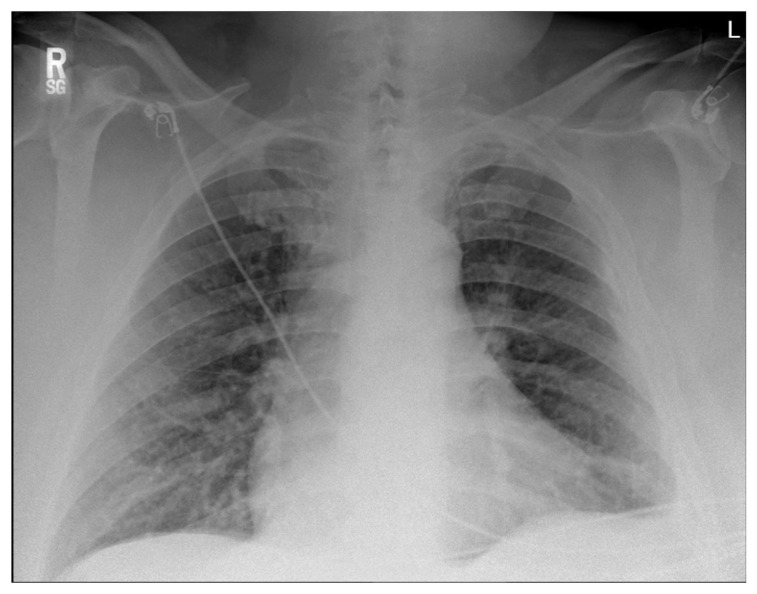
Prior to the TTE, the patient was injected with 5 mL of Lumason reconstitution. Within minutes, he developed severe shortness of breath, cyanosis, tachycardia, diaphoresis, and hypoxia. The patient was immediately given a one-time dose of Solumedrol 125 mg, Albuterol, Diphenhydramine 50 mg, and put on 4 L per minute (lpm) of oxygen through nasal cannula, after which his symptoms improved. On physical examination, the patient had inspiratory and expiratory wheezes in the posterior lung fields, bilaterally. Chest x-ray [Fig. 1] showed mild interstitial prominence at the lung bases but was otherwise unremarkable. Arterial blood gas showed a pH of 7.2, CO2 74.9, O2 387.8, HCO3 28.7. He was then transferred to the ICU for further monitoring. Home medications were reviewed and included Lisinopril 5 mg, Apixaban 5 mg, Sotalol 120 mg, Metformin 500 mg, Furosemide 20 mg, Atorvastatin 40 mg, and Famotidine 20 mg. The patient reported no recent changes in his medication regimen, effectively reducing the possibility of a reaction to another medication that was recently introduced. Later that day, the patient was breathing well on room air with no signs of respiratory distress and was cleared for transfer to the medical floor.
Fig. 1.
Chest x-ray showing mild interstitial prominence at the lung bases.
3. Outcome and follow up
The patient was cleared for transfer to the medical floor after 12 h of observation and continued to improve. During this admission, he developed no further respiratory complications requiring ICU level care.
4. Discussion
There are many types of echocardiography with various indications. The most common type is transthoracic echocardiography (TTE). Common conditions easily evaluated with TTE include congestive heart failure (CHF; both systolic and diastolic), cardiomyopathy, pericardial effusion, valvulopathies, and thromboembolic disease. Other modalities include (3D) echocardiography, bedside echocardiography, and transesophageal echocardiography (TEE). TEE is the most invasive of the modalities but provides a higher degree of sensitivity compared to TTE and is used to identify valvular vegetations in suspected infective endocarditis.7,8 Agitated saline contrast echocardiography is yet another essential modality utilized in identifying intracardiac versus intrapulmonary shunts.9
The complications associated with non-contrast TTE are limited, however with the use of ultrasound-enhancing agents (such as Lumason), the risk of complications increases, as demonstrated in our patient. Additionally, it is important to note that the use of TEE has been associated with the risk of esophageal perforation and bleeding, and the use of agitated saline contrast echocardiography poses a potential risk for air microembolism.10 Therefore, it is essential that both TEE and agitated saline contrast echocardiography be performed by physicians with extensive training to avoid unnecessary harm to the patients and enable decisive action in the event of a complication.8
Presently, two ultrasound-enhancing agents are approved for cardiac imaging in the United States alternative to Lumason; Optison and Definity.11 The FDA is currently aware of 11 cases of anaphylaxis and two deaths, related to an immediate hypersensitivity reaction after the administration of Definity or Lumason among patients with reported allergy to PEG.12 Table 1 explains the compositions and concentrations in the 3 agents.
Table 1.
Comparison of three ultrasound-enhancing agents approved for cardiac imaging.
| Ultrasound enhancing Agent | Shell | Core | Preparation requirements |
|---|---|---|---|
| Lumason | Phospholipid shell | Sulfur hexafluoride gas | Mixing and hand agitation |
| Optison | Human Serum albumin | Octafluoropropane (perflutren) gas | Resuspension of the bubbles by hand |
| Definity | Phospholipid shella | Octafluoropropane (perflutren) gas | Activation with mechanical agitation |
The phospholipid shell of Lumason and Definity, differ in lipid composition and charge.
Amongst North American and European literature, four other reported cases of anaphylaxis to Lumason are currently available at the time of writing. Table 2 displays all five cases (including this one) and outlines the patient demographics, comorbidities, presentation, and interventions. Our patient had no known drug allergies. He was exposed to Lumason only three weeks prior with no anaphylactic reaction. This event may be explained by allergic sensitization of his immune system towards an agent (or multiple) contained in Lumason. The pathophysiologic mechanism of allergic sensitization is caused by the initial presentation of foreign molecules by antigen-presenting cells (APC) to the T-helper cells of our innate and adaptive immune system. T-helper cells present processed antigens to B-cells and trigger IgE production. Interaction between mast cells and IgE results in immune sensitization to the presented molecule. When re-exposure occurs, the antigen binds IgE on mast cells causing degranulation. This in turn triggers the immune cascade, including the release of histamine and serotonin, leading to an allergic reaction. 13 Other immune mediators involved in the allergic response are Th2 lymphocytes, which secrete IL-4, IL-5, IL-13.14
Table 2.
Comparison of the four published case reports of Lumason-induced anaphylaxis.
| Patient Factors | Olson et al. 201820 | Mansour et al. 202221 | Coudray et al. 201722 | Kerber et al. 202223 | Mikhail et al. 2022 |
|---|---|---|---|---|---|
| Age | 63 | 67 | 62 | 47 | 50 |
| Sex | Male | Male | Male | Female | Male |
| Comorbidities | Myelodysplastic syndrome, systemic mastocytosis | Heart failure with preserved ejection fraction (HFpEF) | Hypertension | Not known | Hypertension, atrial fibrillation, pericardial effusion, obstructive sleep apnea, diabetes mellitus type 2, asthma |
| Other allergies | NKDA | NKDA | NKDA | NKDA | NKDA |
| Diagnostic test | Ultrasound-guided biopsy of hepatic mass | Outpatient echocardiogram to evaluate worsening dyspnea on exertion | Control at 6 months for ongoing surveillance post-EVAR | Echo stress test | Transthoracic echocardiogram for stroke workup |
| Reaction | Apnea and pulseless electrical activity within 30 s of administration of Lumason | Severe anaphylactic shock immediately following administration of Lumason | Limb paresthesia, shortness of breath, rash, profuse sweating, loss of consciousness, urination, and severe hypotension within 30 s of administration of SonoVue | Nausea, vomiting, diarrhea, hypotension and hypoxia within 60 s of administration of Lumason | Shortness of breath, diaphoresis, cyanosis, hypoxia, tachycardia following administration of Lumason |
| Intervention | Immediate initiation of CPR with administration of epinephrine; Return of spontaneous circulation achieved after 4 min | Intravenous antihistamines, methylprednisolone and epinephrine were administered followed by transfer to emergency room for monitoring | Immediate transfer to ICU, injection of corticosteroids and antihistamines, normal saline, oxygen, and epinephrine | Epinephrine, dexamethasone, benadryl, oxygen and eventual transfer to ICU | Solumedrol, Albuterol, Diphenhydramine, and oxygen |
The main component of Lumason contrast-enhancing agent is Polyethylene glycol (PEG) 4000. PEG is a macrogol compound, with molecular weights (MW) that range from 200 to 35,000 g/mol. Many liquid and solid formulations of medications, such as laxatives and colonoscopy preparations, contain PEG with MW between 3350 and 6000. The mechanism of hypersensitivity to macrogols is overall poorly understood. There are four types of hypersensitivity reactions classified by Coombs and Gell.15 Type I hypersensitivity is mediated by IgE antibodies, Type II is a cytotoxic reaction that is mediated by IgG or IgM antibodies, Type III is mediated by immune complexes, and Type IV is mediated by a cellular response. Hypersensitivity Types I, II, and III are considered immediate hypersensitivity, occurring within 24 h of exposure to the allergen. Type IV hypersensitivity is considered a delayed reaction, with a maximal reaction time ranging between 48 and 72 h.16 In a study by Stone Jr et al. (2019)17 skin tests were used with PEG and polysorbate-containing agents to determine clinical reactivity and enzyme-linked immunosorbent assay (ELISA) was used to detect anti-PEG specific IgG and IgE. Findings were suggestive of IgE-mediated Type 1 Hypersensitivity reaction, more prominent in high molecular weight formulations of PEG.17 Other less studied components in Lumason include distearoylphosphatidyl-choline (DSPC), dipalmitoylphosphatidylglycerol sodium (DPPG-Na), and palmitic acid, all of which make up <0.5% of each vial of Lumason, as well as the headspace of each vial which is composed of sulfur hexafluoride.18
Further review of the literature has revealed allergic reactions to PEG in several forms. This may come in the form of a medication, over-the-counter pharmaceutical products, lozenges, and lubricants. It is not clearly understood why polyethylene glycol has the propensity to induce acute reactions in susceptible patients; however recent evidence shows that, although rarely reported, allergic reactions to polyethylene glycol-containing compounds are more common than we have recognized.19
Footnotes
Disclosures
None.
Funding
None.
References
- 1. Hillis GS, Bloomfield P. Basic transthoracic echocardiography. BMJ. 2005;330(7505):1432–1436. doi: 10.1136/bmj.330.7505.1432. [DOI] [PMC free article] [PubMed] [Google Scholar]
- 2. Nanda NC. History of echocardiographic contrast agents. Clin Cardiol. 1997;20(S1):7–11. doi: 10.1002/clc.4960201304. [DOI] [PubMed] [Google Scholar]
- 3.Echocardiography. Texas heart institute. Dec 3, 2021. Retrieved September 20, 2022, from https://www.texasheart.org/heart-health/heart-information-center/topics/echocardiography/
- 4. Bracco Dianogstics Inc. Lumason® (sulfur hexafluoride lipid-type A microspheres): Bracco Imaging. Lumason® (sulfur hexafluoride lipid-type A microspheres) j Bracco Imaging. n.d [Google Scholar]
- 5. Molnar D, Kim I, Gada N. Department of health and human services public health service food and drug administration center for drug evaluation and cesearch rffice of surveillance and opidemiology. Pediatr Postmarket Pharmacovigil Rev. 2019 August 23; [Google Scholar]
- 6.National Institutes of Health. DailyMed - Lumasonsulfur hexafluoride kit lumason-sulfur hexafluoride injection, powder, lyophilized, for suspension. U.S. National Library of Medicine; n.d. [Google Scholar]
- 7. Levine RA, Weyman AE, Handschumacher MD. Threedimensional echocardiography: techniques and applications. Am J Cardiol. 1992;69(20):H121–H130. doi: 10.1016/0002-9149(92)90656-j. [DOI] [PubMed] [Google Scholar]
- 8.Omerovic S, Jain A. StatPearls. StatPearls Publishing; 2022. Echocardiogram. [Internet] [PubMed] [Google Scholar]
- 9. Wang Y, Zhang Y. Fetal vascular rings and pulmonary slings: strategies for two-and three-dimensional echocardiographic diagnosis. J Am Soc Echocardiogr. 2021;34(4):336–351. doi: 10.1016/j.echo.2020.10.013. [DOI] [PubMed] [Google Scholar]
- 10. Bassett GC, Lin JW, Tran MM, Sistino JJ. Evaluating the potential risks of bubble studies during echocardiography. Perfusion. 2015;30(3):219–223. doi: 10.1177/0267659114539182. [DOI] [PubMed] [Google Scholar]
- 11. Lindner JR, Hodovan J, Mulvagh S, et al. Ultrasound enhancing agents: recommended laboratory practices from ASE. Am Soc Echocardiogr. 2021 March 10; [Google Scholar]
- 12.In the Aug 13, 2022 issue j American College of Radiology- ACR jFDA Warns Against Some Ultrasound Contrast Agents in Patients With PEG Allergy, n.d. American College of Radiology. (n.d.). From https:/www.acr.org/Advocacy-and-Economics/Advocacy-News/Advocacy-News-Issues/In-the-Aug-13-2022-Issue.
- 13.Molnar C, Gair J. Concepts of biology – 1st Canadian edition. BC campus; 2015. Retrieved from https://opentextbc.ca/biology/ [Google Scholar]
- 14. de Lafaille MAC, Lafaille JJ, Graça L. Mechanisms of tolerance and allergic sensitization in the airways and the lungs. Curr Opin Immunol. 2010;22(5):616–622. doi: 10.1016/j.coi.2010.08.014. [DOI] [PMC free article] [PubMed] [Google Scholar]
- 15. Coombs RRA. Classification of allergic reactions responsible for clinical hypersensitivity and disease. Clin Aspect Immunol. 1968:xxvi+883. [Google Scholar]
- 16.Khaled M, Kondamudi NP. Type IV hypersensitivity reaction. National Library of Medicine; 2022. Retrieved from https://www.ncbi.nlm.nih.gov/books/NBK562228/ [Google Scholar]
- 17. Stone CA, Jr, Liu Y, Relling MV, et al. Immediate hypersensitivity to polyethylene glycols and polysorbates: more common than we have recognized. J Allergy Clin Immunol Pract. 2019;7(5):1533–1540. doi: 10.1016/j.jaip.2018.12.003. [DOI] [PMC free article] [PubMed] [Google Scholar]
- 18.RxList. Lumason (sulfur hexafluoride lipid-type Amicrospheres injectable suspension): uses, dosage, side effects, interactions, warning. RxList; 2022. Aug 26, Retrieved November 7, 2022, from https://www.rxlist.com/lumason-drug.htm. [Google Scholar]
- 19. Wylon K, Dölle S, Worm M. Polyethylene glycol as a cause of anaphylaxis. Allergy Asthma Clin Immunol. 2016;12(1):1–3. doi: 10.1186/s13223-016-0172-7. [DOI] [PMC free article] [PubMed] [Google Scholar]
- 20. Olson MC, Atwell TD, Knudsen JM. Anaphylactic reaction to an ultrasound contrast agent (Lumason) in a patient with systemic mastocytosis. J Clin Ultrasound. 2018;46(8):533–535. doi: 10.1002/jcu.22585. [DOI] [PubMed] [Google Scholar]
- 21. Mansour A, Shinn A, Bataineh MA, Aggarwal KB. Rare anaphylactic reaction to an echocardiography contrast agent. J Am Coll Cardiol. 2022;79(9_Supplement):3116. [Google Scholar]
- 22. Coudray S, Fabre C, Aichoun I, Perez-Martin A. Anaphylactic shock with an ultrasound contrast agent. JMV-Journal de Médecine Vasculaire. 2017;42(6):384–387. doi: 10.1016/j.jdmv.2017.10.001. [DOI] [PubMed] [Google Scholar]
- 23. Kerber A, Li JT. Anaphylaxis to ultrasound-enhancing agent with transient elevation in serum tryptase. Ann Allergy Asthma Immunol. 2022 doi: 10.1016/j.anai.2022.04.015. [DOI] [PubMed] [Google Scholar]


