A 73‐year‐old man presented for lumbar decompression and fusion. His past medical history included hypertension, hypothyroidism and chronic kidney disease. His medications included telmisartan 40 mg and levothyroxine 50 mcg, both taken once daily. He had previously undergone surgery for bilateral carpal tunnel syndrome and a right eye cataract, under regional anaesthesia. Pre‐operative airway examination revealed an unusually large tongue but no other features of concern for airway management. The pre‐operative full blood count and coagulation parameters were within normal range.
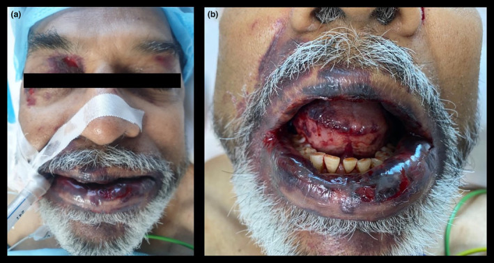
We induced general anaesthesia uneventfully with fentanyl 100 mcg, propofol 100 mg and cisatracurium 14 mg. We intubated the trachea with an 8.5‐mm tracheal tube. Anaesthesia was maintained with sevoflurane (end tidal concentration of 1.2%) in an air/oxygen mixture. While fixing the tracheal tube, we noticed that ecchymoses had developed over the right upper eyelid and right angle of the mouth since induction of anaesthesia. The tracheal tube was changed over a bougie due to cuff malfunction. Following this, new periorbital ecchymoses were noted to have developed bilaterally and well as on the lips (Fig. 1a). We also observed swelling of the tongue with haemorrhagic patches (Fig. 1b). During this time, observations remained stable.
Figure 1.

(a) periorbital and perioral ecchymoses, (b) swelling of the tongue with haemorrhagic patches.
We considered anaphylaxis, an adverse drug reaction or coagulopathy as possible causes but these were ruled out clinically. The surgery was abandoned. We spoke to the family after this adverse event. They told us that the patient had developed bruising around his eye during cataract surgery and had excessive bleeding from his gums after a tooth extraction. The patient was transferred to a critical care area and extubated after an hour. Diagnosis of systemic amyloidosis was made following referral to the immunology team in view of the history of chronic kidney disease, carpal tunnel syndrome and bruising at pressure areas. The diagnosis was confirmed with a skin biopsy.
Pinch purpura is a cutaneous manifestation of systemic amyloidosis [1, 2]. It typically occurs above the nipple line and is often seen in the webbing of the neck, peri‐orbital areas and the eyelids. Amyloid deposition in the skin and blood vessel wall causes capillary fragility and leads to intracutaneous micro‐ and macro‐haemorrhages [1, 2]. Although the diagnosis of systemic amyloidosis under general anaesthesia is rare, items adhering to the skin (e.g. cutaneous electrodes and tape) have been reported as contributing factors [3, 4]. We have reported our findings here with the images of pinch purpura; the recognition of this sign is valuable because early diagnosis of systemic amyloidosis improves the prognosis of the disease.
1. Associate Consultant 2. Principal Consultant 3. Associate Director 4. Senior Director, Department of Neuroanaesthesia and Critical Care 5. Head of Department, Department of Neurosurgery, Max Super Speciality Hospital, Press Enclave Road, Saket, New Delhi, Delhi 110017, India
References
- 1. Apalla Z, Kirtsios T, Katodritou E, Lallas A, Konstantinou D, Karamitsos T. Pinch purpura unmasking systemic amyloidosis. International Journal of Dermatology 2019; 58: e195–e196. [DOI] [PubMed] [Google Scholar]
- 2. Ashwini M. Pinch purpura: a clinical clue for primary systemic amyloidosis. Turkish Journal of Dermatology 2020; 14: 1. [Google Scholar]
- 3. Campos JH. A reaction to tape after tracheal extubation in a patient with systemic amyloidosis. Journal of Clinical Anesthesia 1999; 11: 126–128. [DOI] [PubMed] [Google Scholar]
- 4. Dixon J. Primary amyloidosis and skin damage. Anaesthesia 1987; 42: 218. [DOI] [PubMed] [Google Scholar]


