

Summary
Unilateral congenital cataracts lead to deprivation amblyopia, which can be severe. Until the 1970s, they were believed to be always associated with poor visual outcomes. However, advances in our understanding of the plasticity of the infant brain and the development of better surgical techniques allowed good visual outcomes to be obtained in a few of these patients. The Infant Aphakia Treatment Study (IATS) was conducted to provide empirical evidence regarding the best type of optical correction to be used following surgical extraction of the cataract. Specifically, infants were randomly assigned to either be left aphakic and to wear contact lenses or an intraocular lens (IOL) was implanted and the residual refractive error was corrected with spectacles. The study found that good visual acuity and stereopsis could be achieved in some patients in both treatment groups. Early cataract surgery, consistent optical correction and part-time patching of the fellow eye are important elements needed to achieve good visual outcomes. However, excess patching of the fellow eye may interfere with the development of stereopsis. More adverse events occurred after IOL implantation, particularly visual axis opacification, compared with the infants who were left aphakic. Glaucoma-related adverse events occurred in 40% of eyes after a 10-year follow-up and were not associated with IOL implantation. Further research is needed to increase the percentage of children with unilateral congenital cataracts who achieve good visual outcomes.
I never had the privilege of meeting Dr. Costenbader, but for many years I have been interested in a paper he published in 1957.1 In this paper he summarized the poor visual results and increased risks of glaucoma, pupillary membranes, and retinal detachment after congenital cataract surgery. He emphasized that the visual outcome is particularly poor for children with a unilateral congenital cataract and concluded, “Since visual acuity is not improved, strabismus is not favorably influenced, and photophobia is not alleviated, we would unequivocally advise against surgery in unilateral congenital cataract unless the cataract is becoming hypermature.” There have been many advances in pediatric cataract surgery since the publication of this paper. They include the development of the pars plana vitrectomy,2 improved contact lenses,3,4 foldable intraocular lenses (IOLs)5 and refinements in surgical instruments.6,7 In addition, research on visual deprivation by Hubel and Wiesel has helped us to understand the importance of early surgery for children with congenital cataracts and the requisite penalization of the fellow eye required to allow cortical development of the visual pathways for the formerly cataractous eye.8,9
In the 1970s, there began to be reports of good visual outcomes in children with unilateral congenital cataracts. In 1973, Frey and colleagues10 reported 20/40 visual acuity in the treated eye of 2 children with unilateral congenital cataracts after early surgery, contact lens correction and part-time patching of the fellow eye. In 1981, Beller and colleagues11 reported favorable visual outcomes in the treated eyes of 8 children with unilateral congenital cataracts after early cataract surgery, contact lens correction and part-time patching titrated based on results from visual evoked responses. Despite the good visual outcomes in these cases series, clinicians continued to report poor visual outcomes in most children with unilateral congenital cataracts.12
Non-human Primate Studies
In 1996 Edward Cotlier, David Taylor and I organized a symposium on congenital cataracts that was held in London, England. At the symposium, I presented my research on unilateral congenital cataracts using a nonhuman primate model. I simulated a unilateral congenital cataract in newborn monkeys by placing an opalescent contact lens on their right eyes (Figure 1). When the monkeys were 2 weeks old, I performed a lensectomy on their right eyes and either implanted a custom made IOL into the capsular bag or left the eye aphakic followed by contact lens correction. The fellow left eyes were either occluded part-time with an occluder contact lens or left untreated. Grating acuities were assessed using preferential looking and operant testing methods. The visual outcomes were generally quite good in the treated eyes if the fellow eyes were occluded part-time. However, there was a high incidence of postoperative complications in the pseudophakic eyes, including fibrin encapsulation of the IOL optic, lens reproliferation into the pupillary space, and glaucoma.13,14
FIG 1.
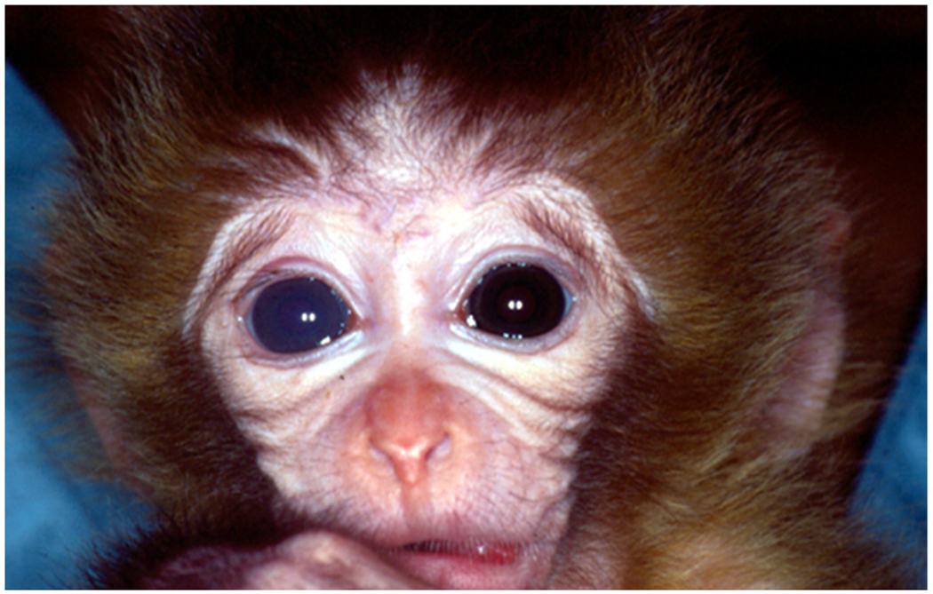
An infant Rhesus monkey wearing an opalescent contact lens on his right eye to simulate a unilateral congenital cataract.
Genesis of The Infant Aphakia Treatment Study
At the symposium, Edward Cotlier proposed that a clinical trial be initiated to compare different treatments for congenital cataracts so their management could become more evidence based. Later that year at the annual meeting of the American Academy of Ophthalmology, a small group of pediatric cataract surgeons and I discussed different ideas for a clinical trial pertaining to congenital cataracts. The consensus was to focus our efforts on treatments for unilateral congenital cataracts given the high frequency of poor visual outcomes with this disorder. We decided that the first question we would study would be whether IOL implantation is associated with better visual outcomes for infants with unilateral congenital cataracts. We began by reviewing outcomes of 25 infants who underwent unilateral congenital cataract surgery with or without an IOL. Eyes that were left aphakic were corrected to a near point with a contact lens. While the grating acuities were better in the pseudophakic eyes at age 12 months, the optotype acuities were similar in the pseudophakic and aphakic eyes after a longer follow-up.15,16 In addition, we noted that the pseudophakic eyes experienced more adverse events.
Survey of AAPOS Membership and Pilot Studies
We next surveyed the AAPOS membership on two different occasions (1997 and 2001) to determine whether clinicians were equipoised on the issue of IOL versus contact lens correction following unilateral congenital cataract surgery.17 In 1997, only 4% of respondents reported that they had implanted an IOL in an infant <7 months of age, whereas the percentage had increased to 21% by 2001. Although most respondents favored contact lens correction after unilateral congenital cataract surgery, 61% indicated that they would be willing to randomize an infant with a unilateral congenital cataract to one of these treatments. Their main concerns with IOL implantation were the poor predictability of the myopic shift, the higher incidence of postoperative complications, and the technical difficulty of implanting an IOL in an infant’s eye. Their primary concerns with contact lens correction were poor compliance with lens wear, a high lens loss rate, the high cost of contact lenses and the increased risk of keratitis associated with contact lens wear.
When we applied for funding for our proposed clinical trial, reviewers were skeptical that parents would be willing to let their child be randomized to IOL implantation versus contact lens correction. To address this concern, we conducted a small pilot study at nine clinical sites to assess the willingness of parents to randomize their child to IOL implantation after unilateral congenital cataract surgery. We identified 24 infants who met our inclusion and exclusion criteria and the parents of 17 (71%) of these children agreed to have their infant randomized to IOL implantation. We used this data to extrapolate the number of patients we would need to recruit to attain the requisite sample size.
Visual Outcomes
In 2004 we received funding from the National Eye Institute to conduct a multicenter randomized clinical trial comparing the visual outcomes and adverse events associated with IOL implantation versus contact lens correction in infants ≤6 months of age following unilateral congenital cataract surgery. Based on what had been previously reported in the literature and our pilot studies, we hypothesized that eyes randomized to IOL implantation would have better visual outcomes, but more adverse events. Enrollment in the Infant Aphakia Treatment Study (IATS) was initiated in 2004 and ended in 2009. In 2010, we reported that the grating acuity, assessed by a traveling tester, was similar in the two treatment groups at age 12 months.18,19 We subsequently received additional funding from the National Eye Institute to evaluate their optotype acuity using the Amblyopia Treatment Study HOTV test at age 4.5 years.20 While the median acuity was similar in the two treatment groups, the visual outcomes were highly variable; about one-quarter of the patients had visual acuity of 20/40 or better in their treated eye, but nearly one-half had a visual acuity of 20/200 or worse. When we retested their acuity at age 10.5 years using the electronic Early Treatment Diabetic Retinopathy Study (E-ETDRS) testing protocol, we found that the median visual acuity was similar in the two treatment groups and about 25% of patients had an acuity of 20/40 or better and 44% had an acuity of 20/200 or worse in their treated eyes.21
Treatment Parameters Correlating with Visual Outcomes
Given the highly variable visual results among the participants in the IATS, we analyzed many baseline and treatment parameters to determine whether they correlated with the visual outcomes. Private insurance, a proxy for socioeconomic status, correlated most closely with visual outcomes.22 Younger age at the time of cataract surgery was also associated with a better vision outcome (logMAR 0.5 vs 1.1; Spearman = 0.19). Within the aphakic group, better contact lens wear was also associated with a better visual outcome.23 Children who wore their spectacles for at least 80% of waking hours were also more likely to have visual acuity of 20/40 or better in their treated eyes.24 We prescribed the same patching regimen for the fellow eye for both treatment groups—one hour per day per month of life until age 8 months followed by 50% of waking hours to age 5 years. Patching adherence was assessed to age 5 years with 48-hour recall interviews every 3 months and prospective 7-day diaries annually (eSupplement 1). Patching adherence during the first year of life and from ages 1 to 4 years correlated with the visual outcome at age 4.5 years, although it only accounted for 15% of the variance.25
Stereopsis
Surprisingly, we found that about one half of the patients with 20/40 or better visual acuity at age 4.5 years manifested at least gross stereopsis. Stereopsis could be detected more often in children who underwent cataract surgery at a younger age, had good visual acuity in their treated eye, and were orthotropic at age 4.5 years.26 On average, children with good visual acuity and stereopsis were patched for 3.4 hour/day during the first year of life and for shorter periods of time during subsequent years. Children with good visual acuity and stereopsis were patched less than children with good visual acuity and no stereopsis (Figure 2). We concluded that there is likely a threshold for patching that will allow a child to achieve good visual outcomes in their treated eye. However, the threshold is probably different for each child based on the severity of their cataract and the age they underwent cataract surgery. Exceeding this threshold may interfere with the development of stereopsis without further improvements in vision.
FIG 2.

A boxplot of the average reported hours of patching per day as a function of age for patients with 20/40 or better visual acuity in the Infant Aphakia Treatment Study.
By age 10 years, strabismus had developed in nearly 90% of the children enrolled in the IATS.27 Esotropia was the most common type of strabismus up to age 5 years, whereas exotropia was most common type at age 10 years. IOL implantation was not associated with the incidence of strabismus. There was trend toward a greater likelihood of orthophoria at age 5 years with cataract surgery performed between 4 to 6 weeks of age than with surgery performed after 6 weeks of age, but this did not reach statistical significance (27% vs 13%; P = 0.085).28
Adverse Events
When we planned the IATS, we hypothesized that children in the IOL treatment group would have more adverse events, which proved to be the case. At age 4.5 years, significantly more patients in the IOL group had had at least one adverse event (81% vs 56%; P = 0.02).29 The most common adverse events in the pseudophakic eyes were lens reproliferation into the visual axis, pupillary membranes and corectopia. A much higher percentage of the children in the IOL group required additional intraocular surgeries (72% vs 16%; P < 0.001).30 Most of these additional intraocular surgeries were performed during the first year of life. Because there were significantly more adverse events and additional intraoperative procedures in the IOL group, we concluded, “When operating on infants younger than 7 months of age with a unilateral cataract, we recommend leaving the eye aphakic and focusing the eye with a contact lens. Primary IOL implantation should be reserved for those infants where, in the opinion of the surgeon, the cost and handling of a contact lens would be so burdensome as to result in significant periods of uncorrected aphakia.”29
The risk of a glaucoma-related adverse event was not significantly different between eyes randomized to IOL implantation versus aphakia in the IATS.31 The number of children who developed a glaucoma-related adverse event rose steadily over time and was similar in both treatment groups. By age 10 years, 22% of patients had developed glaucoma and 40% had either developed glaucoma or were a glaucoma suspect. In nearly all cases the glaucoma was open angle. We noted that the odds of developing a glaucoma-related adverse event was 1.6 times higher for each month earlier than 6 months of age that cataract surgery was performed.32 We also found that a smaller corneal diameter increased the risk of a glaucoma-related adverse event.33 We recommended that the potential for a good visual outcome be weighed against the risk of glaucoma when choosing an age to perform unilateral congenital cataract surgery for a particular patient.34
Myopic Shift
We undercorrected eyes randomized to IOL implantation by 6-8 D in anticipation of a large myopic shift in these eyes as they elongated.18 Despite this under-correction, most of these eyes became myopic and many of them became highly myopic. Treated eyes experienced a mean myopic shift of 0.35 D/month until age 1.5 years and then 0.97 D/year to age 5 years.35 The myopic shift experienced by individual patients was quite variable. An additional mean myopic shift of −3.4 D occurred in pseudophakic eyes without glaucoma between ages 5 and 10 years. An even larger myopic shift occurred during this same time interval in pseudophakic eyes with glaucoma. These large myopic shifts resulted in marked anisometropia in many children randomized to IOL implantation. The median anisometropia in children who didn’t develop glaucoma in their treated eyes was −3.50 D at age 5 years and −5.40 D at age 10 years.36 37 Six pseudophakic eyes underwent an IOL exchange to alleviate their marked anisometropia; four of these eyes had glaucoma or a glaucoma suspect diagnosis. We concluded that the marked variability of the myopic shift after IOL implantation in young children makes it difficult to choose an IOL power for implantation.
We also evaluated the myopic shift occurring in aphakic eyes. Thirty of the 56 eyes randomized to aphakia were still aphakic at age 10 years.38 The mean refractive error for these eyes was 19.01 D immediately after cataract surgery compared to 10.38 D at age 10 years. The mean change in the refractive error was −2.11 D/year to age 1.5 years, −0.68 D/year from ages 1.5 to 5.0 years and −0.35 D/year from ages 5 to 10 years. At age 10 years, 18 of these patients continued to correct their aphakia with a contact lens, 9 with spectacles and 4 wore no correction. By age 10 years, 21 of these eyes had undergone secondary IOL implantation, mostly between the ages of 5 and 6.5 years.39
Contact Lenses
Most patients randomized to aphakia wore silicone elastomer or gas permeable contact lenses to correct their induced high hyperopia.40 Contact lens wear was generally well tolerated and associated with relatively few adverse events. Most parents became proficient at inserting and removing contact lenses from their child’s eye. Only two patients underwent IOL implantation prior to age 4.5 years due to the inability of the parents to manage their child’s contact lenses. Frequent lens replacements were necessary either because lenses were lost or a change was needed in lens parameters. Gas permeable lenses required more frequent lens replacements than silicone elastomer lenses.41 After 5 years of follow-up, the costs associated with aphakia and contact lens wear were less than the costs associated with primary IOL implantation. While all of the costs of contact lens wear were covered by our grant, in the real world we speculated that more of the costs associated with contact lens wear would be borne by parents because many insurance companies do not provide coverage for contact lenses.42,43
Axial Elongation
Axial lengths were measured preoperatively and at ages 5 and 10 years. We found that mean axial lengths were shorter in eyes with cataracts compared to their fellow eyes and this difference persisted to age 10 years.36 Axial length growth was similar in aphakic and pseudophakic eyes. Eyes with glaucoma experienced more axial growth than those without glaucoma (7.3 mm vs 4.7 mm; P = 0.05).44 In addition, eyes receiving additional surgery grew more than eyes not receiving additional surgery.
Best Practices
The IATS demonstrated that it is possible to achieve a good visual outcome and high-grade stereopsis in children after unilateral congenital cataract surgery. However, most patients in the IATS achieved a suboptimal result. How could a good visual outcome be achieved by a higher percentage of children undergoing unilateral congenital cataract surgery? First, early detection of a congenital cataract and timely referral to a cataract surgeon is paramount.45 Second, while the timing of cataract surgery is important, it is probably less important than the care provided after cataract surgery. The optimal age for unilateral congenital cataract surgery is believed to be between ages 4 and 8 weeks34 (Table 1). Third, patching therapy of the fellow eye is important, but too much patching may interfere with the development of binocular functioning. I currently recommend patching the fellow eye: 1 hour/day per month of life until age 8 months; 6 hours/day until age 18 months; 5 hours/day until age 2½ years; 4 hours/day until age 3 years; 2 hours/day until age 4 years; 4 hours/every other day until age 5 years; and 4-5 hours/day twice a week until age 7 years.46 Fourth, good adherence with both contact lens and spectacle wear is critical. The power of contact lenses and spectacles should be adjusted on a regular basis to compensate for the large myopic shift that these eyes typically experience. The type of contact lens worn is less important than how consistently it is worn. Finally, it is important to regularly monitor these eyes for glaucoma given the increased risk of elevated intraocular pressure in these eyes after early cataract surgery.
Table 1.
Pros and cons of performing cataract surgery at 4 versus 8 weeks of age for infants with a unilateral congenital cataract
| 4 weeks | 8 weeks | |
|---|---|---|
| Visual acuity | + | − |
| Stereopsis | + | − |
| Glaucoma | − | + |
| Strabismus | + | − |
Conclusions
The IATS has provided empirical evidence for the care of infants with unilateral congenital cataracts. Its findings have changed the way that cataracts are managed in many parts of the world including the United States.47 The IATS has provided evidence that children with unilateral congenital cataracts can achieve a good visual outcome in their cataractous eye. However, further research is needed to achieve good visual outcomes in a higher percentage of these children.
Supplementary Material
Acknowledgments
I would like to thank all of the clinicians, biostatisticians, epidemiologists, and clinical coordinators who participated in the Infant Aphakia Treatment Study (eSupplement 2, available at jaapos.org). I would also like to give special thanks to Don Everett at the National Eye Institute for his continuous support of this project.
Financial support:
National Institutes of Health Grants U10 EY13272, U10 EY013287, UG1 EY013272, 1UG1 EY025553, and P30 EY026877; Research to Prevent Blindness Inc (New York City, NY).
Footnotes
Publisher's Disclaimer: This is a PDF file of an unedited manuscript that has been accepted for publication. As a service to our customers we are providing this early version of the manuscript. The manuscript will undergo copyediting, typesetting, and review of the resulting proof before it is published in its final form. Please note that during the production process errors may be discovered which could affect the content, and all legal disclaimers that apply to the journal pertain.
References
- 1.Costenbader FD, Albert DG. Conservatism in the management of congenital cataract. AMA Arch Ophthalmol 1957;58:426–30. [DOI] [PubMed] [Google Scholar]
- 2.Machemer R, Buettner H, Norton EW, Parel JM. Vitrectomy: a pars plana approach. Trans Am Acad Ophthalmol Otolaryngol 1971;75:813–20. [PubMed] [Google Scholar]
- 3.Levin AV, Edmonds SA, Nelson LB, Calhoun JH, Harley RD. Extended-wear contact lenses for the treatment of pediatric aphakia. Ophthalmology 1988;95:1107–13. [DOI] [PubMed] [Google Scholar]
- 4.Amos CF, Lambert SR, Ward MA. Rigid gas permeable contact lens correction of aphakia following congenital cataract removal during infancy. J Pediatr Ophthalmol Strabismus 1992;29:243–5. [DOI] [PubMed] [Google Scholar]
- 5.Mazzocco TR. Early clinical experience with elastic lens implants. Trans Ophthalmol Soc U K (1962) 1985;104(Pt 5):578–9. [PubMed] [Google Scholar]
- 6.de Juan E Jr., Hickingbotham D. Refinements in microinstrumentation for vitreous surgery. Am J Ophthalmol 1990;109:218–20. [DOI] [PubMed] [Google Scholar]
- 7.Mackool RJ. Small pupil enlargement during cataract extraction. A new method. J Cataract Refract Surg 1992;18:523–6. [DOI] [PubMed] [Google Scholar]
- 8.Hubel DH, Wiesel TN. The period of susceptibility to the physiological effects of unilateral eye closure in kittens. J Physiol 1970;206:419–36. [DOI] [PMC free article] [PubMed] [Google Scholar]
- 9.Wiesel TN, Hubel DH. Comparison of the effects of unilateral and bilateral eye closure on cortical unit responses in kittens. J Neurophysiol 1965;28:1029–40. [DOI] [PubMed] [Google Scholar]
- 10.Frey T, Friendly D, Wyatt D. Re-evaluation of monocular cataracts in children. Am J Ophthalmol 1973;76:381–8. [DOI] [PubMed] [Google Scholar]
- 11.Beller R, Hoyt CS, Marg E, Odom JV. Good visual function after neonatal surgery for congenital monocular cataracts. Am J Ophthalmol 1981;91:559–65. [DOI] [PubMed] [Google Scholar]
- 12.Cheng KP, Hiles DA, Biglan AW, Pettapiece MC. Visual results after early surgical treatment of unilateral congenital cataracts. Ophthalmology 1991;98:903–10. [DOI] [PubMed] [Google Scholar]
- 13.Lambert SR, Fernandes A, Grossniklaus H, Drews-Botsch C, Eggers H, Boothe RG. Neonatal lensectomy and intraocular lens implantation: effects in rhesus monkeys. Invest Ophthalmol Vis Sci 1995;36:300–10. [PubMed] [Google Scholar]
- 14.Lambert SR, Fernandes A, Grossniklaus HE. Haptic breakage following neonatal IOL implantation in a nonhuman primate model. J Pediatr Ophthalmol Strabismus 1995;32:219–24. [DOI] [PubMed] [Google Scholar]
- 15.Lambert SR, Lynn M, Drews-Botsch C, et al. A comparison of grating visual acuity, strabismus, and reoperation outcomes among children with aphakia and pseudophakia after unilateral cataract surgery during the first six months of life. J AAPOS 2001;5:70–75. [DOI] [PubMed] [Google Scholar]
- 16.Lambert SR, Lynn M, Drews-Botsch C, et al. Optotype acuity and re-operation rate after unilateral cataract surgery during the first 6 months of life with or without IOL implantation. Br J Ophthalmol 2004;88:1387–90. [DOI] [PMC free article] [PubMed] [Google Scholar]
- 17.Lambert SR, Lynn M, Drews-Botsch C, et al. Intraocular lens implantation during infancy: perceptions of parents and the American Association for Pediatric Ophthalmology and Strabismus members. J AAPOS 2003;7:400–405. [DOI] [PubMed] [Google Scholar]
- 18.Lambert SR, Buckley EG, Drews-Botsch C, et al. The infant aphakia treatment study: design and clinical measures at enrollment. Arch Ophthalmol 2010;128:21–7. [DOI] [PMC free article] [PubMed] [Google Scholar]
- 19.Lambert SR, Buckley EG, Drews-Botsch C, et al. A randomized clinical trial comparing contact lens with intraocular lens correction of monocular aphakia during infancy: grating acuity and adverse events at age 1 year. Arch Ophthalmol 2010;128:810–18. [DOI] [PMC free article] [PubMed] [Google Scholar]
- 20.Moke PS, Turpin AH, Beck RW, et al. Computerized method of visual acuity testing: adaptation of the amblyopia treatment study visual acuity testing protocol. Am J Ophthalmol 2001;132:903–9. [DOI] [PubMed] [Google Scholar]
- 21.Lambert SR, Cotsonis G, DuBois L, et al. ; Infant Aphakia Treatment Study Group. Long-term effect of intraocular lens vs contact lens correction on visual acuity after cataract surgery during infancy: a randomized clinical trial. JAMA Ophthalmol 2020;138:365–72. [DOI] [PMC free article] [PubMed] [Google Scholar]
- 22.Hartmann EE, Lynn MJ, Lambert SR; Infant Aphakia Treatment Study Group. Baseline characteristics of the infant aphakia treatment study population: predicting recognition acuity at 4.5 years of age. Invest Ophthalmol Vis Sci 2015;56:388–95. [DOI] [PMC free article] [PubMed] [Google Scholar]
- 23.Cromelin CH, Drews-Botsch C, Russell B, Lambert SR; Infant Aphakia Treatment Study Group. Association of contact lens adherence with visual outcome in the Infant Aphakia Treatment Study: a secondary analysis of a randomized clinical trial. JAMA Ophthalmol 2018;136:279–85. [DOI] [PMC free article] [PubMed] [Google Scholar]
- 24.Lambert SR, DuBois L, Cotsonis G, Hartmann EE, Drews-Botsch C; Infant Aphakia Treatment Study Group. Spectacle adherence among four-year-old children in the Infant Aphakia Treatment Study. Am J Ophthalmol 2019;200:26–33. [DOI] [PMC free article] [PubMed] [Google Scholar]
- 25.Drews-Botsch C, Celano M, Cotsonis G, Hartmann EE, Lambert SR; Infant Aphakia Treatment Study Group. Association between occlusion therapy and optotype visual acuity in children using data from the Infant Aphakia Treatment Study: a secondary analysis of a randomized clinical trial. JAMA Ophthalmol 2016;134:863–9. [DOI] [PMC free article] [PubMed] [Google Scholar]
- 26.Hartmann EE, Stout AU, Lynn MJ, et al. Stereopsis results at 4.5 years of age in the infant aphakia treatment study. Am J Ophthalmol 2015;159:64–70.e2. [DOI] [PMC free article] [PubMed] [Google Scholar]
- 27.Bothun ED, Shainberg MJ, Christiansen SP, et al. Long-term strabismus outcomes after unilateral infantile cataract surgery in the Infant Aphakia Treatment Study. J AAPOS 2022;26:174.e1–4. [DOI] [PMC free article] [PubMed] [Google Scholar]
- 28.Bothun ED, Lynn MJ, Christiansen SP, et al. Sensorimotor outcomes by age 5 years after monocular cataract surgery in the Infant Aphakia Treatment Study (IATS). J AAPOS 2016;20:49–53. [DOI] [PMC free article] [PubMed] [Google Scholar]
- 29.Infant Aphakia Treatment Study G, Lambert SR, Lynn MJ, et al. Comparison of contact lens and intraocular lens correction of monocular aphakia during infancy: a randomized clinical trial of HOTV optotype acuity at age 4.5 years and clinical findings at age 5 years. JAMA Ophthalmol 2014;132:676–82. [DOI] [PMC free article] [PubMed] [Google Scholar]
- 30.Plager DA, Lynn MJ, Buckley EG, Wilson ME, Lambert SR, Infant Aphakia Treatment Study G. Complications in the first 5 years following cataract surgery in infants with and without intraocular lens implantation in the Infant Aphakia Treatment Study. Am J Ophthalmol 2014;158:892–8. [DOI] [PMC free article] [PubMed] [Google Scholar]
- 31.Freedman SF, Beck AD, Nizam A, et al. Glaucoma-related adverse events at 10 years in the Infant Aphakia Treatment Study: a secondary analysis of a randomized clinical trial. JAMA Ophthalmol 2021;139:165–73. [DOI] [PMC free article] [PubMed] [Google Scholar]
- 32.Beck AD, Freedman SF, Lynn MJ, Bothun E, Neely DE, Lambert SR. Glaucoma-related adverse events in the Infant Aphakia Treatment Study: 1-year results. Arch Ophthalmol 2012;130:300–305. [DOI] [PMC free article] [PubMed] [Google Scholar]
- 33.Freedman SF, Lynn MJ, Beck AD, et al. Glaucoma-related adverse events in the first 5 years after unilateral cataract removal in the Infant Aphakia Treatment Study. JAMA Ophthalmol 2015;133:907–14. [DOI] [PMC free article] [PubMed] [Google Scholar]
- 34.Lambert SR. The timing of surgery for congenital cataracts: Minimizing the risk of glaucoma following cataract surgery while optimizing the visual outcome. J AAPOS 2016;20:191–2. [DOI] [PMC free article] [PubMed] [Google Scholar]
- 35.Weakley DR Jr., Lynn MJ, Dubois L, et al. Myopic shift 5 years after intraocular lens implantation in the Infant Aphakia Treatment Study. Ophthalmology 2017;124:822–7. [DOI] [PMC free article] [PubMed] [Google Scholar]
- 36.Weakley D, Cotsonis G, Wilson ME, et al. Anisometropia at age 5 years after unilateral intraocular lens implantation during infancy in the Infant Aphakia Treatment Study. Am J Ophthalmol 2017;180:1–7. [DOI] [PMC free article] [PubMed] [Google Scholar]
- 37.Weakley DR Jr., Nizam A, VanderVeen DK, et al. Myopic shift at 10-year follow-up in the Infant Aphakia Treatment Study. Ophthalmology 2022;129:1064–5. [DOI] [PMC free article] [PubMed] [Google Scholar]
- 38.Lambert SR, Nizam A, DuBois L, et al. The myopic shift in aphakic eyes in the Infant Aphakia Treatment Study after 10 years of follow-up. Eye Contact Lens 2021;47:108–12. [DOI] [PMC free article] [PubMed] [Google Scholar]
- 39.VanderVeen DK, Drews-Botsch CD, Nizam A, et al. Outcomes of secondary intraocular lens implantation in the Infant Aphakia Treatment Study. J Cataract Refract Surg 2021;47:172–7. [DOI] [PMC free article] [PubMed] [Google Scholar]
- 40.Russell B, DuBois L, Lynn M, Ward MA, Lambert SR; Infant Aphakia Treatment Study Group. The Infant Aphakia Treatment Study contact lens experience to age 5 years. Eye Contact Lens 2017;43:352–7. [DOI] [PMC free article] [PubMed] [Google Scholar]
- 41.Russell B, Ward MA, Lynn M, Dubois L, Lambert SR. The infant aphakia treatment study contact lens experience: one-year outcomes. Eye Contact Lens 2012;38:234–9.22669008 [Google Scholar]
- 42.Kruger SJ, DuBois L, Becker ER, et al. Cost of intraocular lens versus contact lens treatment after unilateral congenital cataract surgery in the infant aphakia treatment study at age 5 years. Ophthalmology 2015;122:288–92. [DOI] [PMC free article] [PubMed] [Google Scholar]
- 43.Kruger SJ, Vanderveen DK, Freedman SF, et al. Third-party coverage for aphakic contact lenses for children. Transl Vis Sci Technol 2019;8:41. [DOI] [PMC free article] [PubMed] [Google Scholar]
- 44.Wilson ME, Trivedi RH, Weakley DR Jr., Cotsonis GA, Lambert SR; Infant Aphakia Treatment Study Group. Globe axial length growth at age 10.5 years in the Infant Aphakia Treatment Study. Am J Ophthalmol 2020;216:147–55. [DOI] [PMC free article] [PubMed] [Google Scholar]
- 45.Huang LC, Kumar P, Fredrick DR, et al. Referral patterns for infantile cataracts in two regions of the United States. J AAPOS 2022;26:6.e1–5. [DOI] [PMC free article] [PubMed] [Google Scholar]
- 46.Lenhart PD, Lambert SR. Current management of infantile cataracts. Surv Ophthalmol 2022;67:1476–505. [DOI] [PMC free article] [PubMed] [Google Scholar]
- 47.Koo EB, VanderVeen DK, Lambert SR. Global practice patterns in the management of infantile cataracts. Eye Contact Lens 2018;44 Suppl 2:S292–S6. [DOI] [PMC free article] [PubMed] [Google Scholar]
Associated Data
This section collects any data citations, data availability statements, or supplementary materials included in this article.