

Abstract
Background
Virtual dissection provides a digital experience of medical images to visualize anatomy on touchscreen tables. This study aimed to integrate the virtual dissection table (VDT) into the gastrointestinal anatomy course and assess medical students’ intended learning outcomes and satisfaction with this educational technology.
Methods
This quasi-experimental study enrolled second-year undergraduate medical students who studied anatomical sciences in the autumn semester of 2021–2022 at a single medical school. In the intervention and control groups, the participants were randomized to study anatomy by VDT or topographical anatomy textbooks. The knowledge tests evaluated the students’ learning outcomes of gastrointestinal anatomy, and following the course, students completed a satisfaction survey.
Results
The findings indicated that a significant gain occurred, and instructional intervention during which the learning environment was enriched with virtual dissection could enhance the students’ learning (F = 13.33, df = 2, P < 0.01, partial η2 = 0.20) and satisfaction (T = 6.10, df = 54, P < 0.01, Cohen’s d = 1.63, CI95% = 1.02–2.23).
Conclusions
This study demonstrates the potential for virtual dissection to augment anatomical science education. Further research is required to consider the contributing features and apply this educational technology to enhance students’ anatomy learning.
Supplementary Information
The online version contains supplementary material available at 10.1007/s40670-023-01867-z.
Keywords: Anatomy education, Anatomage table, Educational technology, Gastrointestinal anatomy education, Medical education, Technology-enhanced learning, Virtual dissection table (VDT)
Background
Learning anatomy is an essential component of basic medical sciences, providing a basis for procedural and clinical skills in patient care for health professionals [1–4]. Cheung et al. found that final-year medical students considered the abdomen and gastrointestinal tract anatomy the most challenging topics [5]. On the other hand, most hospital physicians and clinicians consider the current teaching methods inadequate and less than the minimum required for clinical practice and require vertical integration of anatomy teaching in the undergraduate curriculum [6].
Furthermore, integrating the basic and clinical sciences resulted in a higher mastery of clinical knowledge [7]. Medical education has seen a significant curriculum reform to bridge basic sciences such as anatomy with clinical sciences [8]. Thus, medical teachers should teach basic sciences in a clinical context to enhance students’ understanding. According to Washmuth et al., “Cadaveric dissection is considered the gold standard”; however, cost and exposure to formaldehyde should be considered as some of its negative aspects [2]. In addition, due to increased curricular emphasis on early students’ clinical exposure and the restrictions of curricular time because of new subjects’ inclusion, there has been a trend away from cadaveric dissection in many medicine curricula [9–11]. So, other methods in human anatomy education, such as skeleton, radiographs, surface anatomy, and 2D images, are frequently used. In this regard, computer-based instruction successfully integrated anatomical and clinical sciences [12]. Cross-sectional and 3D technologies, such as virtual dissection, effectively visualize anatomical structures and enhance understanding of neighborhood relationships [13] and have demonstrated a favorable effect on students’ learning and perception [14].
Computer-assisted learning tools such as electronic anatomy dissection tables are increasingly used to teach anatomy science [15]. In recent years, educational technologies have led to facilities such as virtual dissection tables (VDTs) based on 3D vectorial atlases, which use medical images to create authentic experiences for anatomy teaching and learning [16]. It can be used in medical education to enhance and facilitate learning anatomy [16–18]. Additionally, the emergence of COVID-19 has significantly impacted medical education and teaching and learning outcomes through the shift from the classroom to virtual learning [19]. At the time when medical students had to get online anatomical courses, educational technologies such as the VDT could provide a valuable resource for anatomy learning [20]. However, the effectiveness of incorporating these tools into anatomy teaching is unclear. Scientific evidence for and against new technologies has accumulated over time [21]. Research findings on anatomy teaching strategies show different implications and results. Lombardi et al. studied the impact of commonly used cardiovascular model-assisted activities consisting of organ dissections, virtual dissections, and plastic models on students’ learning and perspectives about science [22]. They explained that participants in the cadaver dissection and model groups performed better. In addition, students who performed organ dissections had higher positive attitudes toward science. Custer and Michael’s findings indicated that 96% of students claimed to have profited from VDT. The students appreciated learning with VDT as a beneficial tool and efficient experience to enter a healthcare profession [14]. Fyfe et al. found understanding the size of organs and neighborhood relationships were the most valuable aspects of the VDT from the student’s point of view [23]. Boscolo-Berto et al. declared that combining virtual with traditional gross dissection significantly improved medical students’ learning outcomes [21]. Generally, virtual dissections have been proposed by scholars [24]. Consequently, to maintain and enhance anatomical knowledge quality, medical schools must enrich students’ learning environments by incorporating newer teaching methods based on clinical imaging to create authentic anatomy learning experiences [25].
As such, the present study hypothesized that virtual dissection could add value to conventional teaching strategies and improve medical students’ anatomy understanding. This study aimed to integrate the VDT into the gastrointestinal anatomy course and assess medical students’ intended learning outcomes and satisfaction with this educational technology.
Methods
Study Design
A quasi-experimental study was conducted at Mashhad University of Medical Sciences (MUMS), Iran, in the autumn semester of 2021–2022 (Fig. 1). The Institutional Review Board approved the study protocol at the MUMS (registration no. 991767). All participants were informed of the research purpose, and conscious consent was obtained from them. Enrolled medical students had no previous experience with gastrointestinal anatomy.
Fig. 1.

Flowchart of study design. N, number of participants; VDT, virtual dissection table
Study Population
Participants consisted of a sample of second-year undergraduate medical students at MUMS. Medical students in this study were in the basic science curriculum phase of general medicine (GM) and had registered for the course of gastrointestinal anatomy. Of the 70 medical students who agreed to enroll and completed the informed consent form, 14 were excluded due to a lack of desired criteria. For example, more than one absence session in six learning sessions was considered an exclusion criterion (Fig. 1).
Randomization
Students were assigned an identification number using a random group generator (developed with JavaScript) and randomly allocated to either the (1) enriched learning environment with VDT (virtual dissection) or (2) conventional education group (textbooks of topographical anatomy). Since the intervention was apparent to the participants, blinding was not done.
Educational Interventions
This study used KALBODNAMA® (version 1.26) (Rayan Teb Pishgame Parmis Knowledge-based Foundation Company Product, www.kalbodnama.ir) as a VDT. Two virtual dissection labs were developed on campus (the Department of Anatomy and Cell Biology at the School of Medicine) and the skill lab (at one of our university’s teaching hospitals). Due to limited resources (the number of VDTs in the labs) and course duration, students were split into groups of 4 or 5 members, rotated to the VDTs labs, and the educator demonstrated virtual dissection.
The study material included handouts from the atlas and textbooks of topographical anatomy covering the gastrointestinal system. “Gray’s Anatomy for Students” by Richard L. Drake et al., translated into Persian by Shirazi et al. [26], and “Snell’s Clinical Anatomy by Regions” by Lawrence E. Wineski, translated into Persian by Hassanzadeh et al. [27], were used as the medical students’ anatomy textbooks. These textbooks allow the students to quickly find and study the desired subject based on any specific body area. They help medical students focus on essential information about human anatomy and become familiar with the most common clinical materials. This course met twice each week, and the participants attended six sessions. Each session included a 90-min lecture (all the students attended the same lectures) and a 20-min virtual dissection component aimed at learning anatomy for the students who participated in the enriched learning environment with the VDT (intervention group). Before the experiment, students in the intervention group completed a 30-min training module with other anatomical content to familiarize themselves with the device and its application.
Virtual Dissection Table Features
The VDT provided a dynamic, visual, and interactive 3D model of the gastrointestinal anatomy. It gave an entire 3D digital cadaver and supported the following capabilities such as (1) the user could rotate the digital cadaver and cut any point in any direction; (2) the user could see through the body from the coronal, sagittal, and transverse planes; (3) the user could move frontal, sagittal, and transverse planes through the digital cadaver and inspect it; (4) possibility of studying whole digital cadaver in detail with accurate color using a real virtualized cadaver; and (5) scribbling on the touch screen display. Students used these VDT features to attain the learning goals (Supplementary Materials 1) through a technology-enhanced learning experience.
Learning Objectives
Students received a handout describing the learning goals (Supplementary Materials 1) and instructions for the six learning sessions. Adapting Bloom’s taxonomy [9], the learning objectives were categorized as illustrated in Fig. 2.
Fig. 2.
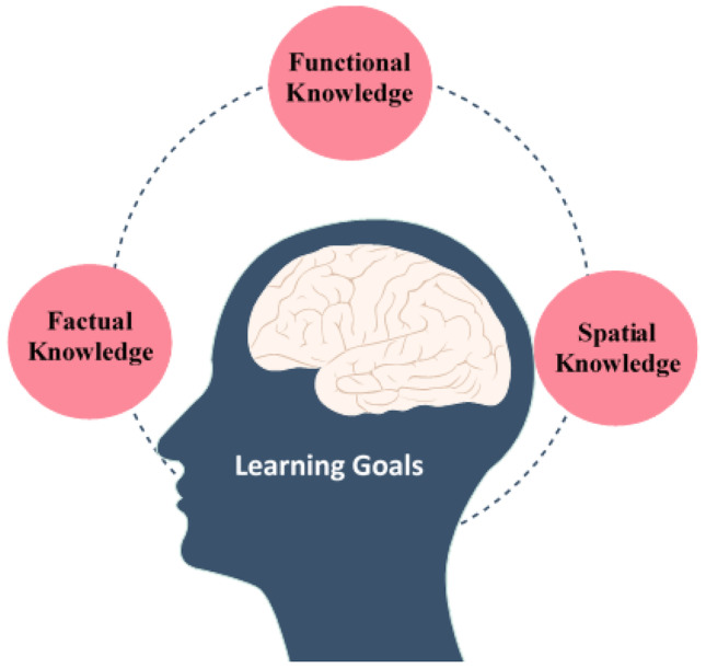
The learning goals of this study
Anatomy Knowledge Assessment
A 20-item knowledge test evaluated the students’ learning outcomes of gastrointestinal anatomy. The tests consisted of scenarios-based questions with four multiple-choice options to answer, and all questions were counted equally (Supplementary Material 2). For each knowledge test, 20 questions were extracted from the question pool. All tests were examined for questions’ repetition, difficulty, and discrimination indexes. Three experts performed content validation in anatomical sciences and medical education for the pretest, posttest, and follow-up tests piloted among 15 medical students for item clarity. The reliability of examinations was good regarding the test using Cronbach’s alpha (Cronbach’s alpha value was in the range of 0.79 to 0.86). All the students completed the same written assessments. The students in both groups attended the pretest (non-announced examination) and posttests. Moreover, they were asked to take the follow-up examination 2 weeks later.
Satisfaction Assessment
The satisfaction questionnaire developed by Borimnejad et al. was used to assess students’ satisfaction. It consisted of 16 items; for each item, students were asked about their agreement with a statement on a 3-point Likert scale (1 = disagree; 2 = neither agree nor disagree; 3 = agree) [28]. The minimum score was 16, and the maximum score was 48. A higher score indicated a higher level of satisfaction. According to Borimnejad et al., the content validity index of the questionnaire was 0.85. They performed the test–retest method to evaluate the tool’s reliability, and the correlation between the two implementation intervals was 0.90.
Statistical Analysis
Participants’ characteristics were summarized using descriptive statistics. The data were analyzed using SPSS, version 27.0 (IBM Corp., Armonk, NY). The Shapiro–Wilk test assessed the normality of data distribution. Student scores for the gastrointestinal anatomy in the pretest, posttest, and follow-up were analyzed with a repeated measure analysis of variance test and subsequently with the Bonferroni post hoc test for pairwise comparisons and the effect size (partial η2). Data on the student’s satisfaction were analyzed using the independent t-test, and the effect size (Cohen’s d) of the differences between the two groups was calculated. The statistical significance was determined at the level of P < 0.05.
Results
A total of 56 medical students participated in this study, with a mean age of 19.94 ± 0.94 years. Most were male (55%), and the mean grade point average (GPA) was 16.70 ± 1.48 out of 20. The control and intervention groups were homogeneous according to their basic characteristics and background knowledge of gastrointestinal anatomy, which was determined based on the student’s pretest scores (Table 1).
Table 1.
Basic characteristics of study participants
| Variable |
Overall (N = 56) |
VDT group (N = 27) |
Textbook group (N = 29) |
p-valuea | |
|---|---|---|---|---|---|
| N (%) | N (%) | N (%) | |||
| Mean ± SD | Mean ± SD | Mean ± SD | |||
| Gender | Female | 25 (44.6) | 13 (48.1) | 12 (41.4) | 0.41 |
| Male | 31 (55.4) | 14 (51.9) | 17 (58.6) | ||
| Age (years) | 19.94 ± 0.94 | 19.85 ± 0.82 | 20.04 ± 1.05 | 0.56 | |
| GPA out of 20 | 16.70 ± 1.48 | 16.84 ± 1.16 | 16.57 ± 1.74 | 0.87 | |
| Background knowledge of gastrointestinal anatomy out of 20 | 6.45 ± 3.6 | 6.22 ± 3.12 | 6.69 ± 3.98 | 0.15 | |
GPA grade point average, SD standard deviation
aChi-square or Mann–Whitney test.
As demonstrated in Fig. 3, in the posttest and follow-up, the mean of students’ learning outcomes of gastrointestinal anatomy in the VDT group was higher than that in the control group.
Fig. 3.

Comparison of students’ learning outcomes of gastrointestinal anatomy between two groups at different times
The repeated measures analysis of the variance test in Table 2 showed a significant difference between the two groups (F = 13.33, df = 2, P < 0.01, partial η2 = 0.20).
Table 2.
Results of univariate analysis of variance in students’ learning outcomes of gastrointestinal anatomy
| Source of changes | Sum of squares | df | Mean of squares | F | p-value | Partial η2 |
|---|---|---|---|---|---|---|
| Timea | 2900.61 | 2 | 1450.30 | 196.84 | 0.001 | 0.79 |
| Groupb | 161.10 | 1 | 161.10 | 7.66 | 0.008 | 0.12 |
| Time × Group | 196.64 | 2 | 98.33 | 13.33 | 0.001 | 0.20 |
aPretest, posttest, and follow-up
bControl and intervention groups
As shown in Table 3, the mean changes in students’ learning significantly increased in both groups, at posttest vs pretest (P < 0.01), follow-up vs pretest (P < 0.01), and follow-up vs posttest (P < 0.01). Comparing the size of the effects showed that the effectiveness of intervention in improving students’ learning outcomes of gastrointestinal anatomy is more than conventional education in the control group. Comparing groups’ satisfaction indicated the intervention group of students who experienced an enriched learning environment with VDT was more satisfied than the control group (T = 6.10, df = 54, P < 0.01, Cohen’s d = 1.63, CI95% = 1.02–2.23).
Table 3.
Comparison of the student’s learning outcomes and satisfaction between groups
| Analysis of students’ learning outcomes | |||||||
|---|---|---|---|---|---|---|---|
| Groups |
Pretest–posttest (mean difference) |
ap-value |
Pretest-follow-up (mean difference) |
ap-value |
Posttest-follow-up (mean difference) |
ap-value | Partial η2 |
| VDT group | − 8.70 | 0.001 | − 11.13 | 0.001 | − 2.43 | 0.007 | 0.84 |
| Textbook group | − 3.45 | 0.001 | − 9.10 | 0.001 | − 5.66 | 0.001 | 0.73 |
| Analysis of students’ satisfaction | |||||||
| Groups | Mean ± SD | Mean difference | df | T | p-value | Partial η2 | |
| VDT group | 45.56 ± 5.03 | 7.53 | 54 | 6.10 | 0.001 | 0.40 | |
| Textbook group | 38.03 ± 4.19 | ||||||
aBonferroni post hoc test
Discussion
The present study results indicated that integrating the VDT into the gastrointestinal anatomy course could enhance the students’ intended learning outcomes and satisfaction. The findings are consistent with several studies that have demonstrated VDT technology’s effectiveness in learning [8–11]. Bartoletti-Stella et al. reported medical students’ great interest and the positive impact of 3D virtual anatomy in teaching gross anatomy [29]. Duraes et al. developed a 4D virtual dissection and declared that most medical students (96.4%) and surgery residents (100%) would recommend virtual dissection to their colleagues. According to their study, the main advantages of that virtual tool were the realistic, 3D features, precise focus feasibility on anatomical structures, interactivity, and entertainment aspect [30]. Tenew et al. found that over 80% of participants were satisfied with using the anatomage table and confirmed its complementary effectiveness in increasing anatomy learning [31]. In Bharati and Rani’s study, 96% of 115 students preferred the images and cross-sections of the dissection table to the pictures from anatomy reference books to understand anatomy. In addition, most students strongly agreed that the VDT is very efficient in visualizing the human body when dissecting. In their opinion, the essential feature of the dissection table was the possibility of reconstructing images [32]. According to Smith et al., the anatomage table helped capture regional anatomy images for lecture presentations and develop textbooks [33].
Nevertheless, our findings contrast with some research regarding the different nature of the study design, learning environment, or additional learning tools. Anand et al. conducted a randomized prospective cross-sectional study of medical students comparing the VDT to conventional dissection. They found the knowledge gained was not different statistically between the groups and inferred that a virtual dissection table could be considered as good as a traditional dissection [34]. Metzler et al. studied undergraduate medical students’ comprehension in a randomized controlled trial. They concluded that training in either 3D or 2D did not affect the students’ correct interpretation of 2D imaging [35].
Cicerchia et al. studied modalities and educational tools across schools and found that anatomy education for most contact time was based upon lecture-based learning and cadaveric dissection. They declared that conventional methods might be more popular as they are integrated and utilized with newer educational technologies [36].
To sum up, the VDT could help to improve students’ understanding of anatomy concepts and organ variation [20]. The addition of educational technologies in anatomy assists medical students in developing deep learning and higher-order thinking, which increases knowledge retention [37, 38]. Moreover, the VDT allows educators to offer a spatial visualization of the human body, facilitate anatomy’s clinical applications [1, 39, 40], and augment the classroom experience of learners [41]. Therefore, it might be assumed that VDT technology is at the top of anatomy education technologies [32, 42, 43]. However, no single or specific method can be prescribed for teaching and learning anatomy. Instead, the findings indicate that students’ learning depends more on how faculty use such techniques [2].
There are some limitations to this research. First, our results arose from a single-center study, and the number of students was restricted. Second, the students’ spatial ability was not considered, while their capacity to understand a 3D structure could interfere with learning. Therefore, measuring students’ spatial ability in future studies is suggested.
Conclusions
Integrating educational technologies will encourage students’ interest, anatomical knowledge retention, and clinical relevance. Findings indicated a positive learning experience, and it is inferred that medical students perceive this approach as an effective method that enhances their understanding and satisfaction with the anatomy curriculum. Using the VDT seems to have an influential and promising role in medical education, and its use is recommended as a learning resource to enrich conventional classrooms based on textbooks. Finally, in addition, its significant role during the COVID-19 pandemic will add great value to medical and health science education in the postmodern era. However, more research is needed to understand this technology’s efficacy and examine its long-term impacts on learning and retaining anatomical concepts. Future works should regularly assess how medical education’s relative use of different teaching tools and educational technologies changes.
Supplementary Information
Below is the link to the electronic supplementary material.
Acknowledgements
This article is an excerpt from the Master’s Thesis on Educational Technology in Medical Sciences. We thank Mashhad University of Medical Sciences for its funding and support. Moreover, we appreciate Tabriz University of Medical Sciences Medical Technology Complex. The authors are grateful to the medical students who contributed to this study.
Author Contributions
Study concept and design: AE and HM; Data acquisition: HE, SM, and ME; Analysis and interpretation of data: HE; Drafting of the manuscript: HM, AE, SM, MY, and HE; Critical revision of the manuscript for important intellectual content: AE, and HM; Study supervision: AE.
Funding
This work was funded by the Mashhad University of Medical Sciences, Iran (Grant Number 991767).
Availability of Data and Materials
The datasets used and/or analyzed during the current study are available from the corresponding author upon reasonable request.
Declarations
Relevant Guidelines and Regulations
All methods and procedures were performed under the relevant guidelines and regulations of the School of Medicine, Mashhad University of Medical Sciences, Iran (approval date was 2021–12-15 with 991767 reference code).
Ethics Approval and Consent To Participate
All procedures were in line with the Research Ethics Committees of the School of Medicine, Mashhad University of Medical Sciences (approval date was 2021–11-30 with IR.MUMS.MEDICAL.REC.1400.607 approval ID), and all participants provided informed consent.
Consent for Publication
Not applicable.
Conflict of Interest
The authors declare no conflict of interests.
Footnotes
Publisher's Note
Springer Nature remains neutral with regard to jurisdictional claims in published maps and institutional affiliations.
Contributor Information
Ali Emadzadeh, Email: EmadzadeA@mums.ac.ir.
Hasan EidiBaygi, Email: EidiH1@mums.ac.ir.
Shabnam Mohammadi, Email: MohammadiSh@mums.ac.ir.
Mohammad Etezadpour, Email: EtezadpourM@mums.ac.ir.
Mehri Yavari, Email: YavariM@mums.ac.ir.
Haniye Mastour, Email: Haniye.Mastour@gmail.com.
References
- 1.Darras KE, Spouge R, Hatala R, Nicolaou S, Hu J, Worthington A, et al. Integrated virtual and cadaveric dissection laboratories enhance first year medical students’ anatomy experience: a pilot study. BMC Med Educ. 2019;19(1):1–6. doi: 10.1186/s12909-019-1806-5. [DOI] [PMC free article] [PubMed] [Google Scholar]
- 2.Washmuth NB, Cahoon T, Tuggle K, Hunsinger RN. Virtual dissection: alternative to cadaveric dissection for a pregnant nurse anesthesia student. Health Prof Educ. 2020;6(2):247–255. [Google Scholar]
- 3.Nyemb PMMA. Anatomy and clinical reasoning. MOJ Anat Physiol. 2018;5(1):19–21. [Google Scholar]
- 4.Elizondo-Omaña RE, Guzmán-López S, García-Rodríguez ML. Dissection as a teaching tool: past, present, and future. Anat Rec B New Anat. 2005;285(1):11–15. doi: 10.1002/ar.b.20070. [DOI] [PubMed] [Google Scholar]
- 5.Cheung CC, Bridges SM, Tipoe GL. Why is anatomy difficult to learn? The implications for undergraduate medical curricula. Anat Sci Educ. 2021;14(6):752–763. doi: 10.1002/ase.2071. [DOI] [PubMed] [Google Scholar]
- 6.Waterston SW, Stewart IJ. Survey of clinicians’ attitudes to the anatomical teaching and knowledge of medical students. Clinical Anatomy: Off J Am Assoc Clin Ana British Assoc Clin Ana. 2005;18(5):380–384. doi: 10.1002/ca.20101. [DOI] [PubMed] [Google Scholar]
- 7.Van der Veken J, Valcke M, De Maeseneer J, Derese A. Impact of the transition from a conventional to an integrated contextual medical curriculum on students’ learning patterns: a longitudinal study. Med Teach. 2009;31(5):433–441. doi: 10.1080/01421590802141159. [DOI] [PubMed] [Google Scholar]
- 8.Khalil MK, Giannaris EL, Lee V, Baatar D, Richter S, Johansen KS, et al. Integration of clinical anatomical sciences in medical education: Design, development and implementation strategies. Clin Ana. 2021;34(5):785–793. doi: 10.1002/ca.23736. [DOI] [PubMed] [Google Scholar]
- 9.Bogomolova K, van der Ham IJM, Dankbaar MEW, van den Broek WW, Hovius SER, van der Hage JA, et al. The Effect of Stereoscopic Augmented Reality Visualization on Learning Anatomy and the Modifying Effect of Visual-Spatial Abilities: A Double-Center Randomized Controlled Trial. Anat Sci Educ. 2020;13(5):558–567. doi: 10.1002/ase.1941. [DOI] [PubMed] [Google Scholar]
- 10.Tan S, Hu A, Wilson T, Ladak H, Haase P, Fung K. Role of a computer-generated three-dimensional laryngeal model in anatomy teaching for advanced learners. J Laryngol Otol. 2012;126(4):395–401. doi: 10.1017/S0022215111002830. [DOI] [PubMed] [Google Scholar]
- 11.Dalgarno B, Lee MJW. What are the learning affordances of 3-D virtual environments? Br J Edu Technol. 2010;41(1):10–32. doi: 10.1111/j.1467-8535.2009.01038.x. [DOI] [Google Scholar]
- 12.Pickering JD, Swinnerton BJ. Exploring the Dimensions of Medical Student Engagement with Technology-Enhanced Learning Resources and Assessing the Impact on Assessment Outcomes. Anat Sci Educ. 2019;12(2):117–128. doi: 10.1002/ase.1810. [DOI] [PubMed] [Google Scholar]
- 13.De Caro R, Macchi V, Porzionato A. Promotion of body donation and use of cadavers in anatomical education at the University of Padova. Anat Sci Educ. 2009;2(2):91–92. doi: 10.1002/ase.69. [DOI] [PubMed] [Google Scholar]
- 14.Custer T, Michael K. The utilization of the anatomage virtual dissection table in the education of imaging science students. J Tomogr Simul. 2015;1(1):102–107. doi: 10.4172/jts.1000102. [DOI] [Google Scholar]
- 15.Vasil'ev YL, Dydykin SS, Kashtanov AD, Molotok EV, Lyakisheva AA, Kytko OV, et al. A comparative analysis of lecturers’ satisfaction with Anatomage and Pirogov virtual dissection tables during clinical and topographic anatomy courses in Russian universities. Anat Sci Educ. 2023;16(2):196–208. doi: 10.1002/ase.2248. [DOI] [PubMed] [Google Scholar]
- 16.Uhl JF, Mogorron J, Chahim M. The Virtual Dissection Table: A 3D Atlas of the Human Body Using Vectorial Modeling from Anatomical Slices. In: Uhl J-F, Jorge J, Lopes DS, Campos PF, editors. Digital Anatomy : Applications of Virtual, Mixed and Augmented Reality. Cham: Springer International Publishing; 2021. pp. 103–120. [Google Scholar]
- 17.Duparc F. 3D-Virtual dissection table: we did not imagine how much it will be useful for teaching anatomy and clinical anatomy. Mesa de Disección Virtual 3D: No imaginamos cuán útil sería para la enseñanza de la Anatomía y la Anatomía Clínica. Revista Argentina de Anatomía Clínica. 2017;9(1):9–10.
- 18.Sadeghi H, Bahramian H, Mosavi-Alkazemi A. Effects of Integrating Applied Material in Traditional Anatomy Education on Medical Students’ Knowledge. J Isfahan Med Sch. 2013;31(225).
- 19.Onigbinde OA, Chia T, Oyeniran OI, Ajagbe AO. The place of cadaveric dissection in post-COVID-19 anatomy education. Morphologie. 2021;105(351):259–266. doi: 10.1016/j.morpho.2020.12.004. [DOI] [PubMed] [Google Scholar]
- 20.Said Ahmed MAA. Use of the anatomage virtual table in medical education and as a diagnostic tool: an integrative review. Cureus. 2023;15(3):e35981. doi: 10.7759/cureus.35981. [DOI] [PMC free article] [PubMed] [Google Scholar]
- 21.Boscolo-Berto R, Tortorella C, Porzionato A, Stecco C, Picardi EEE, Macchi V, et al. The additional role of virtual to traditional dissection in teaching anatomy: a randomised controlled trial. Surg Radiol Anat. 2021;43(4):469–479. doi: 10.1007/s00276-020-02551-2. [DOI] [PMC free article] [PubMed] [Google Scholar]
- 22.Lombardi SA, Hicks RE, Thompson KV, Marbach-Ad G. Are all hands-on activities equally effective? Effect of using plastic models, organ dissections, and virtual dissections on student learning and perceptions. Adv Physiol Educ. 2014;38(1):80–86. doi: 10.1152/advan.00154.2012. [DOI] [PubMed] [Google Scholar]
- 23.Fyfe S, Fyfe G, Dye D, Radley-Crabb H. The Anatomage table: Differences in student ratings between initial implementation and established use. Focus on Health Professional Education: A Multi-Professional Journal. 2018;19(2):41–52. doi: 10.11157/fohpe.v19i2.215. [DOI] [Google Scholar]
- 24.Owolabi J. Protocol development for digisection: making a case for standardizing educational technology use for digital dissection and anatomical studies. Cureus. 2023;15(3):e35766. doi: 10.7759/cureus.35766. [DOI] [PMC free article] [PubMed] [Google Scholar]
- 25.Tretter JT, Gupta SK, Izawa Y, Nishii T, Mori S. Virtual Dissection: Emerging as the Gold Standard of Analyzing Living Heart Anatomy. Journal of Cardiovascular Development and Disease. 2020;7(3):30. doi: 10.3390/jcdd7030030. [DOI] [PMC free article] [PubMed] [Google Scholar]
- 26.Drake RL, Vogl AW, Mitchell AWM. Gray's anatomy for students. Tehran: Andishe Rafi; 2021. [Google Scholar]
- 27.Wineski LE. Snell's clinical anatomy by regions. Tehran: Arjmand; 2019. [Google Scholar]
- 28.Borim Nejad L, Sajadi Hezaveh M, Khosravi S. The effect of learning contract on self-directed learning and satisfaction of nursing students in clinical education. Iran J Med Educ. 2015;14(12):1084–92. http://ijme.mui.ac.ir/article-1-3240-en.html.
- 29.Bartoletti-Stella A, Gatta V, Mariani GA, Gobbi P, Falconi M, Manzoli L, et al. Three-Dimensional Virtual Anatomy as a New Approach for Medical Student’s Learning. Int J Environ Res Public Health. 2021;18(24):13247. doi: 10.3390/ijerph182413247. [DOI] [PMC free article] [PubMed] [Google Scholar]
- 30.Duraes M, Akkari M, Jeandel C, Moreno B, Subsol G, Duflos C, et al. Dynamic three-dimensional virtual environment to improve learning of anatomical structures. Anat Sci Educ. 2022;15(4):754–764. doi: 10.1002/ase.2158. [DOI] [PubMed] [Google Scholar]
- 31.Tenaw B. Teaching gross anatomy: anatomage table as an innovative line of attack. Int J Anat Var. 2020;13(1):76. [Google Scholar]
- 32.Bharati AS, Naslk SK, Rani VS. A study on student perception of virtual dissection table (anatomage) at GSL Medical College, Rajahmundry. 2018;4(2):28. 10.21276/aanat.2018.4.2.8.
- 33.Smith KE, Ruholl HO, Gopalan C. Utilization of Anatomage Table Technology Enhances Knowledge, Comprehension, and Application of Human Anatomy and Physiology in Multiple Settings. The FASEB Journal. 2019;33(S1):598.19.
- 34.Anand MK, Singel TC. A comparative study of learning with “anatomage” virtual dissection table versus traditional dissection method in neuroanatomy. Indian Journal of Clinical Anatomy and Physiology. 2017;4:177–180. [Google Scholar]
- 35.Metzler R, Stein D, Tetzlaff R, Bruckner T, Meinzer H-P, Büchler MW, et al. Teaching on Three-Dimensional Presentation Does Not Improve the Understanding of According CT Images: A Randomized Controlled Study. Teach Learn Med. 2012;24(2):140–148. doi: 10.1080/10401334.2012.664963. [DOI] [PubMed] [Google Scholar]
- 36.Cicerchia G, Lumpkins K, Puche AC. Assessment of anatomy education teaching modalities before and during COVID-19 in US medical schools. Educ Res Int. 2023;2023:2108105. doi: 10.1155/2023/2108105. [DOI] [Google Scholar]
- 37.Turney BW. Anatomy in a Modern Medical Curriculum. The Annals of The Royal College of Surgeons of England. 2007;89(2):104–107. doi: 10.1308/003588407X168244. [DOI] [PMC free article] [PubMed] [Google Scholar]
- 38.Alasmari WA. Medical Students’ Feedback of Applying the Virtual Dissection Table (Anatomage) in Learning Anatomy: A Cross-sectional Descriptive Study. Adv Med Educ Pract. 2021;12:1303–1307. doi: 10.2147/AMEP.S324520. [DOI] [PMC free article] [PubMed] [Google Scholar]
- 39.Paech D, Giesel FL, Unterhinninghofen R, Schlemmer H-P, Kuner T, Doll S. Cadaver-specific CT scans visualized at the dissection table combined with virtual dissection tables improve learning performance in general gross anatomy. Eur Radiol. 2017;27(5):2153–2160. doi: 10.1007/s00330-016-4554-5. [DOI] [PubMed] [Google Scholar]
- 40.Darras KE, Forster BB, Spouge R, de Bruin AB, Arnold A, Nicolaou S, et al. Virtual dissection with clinical radiology cases provides educational value to first year medical students. Acad Radiol. 2020;27(11):1633–1640. doi: 10.1016/j.acra.2019.09.031. [DOI] [PubMed] [Google Scholar]
- 41.Irby DM, Cooke M, O'Brien BC. Calls for reform of medical education by the Carnegie Foundation for the Advancement of Teaching: 1910 and 2010. Acad Med. 2010;85(2):220–227. doi: 10.1097/ACM.0b013e3181c88449. [DOI] [PubMed] [Google Scholar]
- 42.Kažoka D, Pilmane M. Teaching and learning innovation in present and future of human anatomy course at RSU. Paper Anthropol. 2017;26(2):44–52. doi: 10.12697/poa.2017.26.2.05. [DOI] [Google Scholar]
- 43.Fyfe G, Fyfe S, Dye D, editors. Use of Anatomage tables in a large first year core unit. ASCILITE-Australian Society for Computers in Learning in Tertiary Education Annual Conference; 2013: Australasian Society for Computers in Learning in Tertiary Education.
Associated Data
This section collects any data citations, data availability statements, or supplementary materials included in this article.
Supplementary Materials
Data Availability Statement
The datasets used and/or analyzed during the current study are available from the corresponding author upon reasonable request.