Abstract
A 131-day-old male Japanese Black calf presented with a swollen right cheek from birth. Imaging examination revealed a cyst under the right buccal area and debris-containing fluid inside the cyst, and puncture aspiration revealed a mildly cloudy fluid containing hair and tissue fragments. Histological examination of the excised cyst revealed stratified squamous epithelium with skin appendages in the cyst wall, which was diagnosed as a dermoid cyst. In addition, some submandibular gland tissue was found within the cyst wall. After removal of the cyst, there was swelling in the same area, which resolved with steroid administration. Surgical treatment of buccal dermoid cysts should be performed with caution to avoid damage to adjacent salivary gland tissue.
Keywords: calf, cyst, dermoid, malar, mandibular gland
Dermoid cyst is an embryonic developmental anomaly caused by defective epidermal closure along embryonic fissures [23], which isolates an island of ectoderm in the dermis or subcutis [14, 21, 26].
This abnormality has been reported in humans [19, 22, 25, 27, 30], dogs [6, 8, 20, 24, 29, 34, 38], cats [2, 12, 20, 31, 36], horses [18], goats [13], sheep [33], buffalo [9, 39], and camels [26]. Lesions occur in young animals, predominantly on the midline of the head and neck [16, 21]. In cattle, dermoid cysts have been reported in the cornea, conjunctiva, eyelids, and nasolacrimal ducts [3,4,5, 7, 21, 32, 35].
Histologically, the cyst wall is characterized by the presence of skin appendages, such as sebaceous glands, sweat glands, and hair follicles [21]. The cyst itself is often painless and does not present with clinical signs [37]; however, clinical signs may appear due to the internal accumulation of sebaceous secretions, which causes the cyst to enlarge gradually.
In the present case, a cyst was found under the right buccal area in a Japanese Black calf with swelling of the right cheek and mandible from birth. The calf was observed without any specific treatment; however, the cyst gradually enlarged and was surgically removed and diagnosed as a dermoid cyst based on histopathological examination.
At the age of 131 days (first sick day; D1), with a body weight of 122 kg, the calf was admitted to Miyazaki University Veterinary Teaching Hospital for diagnostic and prognostic evaluation and treatment. On initial examination, the calf’s heart rate, respiratory rate, and rectal temperature were 128 beats/min, 52 breaths/min, and 39.1°C, respectively. It was in good general condition, although sialorrhea was seen from the corners of the mouth. The right cheek was swollen to 14 × 12 × 8 cm (Fig. 1A). The swollen area had a rippling sensation; there was no reaction to palpation and no heat or pain clinical signs suggestive of inflammation. Palpation revealed outward displacement of the right mandibular angle.
Fig. 1.
Swelling on the right side of the cheek to the mandible in a Japanese Black calf (A). Lateral radiograph showing the deformity of the right mandible bone (B). Mid-sagittal section computed tomography image showing the cyst present in the right buccal region (C).
The calf’s complete blood cell count was in the normal range (white blood cells: 8,200 cells/µL, red blood cells: 879 × 104 cells/µL, platelets: 45.7 × 10⁴ cells/µL, hematocrit level: 33.0%). The serum examination showed no abnormalities.
Radiographs were taken using a portable X-ray unit (PX-20HF; Kenko, Osaka, Japan) with the following parameters: 70 kV, 1.8 mAs, and 1-m film-focus distance. Images of the head acquired using a computerized radiography unit (Regius Sigma; Konica Minolta, Tokyo, Japan) did not show any osteolysis or periosteal reaction at the site of the cyst (Fig. 1B).
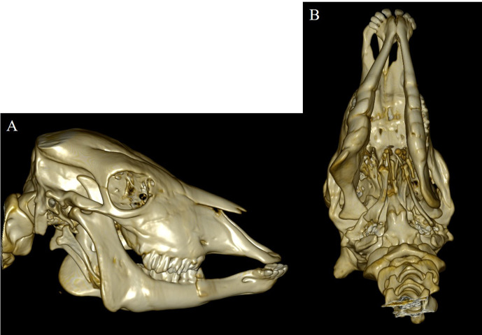
The calf was sedated with intravenous administration of xylazine hydrochloride (0.2 mg/kg) and underwent computed tomography (CT) using a helical 16-row multi-detector CT device (Aquilion Lightning TSX-035A; Canon, Ohtawara, Japan) to evaluate skeletal abnormalities. CT revealed the cyst in the right buccal region (Fig. 1C), as well as outward displacement of the mandibular angle and masseter muscle fossa of the right mandible along the cyst (Fig. 2A) and further displacement of the mandible to the left side, resulting in symmetry loss (Fig. 2B). The CT value of the right buccal cyst was 6–8 HU.
Fig. 2.
Computed tomography (CT) images of the skull bone. CT scan showed outward displacement of the mandibular angle and masseter fossa of the right mandible as if pushed by the cyst (A) and further displacement of the mandible to the left side, resulting in loss of symmetry (B).
Ultrasonography revealed fluid with debris inside the cyst. Aspiration of the cyst fluid yielded a mildly turbid fluid containing small amounts of hair and tissue fragments. The contents of the cyst were aspirated as much as possible; however, the cyst returned to its pre-aspirated state 12 hr later. Bacteriological examination of the aspirated fluid revealed no significant bacterial growth in aerobic and anaerobic cultures.
The clinical diagnosis of buccal dermoid cyst was made based on the condition of the fluid, and cyst excision surgery was performed on D3.
The calf was fasted for 12 hr preoperatively. Then, it received an intravenous injection of cefazolin sodium (5 mg/kg) (Cefazolin-Chu; Fujita Pharmaceutical, Tokyo, Japan) to prevent perioperative infection and flunixin meglumine (2 mg/kg; Forvet50; MSD, Tokyo, Japan) for pain relief. Subsequently, the calf was sedated with intravenous administration of xylazine hydrochloride (0.2 mg/kg) and placed in the left lateral recumbency position. Next, general anesthesia was induced by continuous administration of isoflurane (Isoflu; Zoetis Japan, Tokyo, Japan) at a concentration of 2% in oxygen. Local anesthesia with procaine hydrochloride (Adsan; Riken Vets Pharma, Saitama, Japan) was administered subcutaneously around the incision line of the right cheek.
An approximately 10-cm-long oblique incision was made just above the right buccal swelling in the direction of the mandibular angle from below the ear, and the cyst was found just below the external jugular vein (Fig. 3A). The cyst was adherent to the external jugular vein and mandibular nerve. Notably, the cyst was highly adherent to the mandibular gland. The cyst was carefully removed to avoid damaging these adherent tissues. After removal, the area where the cyst was located became a large cavity (Fig. 3B), but no bleeding or serous leakage was observed.
Fig. 3.
Intraoperative view of cyst removal. A cyst (*) was found just below the external jugular vein (white arrow) (A). After removal of the cyst, the area where the cyst was located became a large cavity (B, white arrow; external jugular vein). The excised cyst was 12 × 11 × 8 cm in size (C); the interior contained translucent fluid containing debris and hair, and the entire inner wall was covered with hair (D).
The subcutaneous tissue was closed with a continuous suture pattern, using a polyglactin 910 synthetic absorbable suture material (coated Vicryl, USP 0). The skin incision was ligatured with intradermal buried sutures using the same synthetic absorbable thread as for the subcutaneous tissue.
The excised cyst was 12 × 11 × 8 cm in size (Fig. 3C), with translucent fluid containing debris and hair inside and hair adhering to the entire inner wall (Fig. 3D). In addition, a simple urine test strip (Labstix, Siemens Medical Solutions USA, Inc., Malvern, PA, USA) showed a pH of 7.5. The cyst was completely removed, and there was no exudate or bleeding from the area around the cyst after removal; therefore, no further treatment was administered. However, 10 hr later, the area from which the cyst had been removed had become swollen to the same degree as before the surgery. Ultrasonography of the area revealed fluid retention in the subcutaneous tissue of the swollen area; edema, not a cyst, was suspected.
Postoperatively, a follow-up CT scan was performed, and the CT value of the right buccal swelling area was 15–30 HU.
Since we could not rule out the possibility that the adjacent mandibular gland was injured when the cyst was removed, we reopened the site on D4 to assess the situation. The subcutaneous tissue was edematous with a small amount of fluid. Considering that the mandibular gland could have been injured during cyst removal, the mandibular gland was removed using a vessel sealing device (Liga-SureTM Smart Jaw; Medtronic PLC, Dublin, Ireland).
The day after the second surgery, the right cheek swelled up again, and ultrasonography showed the same edema-like echo image as the day before. Preoperative ultrasonography of the swollen area showed fluid containing debris, but two postoperative examinations of the swollen area were both suggestive of edema. Prednisolone (50 mg/day, Kyoritsu Seiyaku Corp., Tokyo, Japan) was administered for 4 days to reduce inflammation, and a compound drug containing 200,000 units of benzylpenicillin procaine and 250 mg of dihydrostreptomycin sulfate (0.05 mL/kg, Mycillin Sol; Meiji-Seika Pharma, Tokyo, Japan) was administered intramuscularly for 6 days to prevent postoperative infection.
On the third day of prednisolone administration, the swelling in the right cheek area decreased, and by the 10th postoperative day, the swelling had disappeared completely. Seven months postoperatively, no recurrence was observed.
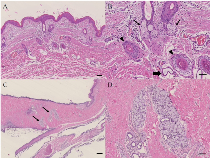
Histopathological examination of the excised cyst revealed that it was lined with stratified squamous epithelium with skin appendages (hair, hair follicles, sweat glands, and sebaceous glands) (Fig. 4A, 4B). These histological findings led to the diagnosis of a dermoid cyst in support of the clinical diagnosis. In addition, some mandibular gland tissue was found within the cyst wall, but there were no findings of an opening into the cyst (Fig. 4C, 4D).
Fig. 4.
Photomicrograph of a section of the dermoid cyst. The cyst was lined with stratified squamous epithelium (H&E; bar=100 μm) (A) and contained widely scattered adnexal structures (arrowheads: follicle; line arrows: sebaceous gland, bold arrow: sweat gland) associated with the cyst wall (H&E; bar=50 μm) (B). Mandibular gland tissue (arrows) was found within the wall of the dermoid cyst (H&E; bar=500 μm) (C). Mandibular gland tissue within the dermoid cyst wall (H&E; bar=100 μm) (D). H&E, hematoxylin and eosin.
A dermoid cyst is the simplest form of teratoma and is usually present from birth [1, 28]. Histologically, they are classified into three categories: epidermoid, dermoid, and teratoid cysts, and dermoid cysts are differentiated from the others by the presence of appendages, such as hair follicles, sweat glands, and sebaceous glands, in the inner wall of the cyst [10, 17, 20, 30]. The acquired form may occur secondary to traumatic displacement of epithelial tissue; however, the incidence is low, approximately 10% [15].
In this calf, hair, hair follicles, and sebaceous glands were observed as skin appendages in the cyst wall, histologically consistent with the description of a dermoid cyst. In addition, there was no history of previous wounds or trauma, the affected area was already swollen at birth, and deformities of the mandible bone were observed along the cyst, suggesting a congenital dermoid cyst.
Surgical excision is recommended for the treatment of dermoid cysts to prevent leakage of the contents [11]. If resection is incomplete and the cyst capsule remains, recurrence is possible.
The differential diagnosis of dermoid cysts includes similar diseases with buccal swelling, such as salivary gland disease, swollen lymph nodes, cellulitis, and odontogenic tumors. In the present case, radiography did not reveal any findings indicating odontogenic tumors, such as osteolysis, lesion periosteal reaction, or the presence of buried teeth. Furthermore, since the lesion was painless and showed no clinical signs, we ruled out inflammatory disease, and since the aspirated contents contained hair, we considered a high possibility of a dermoid cyst and decided to intervene surgically.
At the time of surgery, the dermoid cyst was highly adherent to the mandibular gland. Some mandibular gland tissue was found in the wall of the removed dermoid cyst, but the mandibular gland did not open into the cyst histologically. This suggests that some mandibular gland tissue may have been incorporated into the cyst wall due to the high adherence between the mandibular gland and the dermoid cyst.
However, swelling was observed in the same area after the cyst and mandibular gland removal. Based on the pH of the cyst contents measured preoperatively, the pre- and postoperative CT values of the swollen area, the absence of abnormalities in the tissues of the removed mandibular gland, and the disappearance of the edema after steroid administration, the swelling in the same area in this case was considered to be due to edema of the tissues surrounding the removed site, not to cyst recurrence or damage to the mandibular gland.
Fortunately, in the present case, the mandibular gland tissue identified within the wall of the dermoid cyst was not opened. However, when salivary glands, such as the submandibular gland, are in close proximity to the dermoid cyst, surgical treatment should be performed carefully to avoid damage to salivary gland tissue.
CONFLICT OF INTEREST
The authors declare no conflict of interest.
Acknowledgments
We thank the University of Miyazaki Veterinary Teaching Hospital staff and laboratory student assistants for caring for the calf during hospitalization. This study did not receive financial support.
REFERENCES
- 1.Abou-Rayyah Y, Rose GE, Konrad H, Chawla SJ, Moseley IF. 2002. Clinical, radiological and pathological examination of periocular dermoid cysts: evidence of inflammation from an early age. Eye (Lond) 16: 507–512. doi: 10.1038/sj.eye.6700045 [DOI] [PubMed] [Google Scholar]
- 2.Akhtardanesh B, Kheirandish R, Azari O. 2012. Dermoid cyst in a domestic shorthair cat. Asian Pac J Trop Biomed 2: 247–249. doi: 10.1016/S2221-1691(12)60051-3 [DOI] [PMC free article] [PubMed] [Google Scholar]
- 3.Alam MM, Rahman MM. 2012. A three years retrospective study on the nature and cause of ocular dermoids in cross-bred calves. Open Vet J 2: 10–14. doi: 10.5455/OVJ.2012.v2.i0.p10 [DOI] [PMC free article] [PubMed] [Google Scholar]
- 4.Baird AN, Wolfe DF, Groth AH. 1993. Dermoid cyst in a bull. J Am Vet Med Assoc 202: 298. [PubMed] [Google Scholar]
- 5.Barkyoumb SD, Leipold HW. 1984. Nature and cause of bilateral ocular dermoids in Hereford cattle. Vet Pathol 21: 316–324. doi: 10.1177/030098588402100309 [DOI] [PubMed] [Google Scholar]
- 6.Bornard N, Pin D, Carozzo C. 2007. Bilateral parieto-occipital dermoid sinuses in a Rottweiler. J Small Anim Pract 48: 107–110. doi: 10.1111/j.1748-5827.2006.00171.x [DOI] [PubMed] [Google Scholar]
- 7.Brudenall DK, Ward DA, Kerr LA, Newman SJ. 2008. Bilateral corneoconjunctival dermoids and nasal choristomas in a calf. Vet Ophthalmol 11: 202–206. doi: 10.1111/j.1463-5224.2008.00580.x [DOI] [PubMed] [Google Scholar]
- 8.Burrow RD. 2004. A nasal dermoid sinus in an English bull terrier. J Small Anim Pract 45: 572–574. doi: 10.1111/j.1748-5827.2004.tb00207.x [DOI] [PubMed] [Google Scholar]
- 9.Dhablania DC, Gupta PP. 1979. Cutaneous dermoid cyst in a buffalo heifer. Indian Vet J 56: 798–799. [Google Scholar]
- 10.Edwards JF. 2002. Three cases of ovarian epidermoid cysts in cattle. Vet Pathol 39: 744–746. doi: 10.1354/vp.39-6-744 [DOI] [PubMed] [Google Scholar]
- 11.Ferrari MM, Mezzopane R, Bulfoni A, Grijuela B, Carminati R, Ferrazzi E, Pardi G. 2003. Surgical treatment of ovarian dermoid cysts: a comparison between laparoscopic and vaginal removal. Eur J Obstet Gynecol Reprod Biol 109: 88–91. doi: 10.1016/S0301-2115(02)00510-9 [DOI] [PubMed] [Google Scholar]
- 12.Fleming JM, Platt SR, Kent M, Freeman AC, Schatzberg SJ. 2011. Cervical dermoid sinus in a cat: case presentation and review of the literature. J Feline Med Surg 13: 992–996. doi: 10.1016/j.jfms.2011.08.003 [DOI] [PMC free article] [PubMed] [Google Scholar]
- 13.Gamlem T, Crawford TB. 1977. Dermoid cysts in identical locations in a doe goat and her kid. Vet Med Small Anim Clin 72: 616–617. [PubMed] [Google Scholar]
- 14.Ginn PE, Mansell J, Pakich PM. 2007. Skin and appendages. pp. 592–593. In: Jubb, Kennedy, and Palmer’s Pathology of Domestic Animals, 5th ed. (Maxie MG ed.), Elsevier Saunders, Edinburgh. [Google Scholar]
- 15.Gold BD, Sheinkopf DE, Levy B. 1974. Dermoid, epidermoid, and teratomatous cysts of the tongue and the floor of the mouth. J Oral Surg 32: 107–111. [PubMed] [Google Scholar]
- 16.Goldschmidt MH, Goldschmidt KH. 2016. Epithelial and melanocytic tumors of the skin. pp. 134–138. In: Tumors in Domestic Animals, 5th ed. (Meuten DJ ed.), John Wiley & Sons, New York. [Google Scholar]
- 17.Harada H, Kusukawa J, Kameyama T. 1995. Congenital teratoid cyst of the floor of the mouth--a case report. Int J Oral Maxillofac Surg 24: 361–362. doi: 10.1016/S0901-5027(05)80492-8 [DOI] [PubMed] [Google Scholar]
- 18.Hillyer LL, Jackson AP, Quinn GC, Day MJ. 2003. Epidermal (infundibular) and dermoid cysts in the dorsal midline of a three-year-old thoroughbred-cross gelding. Vet Dermatol 14: 205–209. doi: 10.1046/j.1365-3164.2003.00345.x [DOI] [PubMed] [Google Scholar]
- 19.Hong SW. 1998. Deep frontotemporal dermoid cyst presenting as a discharging sinus: a case report and review of literature. Br J Plast Surg 51: 255–257. doi: 10.1054/bjps.1997.0236 [DOI] [PubMed] [Google Scholar]
- 20.Kiviranta AM, Lappalainen AK, Hagner K, Jokinen T. 2011. Dermoid sinus and spina bifida in three dogs and a cat. J Small Anim Pract 52: 319–324. doi: 10.1111/j.1748-5827.2011.01062.x [DOI] [PubMed] [Google Scholar]
- 21.Mauldin EA, Peters-Kennedy J. 2015. Integumentary system. p. 547.e1. In: Jubb, Kennedy & Palmer’s Pathology of Domestic Animals: Volume 2, 6th ed. (Maxie GM ed.), Elsevier, St. Louis. [Google Scholar]
- 22.Meyer DR, Lessner AM, Yeatts RP, Linberg JV. 1999. Primary temporal fossa dermoid cysts. Characterization and surgical management. Ophthalmology 106: 342–349. doi: 10.1016/S0161-6420(99)90074-X [DOI] [PubMed] [Google Scholar]
- 23.Miller WH, Griffin CE, Campbell K. 2012. Congenital and hereditary defects. pp. 583–584. In Muller & Kirk’s Small Animal Dermatology, 7th ed. (Miller WH, Griffin CE, Campbell K eds.), WB Saunders, Philadelphia. [Google Scholar]
- 24.Motta L, Skerritt G, Denk D, Leeming G, Saulnier F. 2012. Dermoid sinus type IV associated with spina bifida in a young Victorian bulldog. Vet Rec 170: 127. doi: 10.1136/vr.100314 [DOI] [PubMed] [Google Scholar]
- 25.Niederhagen B, Reich RH, Zentner J. 1998. Temporal dermoid with intracranial extension: report of a case. J Oral Maxillofac Surg 56: 1352–1354. doi: 10.1016/S0278-2391(98)90622-X [DOI] [PubMed] [Google Scholar]
- 26.Oryan A, Hashemnia M, Mohammadalipour A. 2012. Dermoid cyst in camel: a case report and brief literature review. Comp Clin Pathol 21: 555–558. doi: 10.1007/s00580-010-1128-9 [DOI] [Google Scholar]
- 27.Parag P, Prakash PJ, Zachariah N. 2001. Temporal dermoid--an unusual presentation. Pediatr Surg Int 17: 77–79. doi: 10.1007/s003830000396 [DOI] [PubMed] [Google Scholar]
- 28.Perry JD, Tuthill R. 2003. Simultaneous ipsilateral temporal fossa and orbital dermoid cysts. Am J Ophthalmol 135: 413–415. doi: 10.1016/S0002-9394(02)01961-X [DOI] [PubMed] [Google Scholar]
- 29.Rahal S, Mortari AC, Yamashita S, Filho MM, Hatschbac E, Sequeira JL. 2008. Magnetic resonance imaging in the diagnosis of type 1 dermoid sinus in two Rhodesian ridgeback dogs. Can Vet J 49: 871–876. [PMC free article] [PubMed] [Google Scholar]
- 30.Rinna C, Reale G, Calafati V, Calvani F, Ungari C. 2012. Dermoid cyst: unusual localization. J Craniofac Surg 23: e392–e394. doi: 10.1097/SCS.0b013e31825ab1e1 [DOI] [PubMed] [Google Scholar]
- 31.Rochat MC, Campbell GA, Panciera RJ. 1996. Dermoid cysts in cats: two cases and a review of the literature. J Vet Diagn Invest 8: 505–507. doi: 10.1177/104063879600800423 [DOI] [PubMed] [Google Scholar]
- 32.Steinmetz A, Locher L, Delling U, Ionita JC, Ludewig E, Oechtering G, Wittek T. 2009. Surgical removal of a dermoid cyst from the bony part of the nasolacrimal duct in a Scottish highland cattle heifer. Vet Ophthalmol 12: 259–262. doi: 10.1111/j.1463-5224.2009.00704.x [DOI] [PubMed] [Google Scholar]
- 33.Stelmann UJP, da Silva AA, De Souza BG, De Oliveira GF, De Mello EFRB, De Souza GCJ, Calderon Gonçalves R, Hess TM. 2012. Dermoid cyst in sheep −a case report. Rev Bras Med Vet 34: 133–136. [Google Scholar]
- 34.Sturgeon C. 2008. Nasal dermoid sinus cyst in a shih tzu. Vet Rec 163: 219–220. doi: 10.1136/vr.163.7.219 [DOI] [PubMed] [Google Scholar]
- 35.Tamilmahan RP, Prabhakar PS. 2018. Surgical management of dermoid cyst in a cross bred calf. J Entomol Zool Stud 6: 2574–2576. [Google Scholar]
- 36.Tong T, Simpson DJ. 2009. Case report: Spinal dermoid sinus in a Burmese cat with paraparesis. Aust Vet J 87: 450–454. doi: 10.1111/j.1751-0813.2009.00487.x [DOI] [PubMed] [Google Scholar]
- 37.Turki IM, Yosr MZ, Hatem R, Hassen T, Sonia N, Sarra BJ, Najla M, Ali A. 2011. A case of large dermoid cyst of the tongue. Egypt J Ear Nose Throat Allied Sci 12: 171–174. doi: 10.1016/j.ejenta.2011.10.001 [DOI] [Google Scholar]
- 38.van der Peijl GJW, Schaeffer IGF. 2011. Nasal dermoid cyst extending through the frontal bone with no sinus tract in a Dalmatian. J Small Anim Pract 52: 117–120. doi: 10.1111/j.1748-5827.2010.01023.x [DOI] [PubMed] [Google Scholar]
- 39.Verma A, Sangwan V, Bansal N, Kaur H, Wangdi N, Mahajan SK. 2022. A rare case of dermoid cyst associated with parotid gland in a male buffalo calf. Large Anim Rev 28: 307–310. [Google Scholar]


