Abstract
Objective
Immune checkpoint inhibitors (ICIs) have revolutionized cancer therapy but come with immune-related adverse events (irAEs) that provide a novel challenge for treating physicians. Neuromuscular irAEs, including myositis, myasthenia gravis (MG), and demyelinating polyradiculoneuropathy, lead to significant morbidity and mortality.
Methods
We present a case of severe myasthenia-myositis-myocarditis overlap in a patient receiving ICIs for breast cancer. Clinical findings were recorded.
Results
A 47-year-old woman developed tetraparesis, dysphagia, and muscle pain during ICI treatment. MG with a thymoma had been diagnosed earlier. Neuromuscular overlap irAEs with cardiac affection was confirmed, and ICI treatment was discontinued. Given a lack of clinical response to standard therapies, a muscle biopsy was performed demonstrating complement deposition. Eculizumab treatment led to rapid improvement in muscle strength and cardiac function.
Discussion
Neuromuscular irAEs are associated with a high in-hospital mortality, and specific treatment strategies remain an unmet need. Here, early muscle biopsy enabled targeted therapy after standard approaches failed, thereby highlighting the value of identifying a specific treatment target. To improve therapeutic outcomes, the development of patient-tailored strategies for neuromuscular irAEs requires further studies.
Introduction
Immune checkpoint inhibitors (ICIs) transformed the therapeutic landscape of oncology.1 However, clinical efficacy comes at the cost of specific immune-related adverse events (irAEs) presenting a novel challenge for treating physicians. Neurologic irAEs develop in an estimated 2%–4 % of patients treated with ICIs with neuromuscular manifestations accounting for most cases.2,3 The latter includes myositis, myasthenia gravis, demyelinating polyradiculoneuropathy, and overlaps thereof.4,5 While the group of neuromuscular irAEs is heterogeneous, patients with myocardial or bulbar affection face a staggering mortality of approximately 60%.6 Consequently, irAEs require comprehensive management to prevent the occurrence of long-term sequelae and death. While mild irAEs may be mitigated by discontinuation of the ICIs and administration of glucocorticoids, severe or refractory cases require specialized treatment strategies. Current viewpoints emphasize the need for personalized treatment algorithms based on the immunopathologic patterns of the affected patients.7 However, patient-tailored approaches require the analysis of the underlying immunopathology. In the field of neurology, the rarity and difficulty of obtaining target tissue often curtail the study of affected organs leaving a knowledge gap for the management of irAEs.
Methods
In this study, we report a severe case of myasthenia-myositis-myocarditis overlap in which muscle biopsy identified substantial complement deposition providing the rationale for complement inhibition as specific rescue therapy.
Results
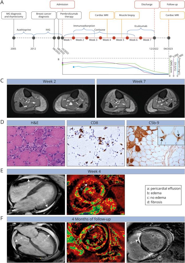
A 47-year-old female patient presented to our clinic with severe tetraparesis. The patient described progressive muscle weakness and difficulties with swallowing and speaking. These symptoms had progressively worsened over the past 2 weeks. MG was diagnosed 17 years earlier based on clinical findings and the detection of antiacetylcholine receptor antibodies (Figure, A). The antiacetylcholine receptor antibody level was 13.2 nmol/L determined approximately 12 months before admission. An associated thymoma had been resected after the diagnosis of MG. The patient had been treated with azathioprine followed by recurrent applications of IV immunoglobulins. The patient experienced only mild muscle weakness of the extremities while receiving this maintenance treatment with her quantitative MG (QMG) score ranging between 2 and 4 points and her MG activities of daily living (ADL) score between 3 and 6 points. Four months before admission, triple-negative breast cancer (cT2, cN, cM0 G3) was diagnosed. Treatment consisted of pembrolizumab, paclitaxel, and epirubicin/cyclophosphamide. Based on the patient history and clinical presentation, we first suspected myasthenic crisis and admitted the patient to our neurologic ward. The QMG score was 17 and the ADL score 18 points on admission. The manual muscle testing (MMT)–8 score was 124/140 with proximal tetraparesis. We monitored the QMG score as readily available clinical parameter throughout the in-hospital stay (Figure, B). Concurrently, laboratory analysis revealed an elevated creatine kinase of approximately 3300 U/I. Because the patient also complained of muscle pain, we considered a neuromuscular overlap of irAEs as underlying pathology. EMG demonstrated abnormal spontaneous activity in both gastrocnemius muscles. ICI was immediately discontinued and the patient started on 2 mg of methylprednisolone/kg. MRI of the lower thigh confirmed active myositis, most pronounced in the gastrocnemius muscle of both sides (Figure, C). The clinical condition worsened with dysphagia as leading symptom requiring parenteral feeding (QMG score: 22 points). We escalated therapy by continuous application of neostigmine and 7 cycles of immunoadsorption. The clinical course remained stable (QMG score 20, MMT8 score 128/140 points), however, without relevant improvements. Elevated troponin (1,071 ng/L) and N-terminal prohormone of brain natriuretic peptide (1,025 ng/L) levels were detected 3 weeks after admission, and pericardial effusion was visible on echocardiography. Cardiac MRI confirmed myocarditis 4 weeks after admission according to the modified Lake Louise criteria 2008.8 In the absence of clinical improvement, we decided to perform a muscle biopsy of the right gastrocnemius muscle. Here, histopathologic analysis demonstrated scattered necrotic and regenerating fibers (Figure, D). Sparse CD8 T-cell infiltration was seen in surrounding of damaged muscle fibers on immunohistochemistry. Concurrently, staining for terminal complement (C5b-9) evidenced substantial complement deposition on scattered muscle fibers and on capillaries. Based on these findings, we considered complement as a therapeutic target and started the patient on the induction cycle of eculizumab 5 weeks after admission. Symptoms improved rapidly as demonstrated by a reduction of the QMG score from 17 points at the start of eculizumab to 6 points 2 weeks later at discharge. Given the clinical improvement, after 6 weeks of treatment, methylprednisolone was tapered until reaching a maintenance dose of 5 mg per day. Follow-up MRI of the lower thigh demonstrated subsiding myositis. Functionally, the patient regained her ability to walk without support. The patient continues to receive eculizumab biweekly. At discharge, serum CK levels declined to 132 U/I. Oncologic therapy was resumed with capecitabine without rechallenging ICI. At follow-up 4 months later, QMG and MMT8 remained stable. The antiacetylcholine receptor antibody level was 11.6 nmol/L. Echocardiography demonstrated a restored ejection fraction. Cardiac MRI was repeated, and myocarditis was ruled out (Figure, E and F).
Figure. Clinical Findings and Course of Disease.
(A) Timeline of preceding events, in-hospital stay and follow-up. (B) Quantitative myasthenia gravis (QMG) score, creatine kinase (CK), and troponin levels during the course of disease. (C) MRI of the lower thigh demonstrating myositis of the gastrocnemius muscle on both sides 2 weeks after admission (left) and 7 weeks after admission (right). T2 sequences with contrast are shown. (D) Hematoxylin and eosin (H&E) stain (left) demonstrating necrotizing myopathy with scattered necrotic and regenerating fibers and numerous capillaries with thickened vessel walls. Immunohistochemistry staining for CD8 (middle). Sparse CD8 positive cells are seen in the vicinity of damaged muscle fibers. Immunohistochemistry staining for C5b-9 (right) displaying complement deposition on scattered muscle fibers and capillaries (marked by black arrows in inlet). 20× magnification was used for image acquisition. (E) Cardiac MRI 4 weeks after admission demonstrating pericardial effusion (a) and substantial edema (b) as signs of myocarditis. T2 sequences are shown (left) and quantification of water content (right; water is indicated in red, healthy tissue in green). (F) Follow-up cardiac MRI 4 months after discharge demonstrating no myocarditis (c) but pericardial effusion (a) and signs of cancer therapy–related cardiac dysfunction with myocardial fibrosis (d). T2 sequences are shown (left) and quantification of water content (middle; water is indicated in red, healthy tissue in green). Furthermore, T2 late gadolinium enhancement is depicted (right).
Discussion
As the use of ICI increases, clinicians will increasingly be confronted with the spectrum of irAEs. Neuromuscular overlap syndromes are of particular interest given their high mortality and morbidity. Indeed, a recent systematic review summarized 60 cases of myasthenia-myositis-myocarditis overlap.6 Here, approximately 70% of patients presented with cardiac arrhythmia and approximately 20% had reduced ejection fraction. Approximately 60% of patients with myasthenia-myositis-myocarditis overlap died in hospital due to acute complications highlighting the urgent need for optimization of diagnostic and therapeutic approaches to combat the associated mortality. One strategy could be the early identification of immunopathologic patterns that might serve as therapeutic target. Indeed, early muscle biopsy proved valuable in our patient because it allowed for the detection of complement, providing the rationale for a tailored treatment strategy after insufficient response to standard approaches. While specific approaches to a number of indications across the spectrum of irAEs were previously suggested,7 the pathophysiologic mechanisms driving irAEs remain insufficiently understood, rendering targeted treatment strategies a persisting challenge. Further scientific effort is needed to understand whether the growing field of complement therapies may be suited to meet the demand for patient-tailored therapeutics for neuromuscular irAEs. A recent study highlighted the value of specific treatment strategies for irAEs with myositis.9 Here, the combination of ruxolitinib and abatacept with supportive management resulted in a substantial reduction of myositis-associated in-hospital mortality.9 Blockade of the interleukin 6 receptor (anti–IL-6R) could offer an alternative strategy for the management of irAEs. Evidence from murine and human studies implicates involvement of T helper 17 (Th17) cells in the development of irAEs.10 The use of anti–IL-6R is motivated by the requirement of IL-6 for naïve CD4 T cells to differentiate into Th17 cells. Indeed, while prospective trials remain an unmet need, in a retrospective analysis of 92 patients, irAEs improved in 73% of patients treated with anti–IL-6R.11 Intriguingly, the complete response rate to ICIs improved in IL-6R-treated patients suggesting that targeting the IL-6 signaling pathway might be effective for managing irAEs without impairing the antitumor immune response.11 Although anecdotal, our report demonstrates that in severe cases with insufficient response to standard therapies, muscle biopsy might enable informed treatment choices based on histopathologic findings. Given the prominent role of complement in the pathophysiology of prototypical MG and the established clinical efficacy of complement inhibition for the treatment of MG,12,13 a similar role might be suspected in irAEs. However, it should be noted that mechanisms of disease are likely distinct because classical MG is antibody mediated while neuromuscular irAEs seem to evolve around T-cell dysfunction.14,15 Further scientific effort and systematic studies are needed to meet the challenge of developing patient-tailored treatment strategies for neuromuscular irAEs.
Appendix. Authors

| Name | Location | Contribution |
| Christopher Nelke, MD | Department of Neurology, Medical Faculty, Heinrich Heine University Dusseldorf, Germany | Drafting/revision of the article for content, including medical writing for content; major role in the acquisition of data; study concept or design; and analysis or interpretation of data |
| Marc Pawlitzki, MD | Department of Neurology, Medical Faculty, Heinrich Heine University Dusseldorf, Germany | Major role in the acquisition of data |
| Ruth Kerkhoff, MD | Department of Neurology, Medical Faculty, Heinrich Heine University Dusseldorf, Germany | Major role in the acquisition of data |
| Christina B. Schroeter, MD | Department of Neurology, Medical Faculty, Heinrich Heine University Dusseldorf, Germany | Major role in the acquisition of data |
| Orhan Aktas, MD | Department of Neurology, Medical Faculty, Heinrich Heine University Dusseldorf, Germany | Major role in the acquisition of data |
| Eva Neuen-Jacob, MD | Institute of Neuropathology, Medical Faculty, Heinrich Heine University Dusseldorf, Germany | Major role in the acquisition of data; analysis or interpretation of data |
| Amin Polzin, MD | Division of Cardiology, Pulmonology, and Vascular Medicine, Medical Faculty, Heinrich Heine University Dusseldorf, Germany | Major role in the acquisition of data |
| Sven G. Meuth, MD, PhD | Department of Neurology, Medical Faculty, Heinrich Heine University Dusseldorf, Germany | Drafting/revision of the article for content, including medical writing for content; study concept or design |
| Tobias Ruck, MD | Department of Neurology, Medical Faculty, Heinrich Heine University Dusseldorf, Germany | Drafting/revision of the article for content, including medical writing for content; major role in the acquisition of data; and study concept or design |
Study Funding
The authors report no targeted funding.
Disclosure
The authors report no relevant disclosures. Go to Neurology.org/NN for full disclosures.
References
- 1.Yan Y-D, Zhao Y, Zhang C, et al. Toxicity spectrum of immunotherapy in advanced lung cancer: a safety analysis from clinical trials and a pharmacovigilance system. eClinicalMedicine. 2022;50:101535. doi: 10.1016/j.eclinm.2022.101535 [DOI] [PMC free article] [PubMed] [Google Scholar]
- 2.Farooq MZ, Aqeel SB, Lingamaneni P, et al. Association of immune checkpoint inhibitors with neurologic adverse events: a systematic review and meta-analysis. JAMA Netw Open. 2022;5(4):e227722. doi: 10.1001/jamanetworkopen.2022.7722 [DOI] [PMC free article] [PubMed] [Google Scholar]
- 3.Martins F, Sofiya L, Sykiotis GP, et al. Adverse effects of immune-checkpoint inhibitors: epidemiology, management and surveillance. Nat Rev Clin Oncol. 2019;16(9):563-580. doi: 10.1038/s41571-019-0218-0 [DOI] [PubMed] [Google Scholar]
- 4.Jordan B, Benesova K, Hassel JC, Wick W, Jordan K. How we identify and treat neuromuscular toxicity induced by immune checkpoint inhibitors. ESMO Open. 2021;6:100317. doi: 10.1016/j.esmoop.2021.100317 [DOI] [PMC free article] [PubMed] [Google Scholar]
- 5.Allenbach Y, Anquetil C, Manouchehri A, et al. Immune checkpoint inhibitor-induced myositis, the earliest and most lethal complication among rheumatic and musculoskeletal toxicities. Autoimmun Rev. 2020;19(8):102586. doi: 10.1016/j.autrev.2020.102586 [DOI] [PubMed] [Google Scholar]
- 6.Pathak R, Katel A, Massarelli E, Villaflor VM, Sun V, Salgia R. Immune checkpoint inhibitor–induced myocarditis with myositis/myasthenia gravis overlap syndrome: a systematic review of cases. Oncologist. 2021;26(12):1052-1061. doi: 10.1002/onco.13931 [DOI] [PMC free article] [PubMed] [Google Scholar]
- 7.Martins F, Sykiotis GP, Maillard M, et al. New therapeutic perspectives to manage refractory immune checkpoint-related toxicities. Lancet Oncol. 2019;20(1):e54-e64. doi: 10.1016/S1470-2045(18)30828-3 [DOI] [PubMed] [Google Scholar]
- 8.Ferreira VM, Schulz-Menger J, Holmvang G, et al. Cardiovascular magnetic resonance in nonischemic myocardial inflammation: expert recommendations. J Am Coll Cardiol. 2018;72(24):3158-3176. doi: 10.1016/j.jacc.2018.09.072 [DOI] [PubMed] [Google Scholar]
- 9.Salem J-E, Bretagne M, Abbar B, et al. Abatacept/ruxolitinib and screening for concomitant respiratory muscle failure to mitigate fatality of immune-checkpoint inhibitor myocarditis. Cancer Discov. 2023;13(5):1100-1115. doi: 10.1158/2159-8290.CD-22-1180 [DOI] [PubMed] [Google Scholar]
- 10.Hailemichael Y, Johnson DH, Abdel-Wahab N, et al. Interleukin-6 blockade abrogates immunotherapy toxicity and promotes tumor immunity. Cancer Cell. 2022;40(5):509-523.e6. doi: 10.1016/j.ccell.2022.04.004 [DOI] [PMC free article] [PubMed] [Google Scholar]
- 11.Fa’ak F, Buni M, Falohun A, et al. Selective immune suppression using interleukin-6 receptor inhibitors for management of immune-related adverse events. J Immunother Cancer. 2023;11(6):e006814. doi: 10.1136/jitc-2023-006814 [DOI] [PMC free article] [PubMed] [Google Scholar]
- 12.Vu T, Meisel A, Mantegazza R, et al. Terminal complement inhibitor ravulizumab in generalized myasthenia gravis. NEJM Evid 2022;1(5):EVIDoa2100066. doi: 10.1056/evidoa2100066 [DOI] [PubMed] [Google Scholar]
- 13.Howard JF, Utsugisawa K, Benatar M, et al. Safety and efficacy of eculizumab in anti-acetylcholine receptor antibody-positive refractory generalised myasthenia gravis (REGAIN): a phase 3, randomised, double-blind, placebo-controlled, multicentre study. Lancet Neurol. 2017;16(12):976-986. doi: 10.1016/S1474-4422(17)30369-1 [DOI] [PubMed] [Google Scholar]
- 14.Touat M, Maisonobe T, Knauss S, et al. Immune checkpoint inhibitor-related myositis and myocarditis in patients with cancer. Neurology. 2018;91(10):e985-e994. doi: 10.1212/WNL.0000000000006124 [DOI] [PubMed] [Google Scholar]
- 15.Gilhus NE, Tzartos S, Evoli A, Palace J, Burns TM, Verschuuren JJGM. Myasthenia gravis. Nat Rev Dis Primers. 2019;5(1):30. doi: 10.1038/s41572-019-0079-y [DOI] [PubMed] [Google Scholar]


