Maternal affiliation behavior by infants is one of the most important social behaviors of mammalians. Attachment to its mother is optimal to ensure nutrition and protection, and therefore essential for an infant’s survival. More importantly, maternal affiliation benefits animals’ mental and physical health in adulthood [1]. Despite the importance of maternal affiliation, its underlying molecular and circuit mechanisms are only partially understood. Ultrasonic vocalizations (USVs) emitted by mice and rats have been extensively studied in infant-mother behaviors since USVs reflected pups’ emotional states and the eagerness to communicate with their mothers [2]. The serotonergic system has been suggested to be implicated in USVs emitted by rodent infants for decades [3]. Mice pups lacking central serotonin (5-HT) showed a reduction of USVs when they were separated from their mother. However, it is not clarified whether this reduction was caused by defective maternal affiliation or damaged body thermoregulation [4]. Therefore, there is a need to clearly probe the relationship between maternal affiliation and the serotonergic system in mammals.
Now, a recent study published in Neuron explored these issues [5]. Liu et al. revealed a conserved role for 5-HT in maternal affiliation from mice and rats to monkeys. Furthermore, they revealed that oxytocinergic neurons in the hypothalamic paraventricular nucleus (PVN) act functionally downstream of serotonergic neurons in the dorsal raphe nucleus (DRN) to regulate maternal preference, which is an important component of affiliative behavior.
To explore the role of 5-HT in maternal affiliative behavior, Liu et al., knocked out the "tryptophan hydroxylase 2" (Tph2) gene to delete 5-HT in three species: mice, rats, and monkeys. Tph2 encodes an enzyme that catalyzes the transformation from tryptophan to 5-hydroxytryptophan (5-HTP) exclusively in the brain, while Tph1 encodes the same enzyme specifically in the peripheral tissues. Since the peripheric serotonin cannot permeate the blood-brain barrier, once Tph2 is deleted, serotonin cannot be expressed in the brain starting from early embryogenesis [6]. Deletion of Tph2 was reported to impair social behaviors in rodents, but it remains unclear how Tph2 contributes to maternal affiliation behavior in infant mammals [7].
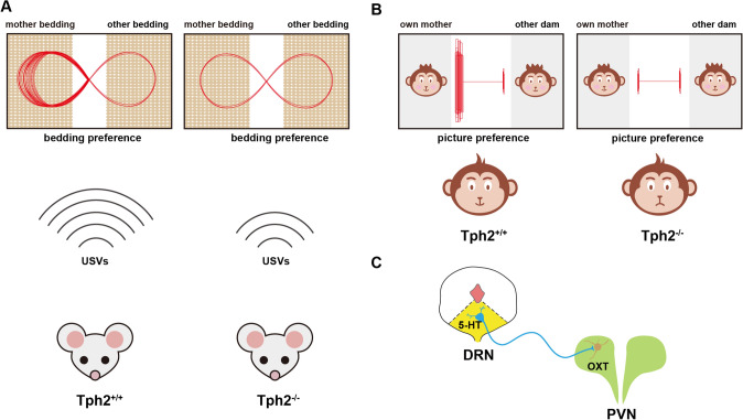
Liu et al., first found that Tph2−/− mouse and rat pups emitted fewer USVs than Tph2+/+ and Tph2+/− littermates did when separated from their mothers no matter at room temperature or at maternal body temperature (Fig. 1A), indicating that Tph2−/− pups were deficient in emitting USVs. Besides, when pups were presented with bedding from their mothers and bedding from other dams or clean bedding, both Tph2+/+ and Tph2+/− pups spent more time with mother bedding than bedding from other dams or clean bedding. In striking contrast, the Tph2−/− pups showed no preference (Fig. 1A). These results suggested that Tph2−/− mouse and rat pups had defects in maternal attachment as compared with controls.
Fig. 1.
Deletion of the Tph2 gene in mammals induced defective maternal affiliation behavior and OXT neurons in the PVN act downstream of 5-HT neurons in the DRN to regulate maternal affiliative preference. A The representative virtual movement traces showing the locations of Tph2+/+ (left) and Tph2−/− (right) rodents in a bedding preference test and representative virtual USVs showing the number of Tph2+/+ (left) and Tph2−/− (right) rodents in an USVs emissions test. B The representative virtual movement traces showing the locations of Tph2+/+ (left) and Tph2−/− (right) monkeys in a mother picture preference test. C Representative illustration showing that OXT neurons in the PVN act downstream of 5-HT neurons in the DRN. DRN, dorsal raphe nucleus; OXT, oxytocin; PVN, hypothalamic paraventricular nucleus; USVs, ultrasonic vocalizations.
Deletion of Tph2 in infant monkeys also resulted in maternal affiliation deficits, as reflected by a reduction in the time spent with their mother. Also, the Tph2−/− infant monkeys exhibited a lack of preference for their mother’s face over that of another dam (Fig. 1B). In addition, in order to explore a direct role of 5-HT in maternal affiliation behavior, the author depleted the 5-HT in Tph2+/+ rat and monkey pups pharmacologically with p-chlorophenylalanine (pCPA). After 5-HT levels decreased, the rat pups showed defective USVs emission and bedding preference for the mother, and the infant monkeys showed a decreased duration to approach awake mothers. On the contrary, after injecting 5-HTP to restore 5-HT levels in the brain, the author found the defective maternal affiliation behavior in Tph2−/− infant rats and monkeys was significantly rescued. Taken together, these findings suggested that 5-HT has an important and conserved role in regulating maternal affiliation from rodents to non-human primates.
Then what is the circuit mechanism underlying 5-HT’s modulation of maternal affiliation? The authors first found that calcium signals in serotonergic neurons of DRN were higher when mouse pups sniffed maternal bedding but not another dam’s bedding. Consistently, the number of c-fos expressions was also increased in these neurons after sniffing mother bedding. Interestingly, except for 5-HT neurons, the c-fos positive oxytocinergic neurons in the PVN increased as well. In line with this observation, the authors found that a supplement of oxytocin (OXT) to Tph2−/− rat pups could significantly rescue the defective choice between maternal bedding over the bedding of other dams. Similarly, administration of OXT to infant Tph2+/+ monkeys whose 5-HT was depleted by pCPA increased the time they spent with their awake mothers. Interestingly, the deletion of the OXT or oxytocin receptor (OXTR) gene in rat pups eliminated the preference between maternal bedding and another dam’s bedding but did not affect on USVs emission and the preference for maternal versus clean bedding, indicating OXT and OXTR were only involved in the maternal preference component of affiliative behavior. These results suggested that OXT acts downstream of 5-HT in only regulating infant maternal preference from rodents to primates. Furthermore, deleting the Tph2 gene specifically in mice DRN 5-HT neurons that project to PVN OXT neurons induced the maternal preference defect, which was rescued by activation of OXT neurons in the PVN. On the contrary, the deficient maternal preference induced by silencing OXT neurons in the PVN could not be rescued by activation of 5-HT neurons in DRN, confirming that OXT acts downstream of 5-HT neurons. Altogether, these findings revealed that the OXT neurons in the PVN act downstream of 5-HT neurons in the DRN to specifically regulate maternal preference (Fig. 1C).
The work of Liu et al. revealed an important and conserved role of 5-HT in regulating maternal affiliation from rodents to non-human primates. What’s more, the authors illustrated that oxytocinergic neurons in PVN act downstream of serotonergic neurons in DRN specifically to modulate maternal bedding preference in mice. The discovery of Liu et al. advanced our understanding of 5-HT and OXT in regulating maternal affiliative behaviors in infant mammals.
Compared to rodents, non-human primates are phylogenetically closer to human beings and are therefore better animal models for studying human behaviors. Indeed, the rhesus macaques have been used in exploring the neurobiological mechanisms involved in caregiver-infant attachment [8]. However, the role of 5-HT in monkeys’ maternal affiliation behavior was rarely studied. Liu et al. knocked out the Tph2 gene in monkeys and found that 5-HT was required for infant monkeys to affiliate with their mothers. This finding not only illustrated the key role of 5-HT in regulating monkeys’ maternal affiliation behavior but also inspired future studies that explore the conserved function of other neuropeptides in mammals’ social behaviors. Nevertheless, although Liu et al., proved that OXT neurons act downstream of 5-HT neurons to specifically regulate the maternal preference component of affiliative behavior in rodents, whether this mechanism exists in primates was not well illustrated. To further elucidate the role of OXT in regulating maternal preference, a rescued experiment that supplements OXT to Tph2−/− infant monkeys and detects the preference between the faces of its mother and another mother should be carried out.
In addition to the serotonergic system, the oxytocinergic system has also been suggested to contribute to maternal affiliation for years [9]. However, previous studies reported a controversial function of OXT in rodent USVs induced by maternal separation. For instance, OXT knockout has been reported to decrease USVs in mouse pups, whereas a supplement of exogenous OXT reduced USVs in rat pups. The work by Liu et al. showed that deletion of the OXT or OXTR gene in rat pups has no effect on USV emission and the preference for maternal versus clean bedding. These results indicated that the role of the oxytocinergic system in regulating maternal affiliation behavior is not conserved in mammals. Furthermore, in order to study the circuit mechanism that regulates maternal preference, Liu et al. deleted the Tph2 gene in the DRN 5-HT neurons that project to PVN OXT neurons in mice and found they showed the maternal preference defect. However, the causal projection from DRN 5-HT neurons to PVN OXT neurons that exclusively regulate maternal preference was not fully established. Directly inhibiting the DRN 5-HT neurons that project to PVN OXT neurons by optogenetics or chemogenetics would provide important evidence that supports the pathway from 5-HT neurons to OXT neurons. Further research is required to verify the anatomical projection from DRN 5-HT neurons to PVN OXT neurons that exclusively regulates maternal preference in the future.
The dysregulation of the serotonin system has been known to be associated with autism spectrum disorder (ASD) for a long time [10]. There are many clinical observations that showed some children with ASD had a failure to develop an attachment to their primary caregivers. The discovery of Liu et al. suggests the potential to improve the deficits of maternal attachment in children with ASD by supplementing 5-HT. Certainly, in the future, further research is required to understand the precise impact of 5-HT on human infants’ maternal attachment behavior.
Acknowledgements
This highlight was supported by the National Natural Science Foundation of China (32125018 and 32071005), the National Key Research and Development Program of China (2021YFA1101701), the Fundamental Research Funds for the Central Universities, the MOE Frontiers Science Center for Brain Science & Brain-Machine Integration of Zhejiang University, and Innovative Research Team of High-level Local Universities in Shanghai (SHSMU–ZDCX20211102).
Conflict of interest
The author declares no competing interest.
References
- 1.Liu Y, Sun Y, Zhao X, Kim JY, Luo L, Wang Q, et al. Enhancement of aggression induced by isolation rearing is associated with a lack of central serotonin. Neurosci Bull. 2019;35:841–852. doi: 10.1007/s12264-019-00373-w. [DOI] [PMC free article] [PubMed] [Google Scholar]
- 2.Premoli M, Pietropaolo S, Wöhr M, Simola N, Bonini SA. Mouse and rat ultrasonic vocalizations in neuroscience and neuropharmacology: State of the art and future applications. Eur J Neurosci. 2023;57:2062–2096. doi: 10.1111/ejn.15957. [DOI] [PubMed] [Google Scholar]
- 3.Nemsadze K, Silagava M. Neuroendocrine foundation of maternal-child attachment. Georgian Med News 2010: 21–26. [PubMed]
- 4.Mosienko V, Beis D, Alenina N, Wöhr M. Reduced isolation-induced pup ultrasonic communication in mouse pups lacking brain serotonin. Mol Autism. 2015;6:13. doi: 10.1186/s13229-015-0003-6. [DOI] [PMC free article] [PubMed] [Google Scholar]
- 5.Liu Y, Shan L, Liu T, Li J, Chen Y, Sun C, et al. Molecular and cellular mechanisms of the first social relationship: A conserved role of 5-HT from mice to monkeys, upstream of oxytocin. Neuron. 2023;111:1468–1485.e7. doi: 10.1016/j.neuron.2023.02.010. [DOI] [PubMed] [Google Scholar]
- 6.Walther DJ, Peter JU, Bashammakh S, Hörtnagl H, Voits M, Fink H, et al. Synthesis of serotonin by a second tryptophan hydroxylase isoform. Science. 2003;299:76. doi: 10.1126/science.1078197. [DOI] [PubMed] [Google Scholar]
- 7.Beis D, Holzwarth K, Flinders M, Bader M, Wöhr M, Alenina N. Brain serotonin deficiency leads to social communication deficits in mice. Biol Lett. 2015;11:20150057. doi: 10.1098/rsbl.2015.0057. [DOI] [PMC free article] [PubMed] [Google Scholar]
- 8.Kraemer GW. Psychobiology of early social attachment in Rhesus monkeys clinical implications. Ann NY Acad Sci. 1997;807:401–418. doi: 10.1111/j.1749-6632.1997.tb51935.x. [DOI] [PubMed] [Google Scholar]
- 9.Winslow JT, Hearn EF, Ferguson J, Young LJ, Matzuk MM, Insel TR. Infant vocalization, adult aggression, and fear behavior of an oxytocin null mutant mouse. Horm Behav. 2000;37:145–155. doi: 10.1006/hbeh.1999.1566. [DOI] [PubMed] [Google Scholar]
- 10.Yang G, Geng H, Hu C. Targeting 5-HT as a potential treatment for social deficits in autism. Neurosci Bull. 2022;38:1263–1266. doi: 10.1007/s12264-022-00876-z. [DOI] [PMC free article] [PubMed] [Google Scholar]


