Abstract
Background
Preclinical studies showed metformin reduces exhaustion of tumor-infiltrating lymphocytes and potentiates programmed cell death protein-1 (PD-1) blockade. We hypothesized that metformin with nivolumab would elicit potent antitumor and immune modulatory activity in metastatic microsatellite stable (MSS) colorectal cancer (CRC). We evaluated this hypothesis in a phase II study.
Methods
Nivolumab (480 mg) was administered intravenously every 4 weeks while metformin (1000 mg) was given orally, two times per day following a 14-day metformin only lead-in phase. Patients ≥18 years of age, with previously treated, stage IV MSS CRC, and Eastern Cooperative Oncology Group 0–1, having received no prior anti-PD-1 agent were eligible. The primary endpoint was overall response rate with secondary endpoints of overall survival (OS) and progression-free survival (PFS). Correlative studies using paired pretreatment/on-treatment biopsies and peripheral blood evaluated a series of immune biomarkers in the tumor microenvironment and systemic circulation using ChipCytometry and flow cytometry.
Results
A total of 24 patients were enrolled, 6 patients were replaced per protocol, 18 patients had evaluable disease. Of the 18 evaluable patients, 11/18 (61%) were women and the median age was 58 (IQR 50–67). Two patients had stable disease, but no patients had objective response, hence the study was stopped for futility. Median OS and PFS was 5.2 months (95% CI (3.2 to 11.7)) and 2.3 months (95% CI (1.7 to 2.3)). Most common grade 3/4 toxicities: Anemia (n=2), diarrhea (n=2), and fever (n=2). Metformin alone failed to increase the infiltration of T-cell subsets in the tumor, but combined metformin and nivolumab increased percentages of tumor-infiltrating leukocytes (p=0.031). Dual treatment also increased Tim3+ levels in patient tissues and decreased naïve CD8+T cells (p=0.0475).
Conclusions
Nivolumab and metformin were well tolerated in patients with MSS CRC but had no evidence of efficacy. Correlative studies did not reveal an appreciable degree of immune modulation from metformin alone, but showed trends in tumorous T-cell infiltration as a result of dual metformin and PD-1 blockade despite progression in a majority of patients.
Keywords: Immunotherapy; Translational Medical Research; Drug Therapy, Combination; Clinical Trials, Phase II as Topic
WHAT IS ALREADY KNOWN ON THIS TOPIC
Metformin has demonstrated ability to act via complex adenosine monophosphate-activated protein kinase-dependent and independent mechanisms that may impact response to programmed cell death protein-1 (PD-1) blockade in solid tumors. A majority of these data were derived from preclinical studies. We were interested in testing the efficacy of metformin and nivolumab in a population of patients with microsatellite stable metastatic colorectal cancer (MSS CRC) and interrogating the immunomodulatory activity in vivo.
WHAT THIS STUDY ADDS
Preclinical studies showed metformin improves tumor-infiltrating lymphocyte exhaustion and potentiates PD-1 blockade. However, most of these studies have been performed in mice. Our study is the first in-human trial investigating the combination of metformin and nivolumab in patients with MSS CRC. Although the treatment showed limited clinical efficacy in the studied population, the metformin and nivolumab combination displayed immunomodulatory activity in both patient tissues and peripheral blood mononuclear cells. Interestingly, metformin and nivolumab (but not single-agent metformin) also elicited an increase in Tim3+ levels in patient tissues, delineating an area for future studies related to potential mechanisms of resistance to the combination treatment.
HOW THIS STUDY MIGHT AFFECT RESEARCH, PRACTICE OR POLICY.
Given the wide clinical interest in metformin in the context of cancer treatment, our findings offer valuable clinical insights into the effects of metformin as a single agent and in combination with immunotherapy in patients with MSS CRC. These findings establish metformin as a safe approach that can be combined with PD-1/programmed death-ligand 1 pathway blockade, but may temper expectations of eliciting efficacy in patients with solid tumors that are traditionally refractory to immunotherapy.
Introduction
Colorectal cancer (CRC) is the third most common cancer worldwide and second leading cause of cancer death in the USA.1 In 2020, CRC accounted for nearly 10% of global cancer incidence and 9.4% of cancer-related deaths.2 The global incidence of new CRC cases is predicted to reach around 3.2 million by 2040 due to poor diet and environmental risks.2 Approximately 21% of patients with CRC have stage 4 disease on initial presentation, while an additional 25–50% are diagnosed at earlier stages, but later develop metastatic disease.1 3 Selected patients with limited liver and/or lung metastases may undergo potentially curative surgical resection, but the majority of patients will receive palliative systemic chemotherapy.4 After decades of fluorouracil as the sole active agent for CRC, the therapeutic landscape has expanded, with the incorporation of agents including oxaliplatin, irinotecan, and monoclonal antibodies against vascular endothelial growth factor and epidermal growth factor receptor.5–8 Despite the significant advances in systemic therapy, only 15% of patients with metastatic CRC (with Stage IV disease) are alive at 5 years, highlighting the need to explore alternative therapeutic options.1
Due to recent advances, distinct molecular and immune subtypes of CRC have been described.9 Microsatellite instability-high (MSI-H) tumors represent about 5% of metastatic CRC and have higher expression of neoantigens and tumor-infiltrating lymphocytes (TILs) in the tumor microenvironment (TME) as compared with microsatellite stable (MSS) tumors.9 10 Other data demonstrate patients with CRC with higher CD8+ and phenotypically-defined memory T cells infiltrating tumors have better survival outcomes, underscoring the potential role of immunotherapy in CRC.9 11 12 Multiple clinical trials have since demonstrated that immune checkpoint inhibitors (ICIs), which augment the antitumor immune response, improve clinical outcomes compared with chemotherapy in patients with MSI CRC, whereas immunotherapy in MSS CRC is typically ineffective.13–17 Mechanisms contributing to lack of benefit from ICIs in MSS CRC include low levels of immunostimulatory tumor neoantigens and exhaustion of TILs in part due to a hypoxic TME.3 18 Thus, altering the TME to enhance activity of immune therapy is a significant unmet need in CRC.
Metformin is a biguanide antidiabetic drug with anticancer effects as shown in several epidemiological studies. Various mechanisms of the antitumor effects of metformin have been proposed, including activation of the adenosine monophosphate (AMP)-activated protein kinase (AMPK) pathway.19–21 Metformin directly inhibits mitochondrial oxidative phosphorylation complex I and decreases adenosine triphosphate (ATP) production, leading to an altered AMP/ATP ratio and stimulation of AMPK. In turn, activation of AMPK leads to inhibition of mTOR/pS6 kinase and consequently a reduction in cellular proliferation and survival.22 Metformin has safely been administered in combination with cytotoxic chemotherapy, or chemoradiotherapy in prospective clinical trials in locally advanced rectal cancer,23 locally advanced non-small cell lung cancer24 25 and melanoma.26 One preclinical study demonstrated that metformin restores exhausted CD8+ TILs from immune exhaustion within tumor tissues via AMPK-mTOR signaling.27 Other studies show that metformin can act via AMPK-independent mechanisms.28 For example, it can decrease hypoxia in the TME by inhibiting oxygen consumption by tumor cells, thereby allowing for T cells to obtain adequate metabolic resources to carry out effector functions including tumor clearance.28 Finisguerra et al, on the other hand, found that metformin did not necessarily reduce hypoxia, but rather rescued CD8 T cells form apoptosis in hypoxic niches and enhanced their infiltration, thus improving the effects of immune checkpoint blockade.29 Finally, preclinical studies demonstrate that metformin can regulate the state of the TME through suppression of M2-like polarization of tumor associated macrophages, which promote cancer progression, and inhibition of naïve CD4+ T cell differentiation into regulatory T cells (Tregs) by reducing forkhead box P3.30 31 These distinct immunomodulatory properties of metformin suggest that the antidiabetic drug could complement and increase sensitivity to immune checkpoint blockade. Based on this rationale, we conducted a phase II study with nivolumab and metformin combination in treatment refractory MSS metastatic CRC. A series of correlative laboratory studies were conducted using tumor biopsies and peripheral blood from patients on this trial to gain greater insight into the mechanisms of metformin and nivolumab action in patients and their relationship to clinical outcome.
Methods
Study design and participants
This prospective, non-randomized, phase II trial aimed to study the combination of nivolumab and metformin in patients with stage IV MSS CRC that progressed on prior therapy (NCT03800602). Patients with histologically or cytologically confirmed MSS stage IV CRC were prospectively enrolled in this clinical trial from January 2019 through June 2020. Key inclusion criteria included the following: prior treatment with 5 fluorouracil (or capecitabine), oxaliplatin, and irinotecan containing chemotherapy. Patients were required to have measurable disease, defined as at least one lesion that can be accurately measured in at least one dimension, and an Eastern Cooperative Oncology Group (ECOG) performance status of 0 or 1. Required laboratory cut-off values were as follows: absolute neutrophil count ≥1500 /µL, platelets ≥100 x109 /L, hemoglobin ≥90 g/L, serum creatinine ≤1.5 × upper limit of normal (ULN), serum total bilirubin ≤1.5 × ULN, and serum albumin ≥2.5 mg/dL. Patients with diabetes mellitus were required to receive a stable diabetic treatment regimen for at least 1 month prior to trial enrollment and keep a blood glucose level log at home for the first 4 weeks of the trial. Key exclusion criteria included the following: metformin use in the last 3 months, history of allergic reactions attributed to compounds of similar chemical or biologic composition to nivolumab and metformin, prior therapy with an anti-programmed cell death protein-1 (PD)-1, anti-programmed death-ligand 1 (PD-L1), or anti-PD-L2 agent, pregnancy or breastfeeding. The trial was conducted at the Winship Cancer Institute of Emory University, Atlanta, Georgia, USA.
Treatment and assessments
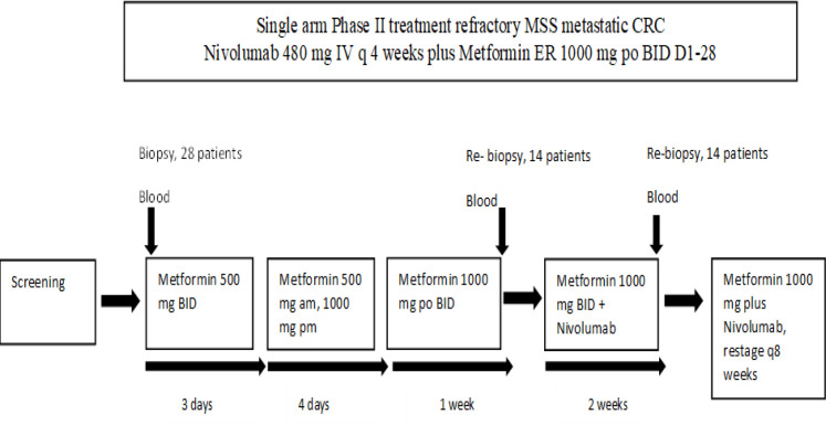
All patients were started on 14 days of a metformin only lead-in period, whereby the dosage of metformin was incrementally increased to ensure tolerability (figure 1). This included a metformin 500 mg tablet two times per day for 3 days, then 500 mg in the morning and 1000 mg in the evening for 4 days, then 1000 mg two times per day for 7 days. After 14 days of the metformin only lead in period, patients continued to receive metformin at 1000 mg orally two times per day and nivolumab at 480 mg intravenously every 4 weeks. Cycles were repeated every 28 days in the absence of disease progression, unacceptable toxicity, or consent withdrawal. Restaging scans were obtained every 8 weeks throughout the study. Adverse events (AEs) were evaluated, recorded, and graded throughout the study and during the follow-up period according to National Cancer Institute Common Toxicity Criteria for Adverse Events V.4.0.
Figure 1.
Clinical trial design. Patient biopsies were isolated from liver metastases prior to and immediately following either the first treatment cycle (metformin) or the second treatment cycle (metformin and nivolumab). They were stained with a panel of 13 markers centered around identifying immune cell populations. Peripheral blood samples were extracted prior to and following each treatment cycle. Flow cytometry was performed to analyze T cell and myeloid markers at different treatment time points. am, in the morning; BID, two times per day; CRC, colorectal cancer; D, day; IV, intravenous; MSS, microsatellite stable; pm, in the evening; q, every.
Collection of biospecimens
All patients underwent research biopsy at baseline prior to start of any systemic therapy, while half of the patients underwent repeat biopsy at the end of metformin only lead-in period, and the other half of the patients underwent a repeat biopsy 14 days after the first dose of nivolumab. The most accessible metastatic disease tumor site was biopsied by an interventional radiologist. The same site was biopsied at biopsy 1 and biopsy 2 if amenable, immediately fixed in formalin embedded in paraffin prior to subsequent multiplex analysis. For analysis of systemic immune parameters, peripheral blood for correlative analysis was collected at baseline, prior to first nivolumab dose, on cycle 1 day 15, on cycle 3 day 1 and at progression. Peripheral blood mononuclear cells (PBMCs) were isolated by density gradient centrifugation with Ficoll-Paque and frozen as previously described.32 Cells were cryopreserved in the vapor phase of liquid nitrogen until analysis, which occurred within 2 years of sample collection. At this point, samples were thawed and immediately subject to flow cytometric analysis as described below.
Outcomes
The primary objective was to evaluate the effect of combined nivolumab and metformin therapy on the overall response rate (ORR) as assessed by Response Evaluation Criteria in Solid Tumors (RECIST) V.1.1 and the rate was calculated as a proportion (responders/total patients). Response-evaluable patients were defined as all enrolled subjects who received at least one dose of study treatment (metformin and nivolumab) and provided at least one post-baseline response assessment. Intention-to-treat (ITT) population was defined as all enrolled subjects who received at least one dose of study treatment. Secondary objectives included determining the effect of the nivolumab and metformin combination on clinical outcomes, progression-free survival (PFS), and overall survival (OS) up to 2 years after study start, and biochemical response (carcinoembryonic antigen, CEA) up to 1 year after study start. Correlative outcomes included comparing the effect of the nivolumab and metformin combination on immune and metabolic biomarkers in the tumor microenvironment and systemic circulation.
Statistical analysis
Simon’s two-stage Minimax design was employed (H0: ORR=4%; H1: ORR=15%; Type I error=0.1; power=80%). If ≥1 objective response was observed in the first evaluable 18 patients, 10 additional patients would be included in the cohort. The initial clinical trial design specified that if no objective response was seen, the study would be stopped for futility. Three or more objective responders in 28 patients would be required to be considered a positive study. Pretreatment and on-treatment research biopsies and correlative peripheral blood specimens were collected. Median follow-up for all eligible patients was estimated using the Kaplan-Meier method. PFS was defined from treatment initiation to progression or death whichever came first and OS was defined from treatment initiation to death. The Kaplan-Meier method was used to estimate PFS and OS time curves, median PFS and OS, and 95% CIs for the cohorts (ITT and per-protocol evaluable for response). Statistical analyses were performed using SAS V.9.4. P value≤0.05 (two-sided) was considered significant.33
Immune profiling and correlative analysis
Formalin-fixed paraffin embedded colon cancer tissue blocks were sectioned using a rotary microtome at 5 µm thickness. The sections were placed in a 40°C water bath, where they were mounted onto Zellsafe coverslips (Canopy Biosciences) and baked in an incubator at 60°C for an hour to allow the excess water to evaporate. Deparaffinization was performed by serial 5-min immersions of the tissue-mounted coverslips in each of the following solutions: (1) xylene, (2) fresh xylene, (3) 100% ethanol, (4) fresh 100% ethanol, (5) 90% ethanol, (6) 70% ethanol, (7) 50% ethanol, and (8) deionized water. Heat-induced antigen retrieval (HIER) was performed in a Coplin jar containing CC1 solution (Ventana, 950–124) heated to 95°C in a circulating water bath for 20 min. After HIER, coverslip-mounted sections were washed in room temperature ZKW Wash Buffer and mounted on ZellSafe Tissue Chips (Canopy Biosciences). The chips were immediately filled with a ZKW storage buffer (Canopy Biosciences) and stored at 4°C until use. The 14-plex immunostaining assay was performed on a ZellScanner One (Canopy Biosciences) using iterative cycles of staining, imaging, and bleaching. This multiplex fluorescence platform was used to examine the phenotypic properties of tumor-infiltrating lymphocytes from paired biopsies obtained from the studies. Briefly, samples were washed with 6 mL of ZKW wash buffer prior to scanning. A scan of background autofluorescence was performed in a single channel (Ex 550/25, Em 605/70) to acquire whole tissue images, which were used to assess tissue integrity and select fields of view (area 0.36 mm2) for further scanning. The full 14-plex assay was executed on samples with at least 10 fields of scannable tissue. The assay was executed in seven cycles, with each cycle beginning with a 10–20 s photobleach and subsequent background measurements in each channel to be measured in that cycle. This was followed by staining with one or multiple antibodies diluted in a ZKW storage buffer for a total volume of 600 µL. Staining was performed by pipetting antibody cocktail working solutions directly into ZellSafe Tissue Chips, incubating for 1 hour, and washing with a 12 mL ZKW wash buffer. For unconjugated antibodies, this step was followed by secondary antibody staining for 1 hour and a second wash step prior to imaging, along with a 1-hour block with 5% normal goat serum (BioLegend, 927501) prior to subsequent stain cycles. After imaging cycles containing Alexa Fluor 488-conjugated antibodies, samples were incubated for 15 min with anti-Alexa Fluor 488 in order to quench fluorescence. In the final cycle, samples were incubated for 15 min with Hoechst DNA dye, washed with 12 mL of ZKW wash buffer, and imaged in two channels (Ex 395/2, Em 392/23; Ex 400/30, Em 460/50). After imaging all cycles, analysis was performed using Canopy Biosciences’ ZellExplorer software. Cells were segmented based on the Hoechst nuclear stain and background-subtracted fluorescence values for each marker were calculated with a proprietary algorithm. A bivariate hierarchical gating strategy was then used to phenotype each cell and quantify populations of interest according to their respective biomarker definition.
Flow cytometry
Prior to and while undergoing therapy, phenotypic analysis of peripheral immune cells was conducted via multicolor flow cytometry. Antibodies are listed in the online supplemental table 1. Cryopreserved peripheral blood mononuclear cells (PBMCs) were thawed at 37оC, washed, centrifuged, and resuspended in the fluorescence activated cell sorting (FACS) buffer (phosphate buffered saline, PBS, 3% fetal bovine serum, FBS, 0.05 mmol/L EDTA). Cells were then incubated with intracellular antibodies for 1 hour at room temperature, washed, and resuspended in the FACS buffer for analysis. Flow cytometric analysis was performed on a Cytek Aurora (Cytek Biosciences). Compensation controls were obtained using UltraComp eBeads Compensation Beads (01–2222–41, Invitrogen). Data were analyzed using FlowJo software version 10.7.2 (FlowJo, LLC). Differences between post and pre time points for each of metformin alone and metformin+nivolumab groups for each immune profile marker was assessed using a Wilcoxon signed-rank test. Differences in immune profile markers between (post and pre) time points among metformin alone and metformin+nivolumab groups was performed using Kruskal-Wallis test. For the flow cytometric analysis of immune populations in the blood of patients following treatment with metformin and metformin and nivolumab, immune markers were log10 transformed. Differences in means between log transformed markers at each time point were compared using repeated measures analysis of variance. Immune profile analysis was performed using SAS V.9.4. Statistical significance was set at 0.05. The reported p values from the immune marker correlative analysis were two-sided and should be considered as exploratory; we regard estimates and CIs as more relevant for clinical decision-making. Additionally, the cryopreservation process should be taken into account as the potential surface protein expression could be considered an artifact from the freezing and defrosting process and may influence ex vivo protein expression.
jitc-2023-007235supp001.pdf (44.2KB, pdf)
Results
Baseline demographics and treatment
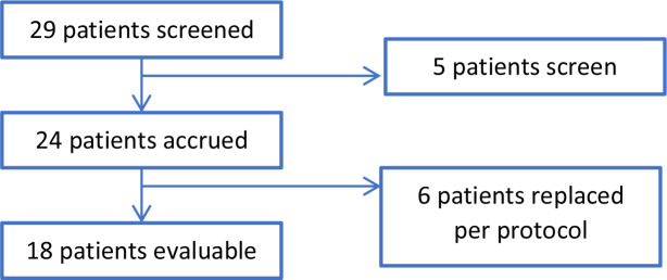
For this clinical study, a total of 29 patients were screened between January 2019 and June 2020. From those screened, 5 patients were ineligible, and 24 patients were enrolled in the study, 6 patients were replaced per protocol, and 18 patients had evaluable disease (figure 2). Those patients who received at least one dose of study treatment were included in the analysis. Considering the nature of patient enrollment in early phase studies, separate analyses were performed for 24 total patients (ITT analysis) and for 18 patients (per-protocol analysis). Table 1 shows the baseline demographic data for each group. Of the 18 evaluable patients, 11 (61.1%) were women, 11 were Caucasian, median age was 58 (IQR 50–67), and 11 had an ECOG of 1 (73.3%). Of the 18 evaluable patients, 83.3% had left-sided tumors, 16.7% received prior adjuvant chemotherapy, 50.0% received four or more prior lines of systemic therapy excluding prior adjuvant chemotherapy, 11.1% had a history of diabetes mellitus, 61.1% had KRAS mutant tumors, and 11.1% had BRAF 600E mutations.
Figure 2.
Consolidated Standards of Reporting Trials flow diagram.
Table 1.
Baseline patient characteristics and clinicopathological features (n=24)
| Intention-to-treat analysis (n=24) | Per-protocol analysis (n=18) | |
| Age, median (range) | 56.5 (32–74) | 58.0 (39–74) |
| Gender, n (%) | ||
| Female Male |
14 (58.3) 10 (41.7) |
11 (61.1) 7 (38.9) |
| Race, n (%) | ||
| Caucasian African-American Asian |
17 (70.8) 5 (20.8) 2 (8.3) |
14 (77.8) 3 (16.7) 1 (5.6) |
| ECOG performance status, n (%) | ||
| 0 1 |
9 (37.5) 15 (62.5) |
6 (33.3) 12 (66.7) |
| Primary tumor location*, n (%) | ||
| Right side Left side |
4 (16.7) 20 (83.3) |
3 (16.7) 15 (83.3) |
| Received adjuvant chemotherapy | 5 (20.8) | 3 (16.7) |
| Number of prior systemic therapies, n (%) | ||
| 2 3 ≥4 |
5 (20.8) 7 (29.2) 12 (50.0) |
3 (16.7) 6 (33.3) 9 (50.0) |
| Diabetes history, n (%) | 3 (12.5) | 2 (11.1) |
| Molecular testing, n (%) | ||
| BRAF 600E positive KRAS mutant KRAS wild type |
2 (8.3) 14 (58.3) 10 (41.7) |
2 (11.1) 11 (61.1) 7 (38.9) |
*In the entire cohort, right-sided included tumors in descending colon (1) and cecum (3); left-sided included tumors in ascending colon (6), sigmoid (7), rectosigmoid (2), and rectum (5).
ECOG, Eastern Cooperative Oncology Group.
Treatment outcomes
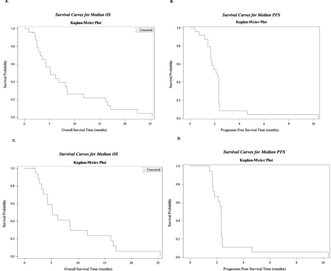
Treatment outcomes were assessed in the 18 evaluable patients, whereby 2 patients had stable disease (SD), characterized by a range encompassing <30% tumor reduction to <20% tumor enlargement. According to RECIST V.1.1, SD is defined as “neither sufficient shrinkage compared to baseline to qualify for partial or complete response nor sufficient increase to qualify for progressive disease”,34 which is the criteria we followed in this study. In the evaluable patients, two patients had stable disease and received 4 and 10 cycles of systemic therapy, respectively (table 2). In the evaluable patients, no objective responses were seen; therefore, the study did not proceed with the second stage of enrollment. The median duration of follow-up for the overall study population was 2.17 (1.67–2.33) months. The median OS and PFS was 5.2 months (95% CI (3.2 to 8.4)) and 2.2months (95% CI (1.7 to 2.3)), respectively, in the ITT cohort (figure 3A,B). The median OS and PFS were 5.2 months (95% CI (3.2 to 11.7)) and 2.3 months (95% CI (1.7 to 2.3)), respectively, in the 18 evaluable patients. (figure 3C,D).
Table 2.
Efficacy assessment
| Intention-to-treat analysis (n=24) | Per-protocol analysis (n=18) | |
| RR, % (95% CI) | 0 (0) | 0 (0) |
| Best response, n (%) | ||
| CR PR SD PD Not evaluable |
0 (0) 0 (0) 2 (8.3) 16 (66.7) 6 (25.0) |
0 (0) 0 (0) 2 (11.1) 16 (88.9) 0 (0) |
| Median PFS, months (95% CI) | 2.2 (1.7 to 2.3) | 2.3 (1.7 to 2.3) |
| Median OS, months (95% CI) | 5.2 (3.2 to 8.4) | 5.2 (3.2 to 11.7) |
CR, complete response; OS, overall survival; PD, progressive disease; PFS, progression-free survival; PR, partial response; SD, stable disease.
Figure 3.
(A and B) Median OS and PFS was 5.2 months (95% CI (3.2 to 8.4)) and 2.2 months (95% CI (1.7 to 2.3)) in intention-to-treat cohort. (C and D) Median OS and PFS was 5.2 months (95% CI (3.2 to 11.7)) and 2.3 months (95% CI (1.7 to 2.3)) in evaluable patients. OS, overall survival; PFS, progression-free survival.
Safety and toxicity
The occurrence of AEs is summarized in table 3 for both ITT and per-protocol analysis. In this study, AEs did not lead to the discontinuation of treatment in any patient and the combination therapy was well tolerated. Our analysis indicated the most common grade 3 and 4 AEs observed in both groups were anemia (n=2), diarrhea (n=2), and fever (n=2). For any grade, diarrhea, nausea, and abdominal pain, fatigue and vomiting were most commonly seen in both ITT and per-protocol analysis groups.
Table 3.
Most common adverse events
| Toxicity, n | Intention-to-treat analysis (n=24) | Per-protocol analysis (n=18) | ||
| Any grade | Grade 3/4 | Any grade | Grade 3/4 | |
| Diarrhea | 10 | 2 | 10 | 2 |
| Nausea | 8 | 0 | 6 | 0 |
| Vomiting | 6 | 1 | 5 | 1 |
| Abdominal pain | 7 | 1 | 5 | 0 |
| Fatigue | 5 | 1 | 5 | 1 |
| Anemia | 3 | 2 | 3 | 2 |
| Fever | 3 | 2 | 3 | 2 |
Metformin and nivolumab treatment increases leukocyte percentages in patient tissues
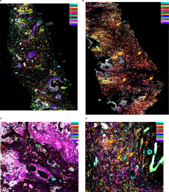
Eighteen patients had pretreatment biopsies, nine had biopsies after metformin only lead in, and nine had biopsies after metformin and nivolumab combination treatment obtained at cycle 1 day 15 (C1D15). Tissue analysis was performed to evaluate the immune landscape in the biopsies of metastatic CRC patients following treatment with metformin as a single agent, versus the metformin and nivolumab combination. Multiplex spatial immune profiling analysis encompassed 13 phenotypically distinct populations including leukocytes, effector CD4+/CD8+ T cells (CD45+, CD4+/CD8+, CD38+, CD45RO−), naïve CD4+/CD8+ T cells (CD45+, CD4+/CD8+, CD38−, CD45RO−), central memory CD4+/CD8+ T cells (CD45+, CD4+/CD8+, CD38−, CD45RO+), effector memory CD4+/CD8+ T cells (CD45+, CD4+/CD8+, CD38+, CD45RO+) and others (figure 4 and online supplemental tables S2 and S3)
Figure 4.
Immune profiling of patient tissues. (A–B) Patient 004, metformin only. Right: pretreatment; Left: post-treatment. 13-plex marker staining suggests that metformin treatment increased the infiltration of T-cell subsets into patient tumor microenvironment. (C–D) Patient 013, metformin and nivolumab. Right: pretreatment; Left and bottom: post-treatment. 13-plex marker staining suggests that metformin and nivolumab combination treatment increased the infiltration of T-cell subsets into patient tissue tumor microenvironment, illustrating the immunomodulatory effect of this treatment on the tumor microenvironment of this individual patient (013). Arrows in orange indicate populations of interest that have increased post-treatment, mostly CD4+ and CD45RO+ populations.
The ability of metformin to modulate previously characterized biologic targets was assessed using pAMPK staining as one potential surrogate pharmacodynamic biomarker.35 This analysis revealed no significant changes in the percentages of pAMPK+ cells in metformin only and metformin and nivolumab patient tissues. There was no evidence of differences in CD4+ T-cell recruitment into patient tissues following treatment with metformin alone, as compared with baseline. However, metformin treatment was associated with significantly lower percentages of effector CD4+ T cells, defined as CD45+ CD4+ CD38+ CD45RO− populations, in these patients (p=0.031, figure 5). Statistical comparison between the two treatment groups (metformin vs metformin and nivolumab) revealed that there was a statistically significant difference in treatment-induced changes in CD4+ effector T-cell percentages between the two groups (p=0.004). Namely, biopsies obtained from patients treated with a lead-in containing metformin only had a lower percentage of CD4+ effector T cells, whereas tumors from patients treated with both metformin and nivolumab had no change of CD4+ effector T cells. In contrast, treatment with the metformin and nivolumab combination resulted in significantly higher percentages of CD45+ leukocytes in patient tissues (p=0.031; figure 5). Furthermore, biopsies from patients undergoing dual treatment with metformin and nivolumab had higher percentages of Tim3+ (CD366+) T cells compared with baseline (p=0.031; Online supplemental table S3). Biopsies obtained from the metformin and nivolumab combination showed a significant increase in PDL1−Tim3+ cells compared with metformin alone group (p=0.022; figure 5).
Figure 5.
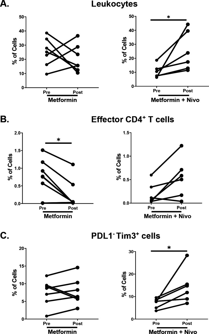
Treatment-induced changes in immune profiles of metastatic colorectal tumors. 13-plex immunohistochemistry analysis revealed changes in immune cell phenotypes in pretreatment versus post-treatment liver biopsy tissue between cohorts of patients receiving a lead in with metformin (n=7) only versus metformin and nivolumab (n=6). Changes were identified in (A) lymphocytes, (B) percentages of effector CD4 T cells and (C) PDL1−Tim3+ cell populations. Each line represents matched pretreatment and post-treatment biopsies from the same patient. *p<0.05 via Wilcoxon signed-rank test.
jitc-2023-007235supp002.pdf (106.6KB, pdf)
Treatment with metformin and nivolumab decreases percentages of naïve CD8+ T cells in patient blood samples
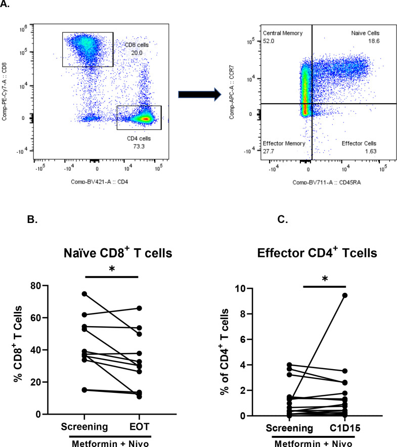
Flow cytometric analysis of PBMCs was performed to characterize potential changes in immune populations in the blood of patients following treatment with metformin and metformin and nivolumab. Our analysis encompassed 13 phenotypically distinct populations including CD3+ T cells, naïve CD4+/CD8+ T cells (CD3+, CD4+/CD8+, CCR7+, CD45RA+), effector CD4+/CD8+ T cells (CD3+, CD4+/CD8+, CCR7−, CD45RA+), central memory CD4+/CD8+ T cells (CD3+, CD4+/CD8+, CCR7+, CD45RA−), effector memory CD4+/CD8+ T cells (CD3+, CD4+/CD8+, CCR7−, CD45RA−) and activated 137+ CD4+/CD8+ T cells (figure 6). We found that two phenotypically defined T-cell populations were statistically significantly different following treatment with metformin and nivolumab. Percentages of circulating effector CD4+ T cells were higher overall at C1D15, that is, following treatment with metformin then metformin plus nivolumab compared to pre-screening levels (p=0.0316). However, these data must be interpreted with caution, given that a single outlier sample likely influenced the statistical result. In contrast, percentages of naïve CD8+ T cells were lower at the end of treatment time point compared with pre-screening (p=0.0475). Analysis also revealed that there was no statistically significant difference in the percentages of naïve CD4+ cells, effector CD8+ T cells, central memory CD4+/CD8+ T cells, effector memory CD4+/CD8+ T cells and activated CD137+ CD4+/CD8+ T cells (online supplemental table S4).
Figure 6.
Treatment-induced changes in peripheral blood immune cells of patients with metastatic colorectal cancer. (A) Representative gating scheme for phenotypic analysis of naïve CD3+ CD4+/8+ CD45RA+CCR7+ T cells, effector CD4+/8+ CD45RA+CCR7 T cells, central memory CD4+/8+ T CD45RA− CCR7+ cells and effector memory CD4+/8+ CD45RA− CCR7− T cells (left figure shows the gating for CD8 and CD4 while the right figure shows the gating for CCR7 and CD45RA). (B) Changes in populations of effector CD3+ CD4+ CD45RA+ CCR7− T cells from screening to C1D15 (n=17). (C) Changes in populations of naïve CD3+ CD8+ CD45RA+ CCR7+ T cells from screening to end of treatment (n=17). Each line represents matched peripheral blood samples from the same patient. *p<0.05 via Kruskal-Wallis statistical test.
Discussion
This report represents the first study combining metformin and nivolumab in patients with treatment-refractory MSS metastatic CRC. Unfortunately, there were no objective responses, but two patients achieved stable disease. This population was heavily pretreated as 50% of patients received four or more prior lines of systemic therapy in the metastatic setting. The combination of metformin and nivolumab was well tolerated, safety of the combination was consistent with those of the individual drugs, and few patients experienced grade 3 or 4 adverse events.
Immunotherapy with checkpoint inhibitors alone is ineffective in MSS metastatic CRC.9 15 Concerted efforts are underway in MSS CRC to improve efficacy of ICIs by combining them with other strategies to impact the TME.36 37 Metabolic reprogramming from metformin represents one such approach based on efficacy in the preclinical setting when combined with immune checkpoint inhibitors.38 At a mechanistic level, metformin elicits metabolic changes that favor decreased intratumoral oxygen consumption in vitro and in vivo. Furthermore, metformin increases oxygen consumption by CD8+ TILs and improves the effector cytokine production by CD8+ TILs in B16 melanoma and MC38 colon adenocarcinoma tumor models.28 Other data indicate expression of co-inhibitory molecules PD1 and Tim3 are modulated in metformin-treated mice compared with controls.28 Finally, the combination of metformin and a PD-1 inhibitor improved intratumoral T-cell function and increased antitumor activity.28
Other features of metformin suggest it is a viable agent for enhancing immunotherapy efficacy. For instance, a study by Eikawa et al 27 showed that metformin increased CD8+ TILs and prevented apoptosis and immune exhaustion of TILs as characterized by reduced tumor necrosis factor alpha, interferon gamma and interleukin 2 production in in BALB/c and C57BL/6 (B6) murine tumor models including BALB/c mice bearing Colon 26 tumors. Additionally, metformin treatment restored the multifunctionality of exhausted PD1− Tim3+ CD8+ TILs through a shift from central memory to effector memory phenotype, indicating that the effect of metformin was linked to significant alterations of CD8+TIL characteristics in the TME.27 In head and neck squamous cell cancer, metformin increased effector CD8+ T cells and Tregs in the TME.22 In patients with diabetes with non-small cell lung cancer, metformin activates AMPK, which decreases micro-RNA-107 expression, thus enhancing Eomesodermin expression. This suppresses the PDCD1 (encodes PD-1) transcription in metformin treated CD8+ T cells, hence improving CD8+ memory stem and central memory T cell differentiation through the AMPK-miR-18-Eomes-PD-1 pathway.39 Metformin reduces Tregs in the TME and contributes to a more immune favorable state.30 The basis for co-administration of metformin with anti-PD-1 therapy relies on inhibiting mitochondrial complex I activity in tumor cells, reducing oxygen consumption rates (OCR) in tumor cells while concomitantly increasing OCR of the infiltrating T-cell subsets.28
Although these immune modulatory effects of metformin suggest potential for robust metabolic modulation, most of these studies, however, were conducted in murine models. We were interested in testing whether the immune effects of metformin, previously observed in mouse models, could be translated to human patients to improve immunotherapy outcomes. Correlative analysis of the paired tumor samples of the 18 patients in our cohort did not reveal any statistically significant difference in the percentages of pAMPK+ cells following treatment with metformin as a single agent or metformin plus nivolumab compared with baseline. Thus, metformin at the prescribed dose was not potent enough to significantly impact AMPK as a pharmacodynamic target in treated patient tissues. The AMPK-independent mechanism of action of metformin such as reactive oxygen species (ROS) production which was previously demonstrated in colon cancer cells was not evaluated in this study.40 Unlike preliminary studies which showed that metformin rescued CD8+ T cells from exhaustion41 and increased the infiltration of TILs39 42 43 in colorectal and other cancer models, our tissue analysis indicated that metformin alone did not significantly alter the percentages of leukocytes and effector or central memory CD8+ T cells in patient tissues. In fact, metformin as a single agent decreased the percentages of effector CD4+ T cells in treated patient tissues. This discrepancy in the effects of metformin in mouse models versus human patients could be due to the higher doses of metformin administered to mice compared with humans or alternatively, resistance pathways in the TME of patient tissues may also explain these differences. Alternatively, 2 weeks of metformin may be insufficient to induce meaningful changes, which might represent a potential limitation in interpreting these biomarker data.
Treatment with metformin and nivolumab significantly increased the percentages of leukocytes in the tissue and induced a trend towards higher percentages of phenotypically defined effector memory CD8+ T cells, which have been linked to decreased metastasis and improved survival in CRC.11 Hence, the combination treatment influenced the distribution of immune cell populations although this reprogramming action did not translate into improved OS. Interestingly, the combination treatment also increased percentages of Tim3+ cells in the TME. Previous research has shown that the interaction between Tim3 and Gal9 suppresses the adaptive immune system, thereby repressing antitumor immunity in both solid and blood tumors.44 While intriguing, upregulated Tim3+ cells were not evident in response to metformin alone. Based on these findings, further research to define the contribution from metformin in the observed Tim3 increase is needed to guide any subsequent therapeutic approaches.
Peripheral blood analysis showed that the percentages of circulating effector CD4+ T cells were higher at C1D15 compared with pre-screening levels. This increase was in agreement with the trends observed in patient tissues following metformin and nivolumab treatment, indicating that combination treatment seems to increase the proportion of CD4+ T cells in both patient tissues and peripheral blood. One of the limitations of these findings is that it is driven by one pair of samples, while the majority of paired sets displayed no post-treatment change or a slight decrease. A larger cohort of patients might be required to verify this trend. In contrast, combination treatment decreased the percentages of naïve CD8+ T cells coupled with a trend towards increased percentages of circulating effector CD8+ at end of treatment. This reduction in naïve CD8+ T cells in theory may counter any immunomodulatory activity. Unfortunately, our exploratory analysis of data from both tissue and peripheral blood did not identify any trends evident in the two patients who experienced stable disease, as compared with the remainder of the cohort who progressed on therapy (online supplemental table S5 and S6). Finally, we acknowledge this study had other potential limitations including the small cohort size, as well as the handling process including freezing and thawing, which could impact the expression of immune surface markers, even despite our care to ensure sample collection was performed using published Standard Operating P.32
Conclusion
In conclusion, metformin and nivolumab treatment was well-tolerated and displayed immune modulator activity in patient tissues and PBMCs. Despite this, limited efficacy was observed in this population of patients with treatment refractory MSS metastatic CRC. Ongoing studies will help uncover potential mechanisms of resistance to metformin and combination therapy. These could include Tim3 and other pathways that deserve further investigation.
Acknowledgments
The clinical trial was funded by Bristol-Myers Squibb (BMS). Research reported in this manuscript was supported by BMS funds (principal investigator: MA, NCT03800602) and in part by Developmental Funds from Biostatistics Shared Resource of Winship Cancer Institute of Emory University under award number P30CA138292. The content is solely the responsibility of the authors and does not necessarily represent the official views of the National Institutes of Health. The spatial immune profiling of patient tissue was performed by Canopy Biosciences using ChipCytometry. Additionally, we would like to thank the ICI core of Emory University for assisting with multiplex tissue reconstruction. Multiplex tissue images were analyzed and reconstructed using NIS elements and QuPath (04: Canopy Biosciences/ 013: first authors of this manuscript). Schematic illustrations produced with Paint. Part of the data in this manuscript were presented at the 2020 Gastrointestinal Cancers Symposium (Akce et al, Journal of Clinical Oncology. 2021;39(3_suppl):95-. doi: 10.1200/JCO.2021.39.3_suppl.95.) and 2022 American Association for Cancer Research meeting as poster (Farran et al, 2022;82(12_Supplement):3482-. doi: 10.1158/1538 7445.Am2022-3482).
Footnotes
Twitter: @LesinskiLab
Contributors: MA: conceptualization, methodology, formal analysis, investigation, visualization, writing—original draft, supervision and editing, funding acquisition, guarantor. BF: conceptualization, methodology, formal analysis, investigation, visualization, writing—original draft, supervision, and editing. GBL and BE-R: conceptualization, methodology, formal analysis, investigation, visualization, supervision and editing, guarantor. JMS and MR: methodology, formal analysis, investigation, visualization, writing—original draft. AR-J, BO: methodology, formal analysis, visualization. SK, LK, WLS, CW, OBA, MD: visualization, editing, guarantor.
Funding: The clinical trial was funded by Bristol-Myers Squibb (BMS). Research reported in this manuscript was supported by BMS funds (principal investigator: MA, NCT03800602) and in part by Developmental Funds from Winship Cancer Institute of Emory University under award number P30CA138292.
Competing interests: MA has consulted for Eisai, Ipsen, Exelixis, GSK, QED, Isofol, Curio Science, AstraZeneca and Genentech and has received research funding from Tesaro (Inst), RedHill Biopharma Limited (Inst), Polaris (Inst), Bristol-Myers Squibb-Ono Pharmaceutical (Inst), Xencor (Inst), Merck Sharp & Dohme (Inst), Eisai (Inst), GSK (Inst). BER has consulted for AZ, Ipsen, Exelixis, Seagen and received research funding from BMS, Merck, AZ, Novartis, EUSA, adptamune, Bayer, Exelixis. GBL has consulted for ProDa Biotech, LLC and received compensation. GBL has received research funding through a sponsored research agreement between Emory University and Merck and Co., Bristol-Myers Squibb, Boerhinger-Ingelheim, and Vaccinex. CW has consulted for Array Biopharma, Natera, Seagen, Pfizer, Daiichi Sankyo/Lilly and has received research funding (trial funding to institution) through Pfizer, Hutchmed and Seagen. Employment/leadership/stock and other ownership—nothing to disclose. Honoraria: Cancer Treatment Centers of America, Medscape. WLS is on the advisory board of Mylan, BMS, Esai, Tesaro, Blueprints, Seagen and a member of the speaker’s bureau of Guardant health, Cumberland therapeutics, Seagen. His research has been funded by GSK, Eli Lilly, Tesaro. BO has received research funding through a sponsored research agreement between Emory University and Boehringer Ingelheim. OBA has consulted for Ipsen Pharmaceuticals, Aadi Bioscience, Taiho, Pfizer, Seagen Inc. Bristol-Myers Squibb and AstraZeneca. He has received research funding from Taiho Oncology, Ipsen Pharmaceuticals, GSK, Bristol-Myers Squibb, PCI Biotech AS, ASCO, Calithera Biosciences, Inc., SynCore Biotechnology Co. Ltd., Suzhou Transcenta Therapeutics Co., Ltd, Corcept Therapeutics Inc., Hutchison MediPharma, Boehringer Ingelheim, Xencor Inc., Cue Biopharma, Inc., Merck, Syros Pharmaceuticals Inc., Inhibitex Inc, Arcus Biosciences Inc. and ImmunoGen. MD has consulted for Novartis and Guardant Health. The rest of the authors have no conflicts of interest to disclose.
Provenance and peer review: Not commissioned; externally peer reviewed.
Supplemental material: This content has been supplied by the author(s). It has not been vetted by BMJ Publishing Group Limited (BMJ) and may not have been peer-reviewed. Any opinions or recommendations discussed are solely those of the author(s) and are not endorsed by BMJ. BMJ disclaims all liability and responsibility arising from any reliance placed on the content. Where the content includes any translated material, BMJ does not warrant the accuracy and reliability of the translations (including but not limited to local regulations, clinical guidelines, terminology, drug names and drug dosages), and is not responsible for any error and/or omissions arising from translation and adaptation or otherwise.
Data availability statement
Data are available upon reasonable request.
Ethics statements
Patient consent for publication
Consent obtained directly from patient(s).
Ethics approval
This study involves human participants and was approved by Emory University IRB. IRB # is IRB00106678. Participants gave informed consent to participate in the study before taking part.
References
- 1. Siegel RL, Miller KD, Fuchs HE, et al. Cancer Statistics. CA Cancer J Clin 2022;72:7–33. 10.3322/caac.21708 [DOI] [PubMed] [Google Scholar]
- 2. Xi Y, Xu P. Global colorectal cancer burden in 2020 and projections to 2040. Transl Oncol 2021;14:101174. 10.1016/j.tranon.2021.101174 [DOI] [PMC free article] [PubMed] [Google Scholar]
- 3. Ganesh K, Stadler ZK, Cercek A, et al. Immunotherapy in colorectal cancer: rationale, challenges and potential. Nat Rev Gastroenterol Hepatol 2019;16:361–75. 10.1038/s41575-019-0126-x [DOI] [PMC free article] [PubMed] [Google Scholar]
- 4. Shah SA, Haddad R, Al-Sukhni W, et al. Surgical resection of hepatic and pulmonary metastases from colorectal carcinoma. J Am Coll Surg 2006;202:468–75. 10.1016/j.jamcollsurg.2005.11.008 [DOI] [PubMed] [Google Scholar]
- 5. de Gramont A, Figer A, Seymour M, et al. Leucovorin and fluorouracil with or without Oxaliplatin as first-line treatment in advanced colorectal cancer. J Clin Oncol 2000;18:2938–47. 10.1200/JCO.2000.18.16.2938 [DOI] [PubMed] [Google Scholar]
- 6. Douillard JY, Cunningham D, Roth AD, et al. Irinotecan combined with fluorouracil compared with fluorouracil alone as first-line treatment for metastatic colorectal cancer: a Multicentre randomised trial. Lancet 2000;355:1041–7. 10.1016/s0140-6736(00)02034-1 [DOI] [PubMed] [Google Scholar]
- 7. Hurwitz H, Fehrenbacher L, Novotny W, et al. Bevacizumab plus Irinotecan, fluorouracil, and Leucovorin for metastatic colorectal cancer. N Engl J Med 2004;350:2335–42. 10.1056/NEJMoa032691 [DOI] [PubMed] [Google Scholar]
- 8. Van Cutsem E, Köhne C-H, Hitre E, et al. Cetuximab and chemotherapy as initial treatment for metastatic colorectal cancer. N Engl J Med 2009;360:1408–17. 10.1056/NEJMoa0805019 [DOI] [PubMed] [Google Scholar]
- 9. Basile D, Garattini SK, Bonotto M, et al. Immunotherapy for colorectal cancer: where are we heading Expert Opin Biol Ther 2017;17:709–21. 10.1080/14712598.2017.1315405 [DOI] [PubMed] [Google Scholar]
- 10. Gang W, Wang J-J, Guan R, et al. Strategy to targeting the immune resistance and novel therapy in colorectal cancer. Cancer Med 2018;7:1578–603. 10.1002/cam4.1386 [DOI] [PMC free article] [PubMed] [Google Scholar]
- 11. Pagès F, Berger A, Camus M, et al. Effector memory T cells, early metastasis, and survival in colorectal cancer. N Engl J Med 2005;353:2654–66. 10.1056/NEJMoa051424 [DOI] [PubMed] [Google Scholar]
- 12. Galon J, Costes A, Sanchez-Cabo F, et al. Type, density, and location of immune cells within human colorectal tumors predict clinical outcome. Science 2006;313:1960–4. 10.1126/science.1129139 [DOI] [PubMed] [Google Scholar]
- 13. André T, Shiu K-K, Kim TW, et al. Pembrolizumab in Microsatellite-instability–high advanced colorectal cancer. N Engl J Med 2020;383:2207–18. 10.1056/NEJMoa2017699 [DOI] [PubMed] [Google Scholar]
- 14. Bashir B, Snook AE. Immunotherapy regimens for metastatic colorectal Carcinomas. Human Vaccines & Immunotherapeutics 2018;14:250–4. 10.1080/21645515.2017.1397244 [DOI] [PMC free article] [PubMed] [Google Scholar]
- 15. Diaz LA, Le DT. PD-1 blockade in tumors with mismatch-repair deficiency. N Engl J Med 2015;373:1979. 10.1056/NEJMc1510353 [DOI] [PubMed] [Google Scholar]
- 16. Overman MJ, McDermott R, Leach JL, et al. Nivolumab in patients with metastatic DNA mismatch repair-deficient or Microsatellite instability-high colorectal cancer (Checkmate 142): an open-label, Multicentre, phase 2 study. Lancet Oncol 2017;18:1182–91. 10.1016/S1470-2045(17)30422-9 [DOI] [PMC free article] [PubMed] [Google Scholar]
- 17. Le DT, Kim TW, Van Cutsem E, et al. Phase II open-label study of Pembrolizumab in treatment-refractory, Microsatellite instability-high/mismatch repair-deficient metastatic colorectal cancer: KEYNOTE-164. J Clin Oncol 2020;38:11–9. 10.1200/JCO.19.02107 [DOI] [PMC free article] [PubMed] [Google Scholar]
- 18. Bannoud N, Dalotto-Moreno T, Kindgard L, et al. Hypoxia supports differentiation of terminally exhausted Cd8 T cells. Front Immunol 2021;12:660944. 10.3389/fimmu.2021.660944 [DOI] [PMC free article] [PubMed] [Google Scholar]
- 19. He XK, Su TT, Si JM, et al. Metformin is associated with slightly reduced risk of colorectal cancer and moderate survival benefits in diabetes mellitus: A meta-analysis. Medicine (Baltimore) 2016;95:e2749. 10.1097/MD.0000000000002749 [DOI] [PMC free article] [PubMed] [Google Scholar]
- 20. Mei Z-B, Zhang Z-J, Liu C-Y, et al. Survival benefits of metformin for colorectal cancer patients with diabetes: a systematic review and meta-analysis. PLoS One 2014;9:e91818. 10.1371/journal.pone.0091818 [DOI] [PMC free article] [PubMed] [Google Scholar]
- 21. Meng F, Song L, Wang W. Metformin improves overall survival of colorectal cancer patients with diabetes: A meta-analysis. J Diabetes Res 2017;2017:5063239. 10.1155/2017/5063239 [DOI] [PMC free article] [PubMed] [Google Scholar]
- 22. Curry J, Johnson J, Tassone P, et al. Metformin effects on head and neck squamous carcinoma Microenvironment: window of opportunity trial. Laryngoscope 2017;127:1808–15. 10.1002/lary.26489 [DOI] [PMC free article] [PubMed] [Google Scholar]
- 23. Wong CS, Chu W, Ashamalla S, et al. Metformin with Neoadjuvant Chemoradiation to improve pathologic response in Rectal cancer: A pilot phase I/II trial. Clin Transl Radiat Oncol 2021;30:60–4. 10.1016/j.ctro.2021.07.001 [DOI] [PMC free article] [PubMed] [Google Scholar]
- 24. Skinner H, Hu C, Tsakiridis T, et al. Addition of metformin to concurrent Chemoradiation in patients with locally advanced non-small cell lung cancer: the NRG-Lu001 phase 2 randomized clinical trial. JAMA Oncol 2021;7:1324–32. 10.1001/jamaoncol.2021.2318 [DOI] [PMC free article] [PubMed] [Google Scholar]
- 25. Tsakiridis T, Pond GR, Wright J, et al. Metformin in combination with Chemoradiotherapy in locally advanced non-small cell lung cancer: the OCOG-ALMERA randomized clinical trial. JAMA Oncol 2021;7:1333–41. 10.1001/jamaoncol.2021.2328 [DOI] [PMC free article] [PubMed] [Google Scholar]
- 26. Novik AV, Protsenko SA, Baldueva IA, et al. Melatonin and metformin failed to modify the effect of Dacarbazine in Melanoma. Oncologist 2021;26:364–e734. 10.1002/onco.13761 [DOI] [PMC free article] [PubMed] [Google Scholar]
- 27. Eikawa S, Nishida M, Mizukami S, et al. Immune-mediated antitumor effect by type 2 diabetes drug, metformin. Proc Natl Acad Sci U S A 2015;112:1809–14. 10.1073/pnas.1417636112 [DOI] [PMC free article] [PubMed] [Google Scholar]
- 28. Scharping NE, Menk AV, Whetstone RD, et al. Efficacy of PD-1 blockade is potentiated by metformin-induced reduction of tumor hypoxia. Cancer Immunol Res 2017;5:9–16. 10.1158/2326-6066.CIR-16-0103 [DOI] [PMC free article] [PubMed] [Google Scholar]
- 29. Finisguerra V, Dvorakova T, Formenti M, et al. Metformin improves cancer Immunotherapy by directly rescuing tumor-infiltrating Cd8 T lymphocytes from hypoxia-induced immunosuppression. J Immunother Cancer 2023;11:e005719. 10.1136/jitc-2022-005719 [DOI] [PMC free article] [PubMed] [Google Scholar]
- 30. Kunisada Y, Eikawa S, Tomonobu N, et al. Attenuation of Cd4(+)Cd25(+) regulatory T cells in the tumor Microenvironment by metformin, a type 2 diabetes drug. EBioMedicine 2017;25:154–64. 10.1016/j.ebiom.2017.10.009 [DOI] [PMC free article] [PubMed] [Google Scholar]
- 31. Ding L, Liang G, Yao Z, et al. Metformin prevents cancer metastasis by inhibiting M2-like polarization of tumor associated Macrophages. Oncotarget 2015;6:36441–55. 10.18632/oncotarget.5541 [DOI] [PMC free article] [PubMed] [Google Scholar]
- 32. Fisher WE, Cruz-Monserrate Z, McElhany AL, et al. Standard operating procedures for Biospecimen collection, processing, and storage: from the consortium for the study of chronic Pancreatitis, diabetes, and Pancreatic cancer. Pancreas 2018;47:1213–21. 10.1097/MPA.0000000000001171 [DOI] [PMC free article] [PubMed] [Google Scholar]
- 33. Liu Y, Nickleach DC, Zhang C, et al. Carrying out streamlined routine data analyses with reports for observational studies: introduction to a series of generic SAS (®) Macros. F1000Res 1955;7:1955. 10.12688/f1000research.16866.2 [DOI] [PMC free article] [PubMed] [Google Scholar]
- 34. Schwartz LH, Litière S, de Vries E, et al. RECIST 1.1-update and clarification: from the RECIST committee. Eur J Cancer 2016;62:132–7. 10.1016/j.ejca.2016.03.081 [DOI] [PMC free article] [PubMed] [Google Scholar]
- 35. Rena G, Hardie DG, Pearson ER. The mechanisms of action of metformin. Diabetologia 2017;60:1577–85. 10.1007/s00125-017-4342-z [DOI] [PMC free article] [PubMed] [Google Scholar]
- 36. Golshani G, Zhang Y. Advances in Immunotherapy for colorectal cancer: a review. Therap Adv Gastroenterol 2020;13:1756284820917527. 10.1177/1756284820917527 [DOI] [PMC free article] [PubMed] [Google Scholar]
- 37. Eng C, Kim TW, Bendell J, et al. Atezolizumab with or without Cobimetinib versus Regorafenib in previously treated metastatic colorectal cancer (Imblaze370): a Multicentre, open-label, phase 3, randomised, controlled trial. Lancet Oncol 2019;20:849–61. 10.1016/S1470-2045(19)30027-0 [DOI] [PubMed] [Google Scholar]
- 38. Verdura S, Cuyàs E, Martin-Castillo B, et al. Metformin as an Archetype Immuno-metabolic adjuvant for cancer Immunotherapy. Oncoimmunology 2019;8:e1633235. 10.1080/2162402X.2019.1633235 [DOI] [PMC free article] [PubMed] [Google Scholar]
- 39. Zhang Z, Li F, Tian Y, et al. Metformin enhances the antitumor activity of Cd8(+) T lymphocytes via the AMPK-miR-107-Eomes-PD-1 pathway. J Immunol 2020;204:2575–88. 10.4049/jimmunol.1901213 [DOI] [PubMed] [Google Scholar]
- 40. Khodaei F, Hosseini SM, Omidi M, et al. Cytotoxicity of metformin against Ht29 colon cancer cells contributes to mitochondrial Sirt3 upregulation. J Biochem Mol Toxicol 2021;35:e22662. 10.1002/jbt.22662 [DOI] [PubMed] [Google Scholar]
- 41. Wabitsch S, McCallen JD, Kamenyeva O, et al. Metformin treatment Rescues Cd8+ T-cell response to immune Checkpoint inhibitor therapy in mice with NAFLD. J Hepatol 2022;77:748–60. 10.1016/j.jhep.2022.03.010 [DOI] [PMC free article] [PubMed] [Google Scholar]
- 42. Nishida M, Yamashita N, Ogawa T, et al. Mitochondrial reactive oxygen species trigger metformin-dependent antitumor immunity via activation of Nrf2/Mtorc1/P62 axis in tumor-infiltrating Cd8t lymphocytes. J Immunother Cancer 2021;9:e002954. 10.1136/jitc-2021-002954 [DOI] [PMC free article] [PubMed] [Google Scholar]
- 43. Chung YM, Khan PP, Wang H, et al. Sensitizing tumors to anti-PD-1 therapy by promoting NK and Cd8+ T cells via pharmacological activation of Foxo3. J Immunother Cancer 2021;9:e002772. 10.1136/jitc-2021-002772 [DOI] [PMC free article] [PubMed] [Google Scholar]
- 44. Kandel S, Adhikary P, Li G, et al. The Tim3/Gal9 signaling pathway: an emerging target for cancer Immunotherapy. Cancer Lett 2021;510:67–78. 10.1016/j.canlet.2021.04.011 [DOI] [PMC free article] [PubMed] [Google Scholar]
Associated Data
This section collects any data citations, data availability statements, or supplementary materials included in this article.
Supplementary Materials
jitc-2023-007235supp001.pdf (44.2KB, pdf)
jitc-2023-007235supp002.pdf (106.6KB, pdf)
Data Availability Statement
Data are available upon reasonable request.


