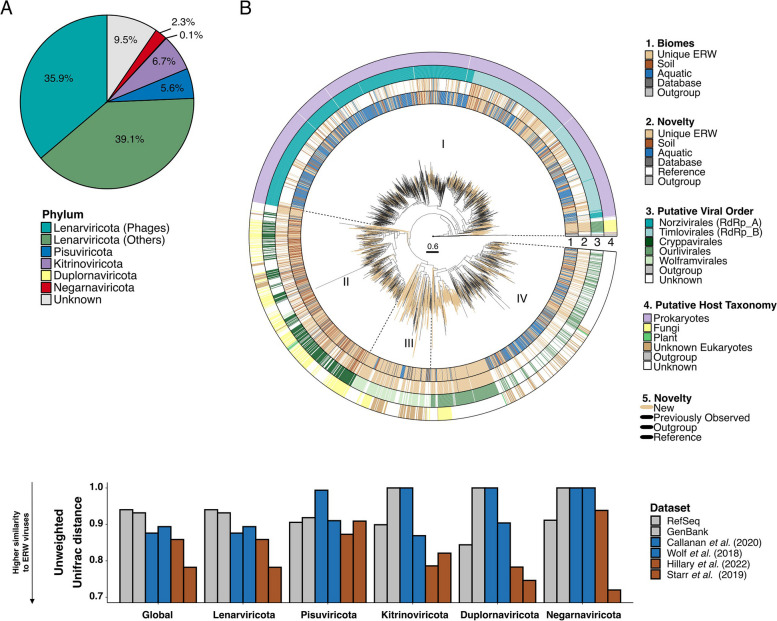
Fig. 2.
Diversity and phylogenetic analyses of ERW RNA viral communities. A Distribution of RNA viral phyla across ERW RNA virus sequences, based on taxonomic assignments from RdRP phylogenies. The Lenarviricota phylum is further divided between bacteria-infecting viruses (“phages,” Leviviricetes) and eukaryote-infecting viruses. B Rooted phylogenetic tree of RdRP sequences belonging to the ssRNA Lenarviricota phylum. The tree is rooted using reverse transcriptases as an outgroup and visualized with ggtree. The tree contains 1331 cluster representatives from ERW soil samples (ring 1, light brown), aligned with those used to construct the RNA global phylogeny from previous metatranscriptomic studies and public databases. Clusters composed exclusively of ERW sequences are colored in brown (ring 2) with branches leading to these clusters highlighted in light brown in the tree, while clusters composed of ERW sequences and existing virus sequences are colored by the environment type of the study (soil: dark brown, aquatic: blue, public databases: dark gray). Virus taxonomy (ring 3) and host (ring 4) are predicted based on the position of reference sequences from the RefSeq database in the tree (see the “Methods” section). C Unweighted UNIFRAC distances between RdRP sequences identified in this study and previously published collections of environmental RNA viruses [14, 29, 78, 80]. ERW and Starr sequences were obtained from RNA shotgun sequencing of bulk samples, while Hillary sequences come from RNA shotgun, sequencing of viral fraction (see [14]). UNIFRAC distances were calculated and are presented separately for each of the 5 RNA virus phyla, with the average distance presented in the first “Global” column. Reference databases are colored in gray, studies from aquatic environments in blue, and soils in dark brown. A distance close to 0 means that the two datasets are phylogenetically similar

