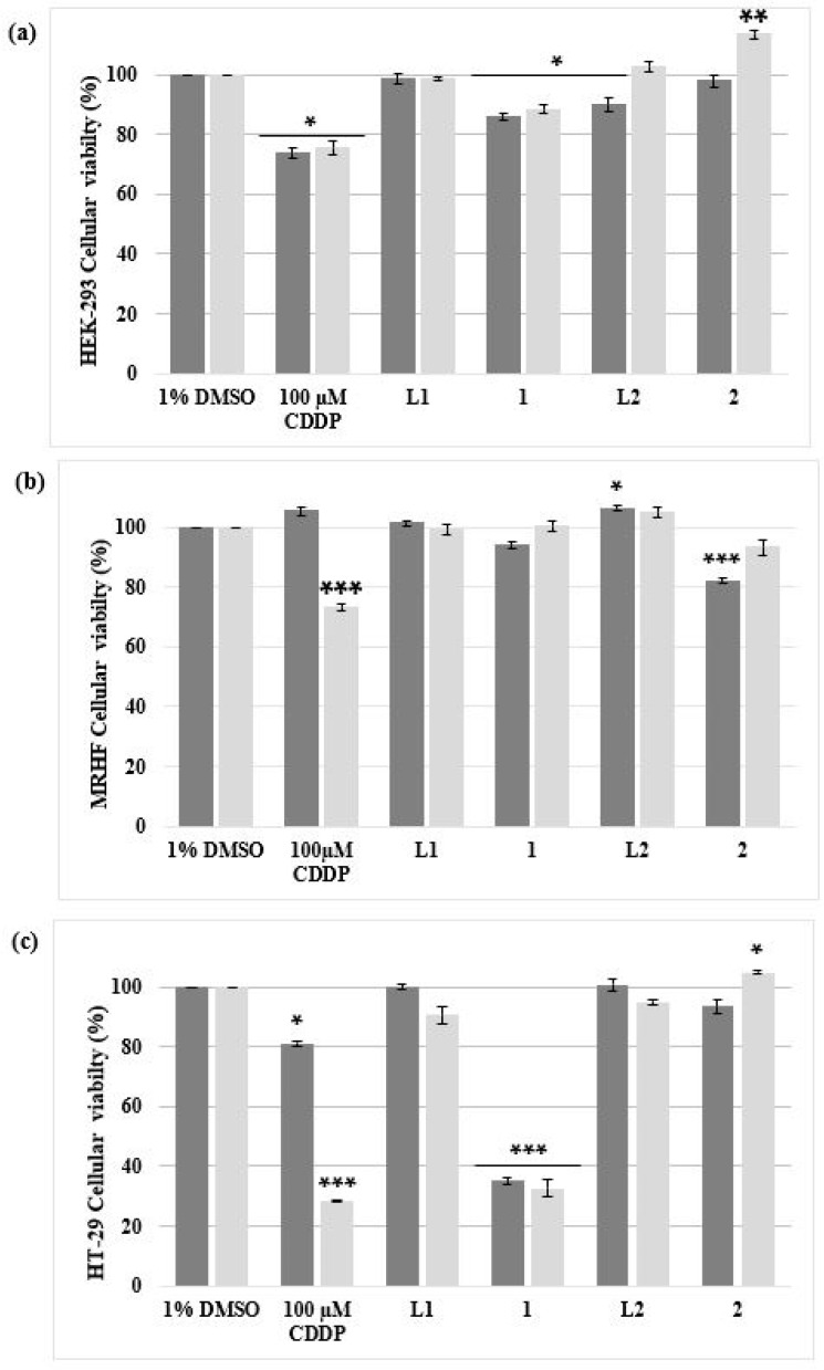
Figure 2.
The percentage cell viability of two non-malignant (a) HEK-293 and (b) MRHF cell lines and (c) one malignant HT-29 cell line analyzed using an alamarBlue® proliferation assay. The cells were treated for 24 h (dark grey) and 48 h (light grey) with 10 µM of complex 1 and 2, including their ligands L1 and L2 dissolved in DMSO (vehicle control; VC). Cells treated with 100 µM CDDP, a positive apoptotic control, were also included. The ± SEM (n = 3) are represented as error bars. Asterisks indicate significant differences between treatments and 1% DMSO (* p < 0.05; ** p < 0.01 and *** p < 0.001).

