

Numerous sources of interference with Bispectral Index™ (BIS) values have been reported, including electrocautery, forced‐air‐warming devices, and pacemakers [1, 2, 3], electrical artefact can be misinterpreted by the BIS algorithm, resulting in misleading values [1].
Quantitative neuromuscular monitoring at the corrugator supercilii muscle is of particular utility when a patient's arms are not accessible due to surgical positioning. However, this site, involving electrodes applied to the patient's forehead, might impair BIS interpretation. We observed these changes during a steady state of general anaesthesia with a BIS Vista sensor (Covidien, Dublin, Ireland) placed on the left forehead of a patient undergoing laparoscopic abdominal surgery.
After achieving a constant effect‐site concentration of propofol and an appropriate depth of anaesthesia according to BIS monitoring, and assuring neuromuscular blockade with a bolus of rocuronium, we set up a train of four (TOF) acceleromyography monitor (ToFscan®, Dräger Medical, Lübeck, Germany) with a stimulating current set at 30 mA and stimulating electrodes placed over the facial nerve, as shown in Figure 1.
Figure 1.
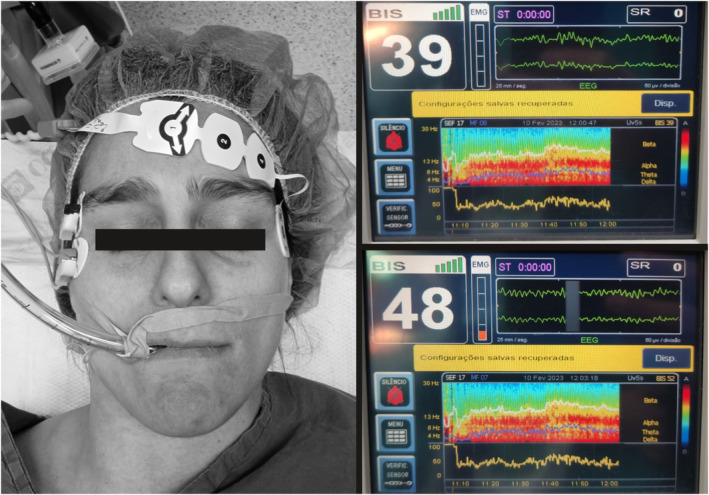
BIS sensor and TOF stimulating electrodes positioning staged for purpose (left). The BIS monitor screen is displayed before (upper right) and after (lower right) turning on the TOF monitor. Note the increase in the BIS numerical value from 39 to 48 when the TOF monitor is turned on (even without TOF measurements) and the slight increments in the electromyogram signal, even though the signal quality indicator remains high.
Within 1 min of placing the TOF electrodes (without obtaining measurements, just with the monitor turned on), a sustained increase of between 5 and 15 points in the BIS value was observed. There were no other indications of a variation in anaesthetic depth, and there were no expected surgically induced variations in anaesthetic requirements. The BIS monitor displayed optimal signal quality (full bars), but the electromyogram (EMG) signal indicator increased slightly. Switching off the TOF monitor (maintaining connector cables applied), caused a reduction to the previously observed BIS values within 2 min.
This unexpected increase in BIS value may be explained by the fact that TOF electrode connector cables, simply attached with the monitor turned on, are a source of electrical noise [1, 3, 4]. When asked about potential interference, the manufacturer of ToFscan suggested that a probable explanation is related to frequent and periodic (every few seconds) impedance checks. Additionally, in accordance with our observations, they reported that this interference is not present when the stimulating electrodes are placed over the ulnar nerve and is no greater than that of an electric scalpel. With that in mind, using a standard digital multimeter, we measured the voltage between the two TOF electrodes and verified repeating brief rises to a maximum of 27 mV (a typical adult human electroencephalogram signal is up to 200 μV), which supports the previous explanation.
When BIS values are exceedingly high and inconsistent with clinical assessment, one should carefully confirm that no sources of interference are present. Subtle changes may go unnoticed by the BIS signal quality indicator [1]. Although variation in‐between the boundaries of the target range of 40 to 60 may be of small clinical importance, increases over 60, particularly in frail patients, could potentially lead to harmful effects associated with unnecessary deepening of the hypnotic state [5].
When assessing TOF count at the corrugator supercilii muscle, the BIS value can be falsely elevated, and one possible solution is to turn off the TOF between readings and avoid timed automatic measurements.
Acknowledgements
Published with the written consent of the patient. No external funding and no competing interests declared.
1 Resident Physician, 2. Consultant, Department of Anaesthesiology, Centro Hospitalar de Setúbal E.P.E., Setúbal, Portugal
References
- 1. Dahaba AA. Different conditions that could result in the bispectral index indicating an incorrect hypnotic state. Anesthesia and Analgesia 2005; 101: 765–773. [DOI] [PubMed] [Google Scholar]
- 2. Hemmerling TM, Desrosiers M. Interference of electromagnetic operating systems in otorhinolaryngology surgery with BISPECTRAL Index Monitoring. Anesthesia and Analgesia 2003; 96: 1698–1699. [DOI] [PubMed] [Google Scholar]
- 3. Hemmerling TM, Fortier JD. Falsely increased Bispectral index values in a series of patients undergoing cardiac surgery using forced‐air‐warming therapy of the head. Anesthesia and Analgesia 2002; 95: 322–323. [DOI] [PubMed] [Google Scholar]
- 4. Chakrabarti D, Surve RM, Bs D, Masapu D. Intraoperative aberrant bispectral index values due to facial nerve monitoring. Journal of Clinical Anesthesia 2017; 37: 61–62. [DOI] [PubMed] [Google Scholar]
- 5. Chan MTV, Cheng BCP, Lee TMC, Gin T. Bis‐guided anesthesia decreases postoperative delirium and cognitive decline. Journal of Neurosurgical Anesthesiology 2013; 25: 33–42. [DOI] [PubMed] [Google Scholar]