Abstract
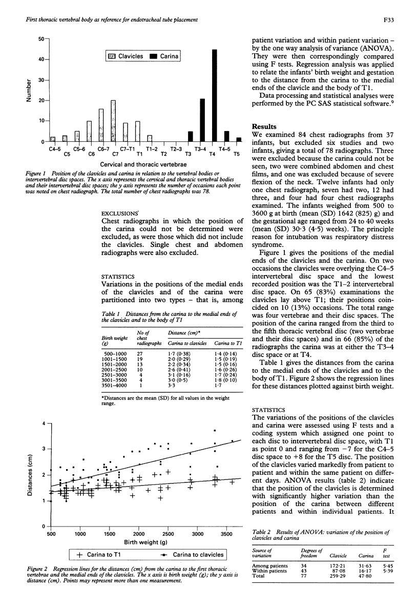
After intubation in newborn infants, the placement of the endotracheal tube in the trachea must be checked by a chest radiograph. The most commonly used reference point for placement is the medial ends of the clavicles. The position of the clavicles may be variable. The present study was carried out to determine whether the body of the first thoracic vertebra should be used instead of the clavicles because its position on chest radiographs is more constant. Seventy eight radiographs obtained from 35 neonates were examined prospectively. The carina was situated between T3 and T5, most commonly at T3-4 or T4 (85%). The position of the clavicles varied markedly from patient to patient and within the same patient on different days, and this variation was significantly higher than that of the carina. On 65 (83%) examinations the clavicles lay above the first thoracic vertebra. It is recommended that, for accurate placement within the trachea, the tip of the endotracheal tube should be placed at the level of the body of the first thoracic vertebra; this could be used as the sole reference point on chest radiographs obtained in the neonatal period.
Full text
PDF


Selected References
These references are in PubMed. This may not be the complete list of references from this article.
- Bednarek F. J., Kuhns L. R. Endotracheal tube placement in infants determined by suprasternal palpation: a new technique. Pediatrics. 1975 Aug;56(2):224–229. [PubMed] [Google Scholar]
- Blayney M., Costello S., Perlman M., Lui K., Frank J. A new system for location of endotracheal tube in preterm and term neonates. Pediatrics. 1991 Jan;87(1):44–47. [PubMed] [Google Scholar]
- Bloch E. C., Ossey K., Ginsberg B. Tracheal intubation in children: a new method for assuring correct depth of tube placement. Anesth Analg. 1988 Jun;67(6):590–592. [PubMed] [Google Scholar]
- Goodman L. R., Conrardy P. A., Laing F., Singer M. M. Radiographic evaluation of endotracheal tube position. AJR Am J Roentgenol. 1976 Sep;127(3):433–434. doi: 10.2214/ajr.127.3.433. [DOI] [PubMed] [Google Scholar]
- Hauser G. J., Pollack M. M., Sivit C. J., Taylor G. A., Bulas D. I., Guion C. J. Routine chest radiographs in pediatric intensive care: a prospective study. Pediatrics. 1989 Apr;83(4):465–470. [PubMed] [Google Scholar]
- Heller R. M., Cotton R. B. Early experience with illuminated endotracheal tubes in premature and term infants. Pediatrics. 1985 Apr;75(4):664–666. [PubMed] [Google Scholar]
- Kuhns L. R., Poznanski A. K. Endotracheal tube position in the infant. J Pediatr. 1971 Jun;78(6):991–996. doi: 10.1016/s0022-3476(71)80429-8. [DOI] [PubMed] [Google Scholar]
- Slovis T. L., Poland R. L. Endotracheal tubes in neonates: sonographic positioning. Radiology. 1986 Jul;160(1):262–263. doi: 10.1148/radiology.160.1.3520649. [DOI] [PubMed] [Google Scholar]


