Abstract
Human papillomavirus type 16 (HPV16) can cause cervical cancer. Expression of the viral E1∧E4 protein is lost during malignant progression, but in premalignant lesions, E1∧E4 is abundant in cells supporting viral DNA amplification. Expression of 16E1∧E4 in cell culture causes G2 cell cycle arrest. Here we show that unlike many other G2 arrest mechanisms, 16E1∧E4 does not inhibit the kinase activity of the Cdk1/cyclin B1 complex. Instead, 16E1∧E4 uses a novel mechanism in which it sequesters Cdk1/cyclin B1 onto the cytokeratin network. This prevents the accumulation of active Cdk1/cyclin B1 complexes in the nucleus and hence prevents mitosis. A mutant 16E1∧E4 (T22A, T23A) which does not bind cyclin B1 or alter its intracellular location fails to induce G2 arrest. The significance of these results is highlighted by the observation that in lesions induced by HPV16, there is evidence for Cdk1/cyclin B1 activity on the keratins of 16E1∧E4-expressing cells. We hypothesize that E1∧E4-induced G2 arrest may play a role in creating an environment optimal for viral DNA replication and that loss of E1∧E4 expression may contribute to malignant progression.
Papillomaviruses are small DNA viruses that infect epithelial tissue (29). Although papillomaviruses have a common genomic organization, they show varied tropisms for epithelial tissue, with different types being highly species specific. Human papillomaviruses (HPVs) are classified as low or high risk (29). Low-risk viruses such as HPVs 1 and 11 are only associated with benign lesions (often called warts), while high-risk viruses such as HPVs 16 and 18 cause lesions that may become malignant. HPV16 is the major risk factor associated with cervical cancer (47).
Papillomavirus infection begins in the basal cells of the epithelium, and as these cells divide, differentiate, and migrate towards the surface of the epithelium, the virus is able to complete its life cycle (23). In the basal cells and in the lower layers of the epithelium, the papillomavirus genome is normally maintained at low copy number. As the cells reach the higher epithelial layers, levels of viral DNA replication increase significantly (viral DNA amplification) to allow the production of new viral genomes, which are packaged into infectious virions (29).
Expression of 16E1∧E4 occurs in the upper layers of the epithelium, i.e., in the later stages of the viral life cycle, where it appears to coincide with the onset of viral DNA amplification (6, 12). However, while viral DNA replication occurs in the nucleus, 16E1∧E4 is a predominantly cytoplasmic protein (12). 16E1∧E4 localizes to the cytokeratin filament networks and can induce the collapse of these networks into bundles both in cell culture (11) and in vivo (48). The significance of this is not clear, but it has been proposed that disruption of the keratin networks could facilitate viral egress. A similar role has been proposed for the 11E1∧E4 protein, which may increase the fragility of the cells by associating with the cornified envelope (4). The 16E1∧E4 protein can associate with mitochondria and alter their membrane potential (40) and can also bind the RNA helicase E4-DBP (9), but the significance of this is unclear. While 16E1∧E4 is expressed abundantly in productive infections, it is not expressed in lesions that have progressed to high-grade neoplasia (31). When it is expressed, the 16E1∧E4 protein is translated from a spliced mRNA, the first five amino acids being derived from the E1 open reading frame and the remainder from the E4 open reading frame (13).
We have previously shown that 16E1∧E4 induces a G2 cell cycle arrest (7), and this has subsequently also been shown for 18E1∧E4 (35). For both 16E1∧E4 and 18E1∧E4, arrest appears to be mediated by a central region of the protein (between amino acids 17 and 45 for 16E1∧E4 and between amino acids 21 and 59 for 18E1∧E4). Mutation of residues T22 and T23 to alanines inhibits the ability of the 16E1∧E4 protein to induce G2 arrest (7).
G2 arrest has been shown to be a feature of several viral proteins. These include the HPV E2 protein (15), the human immunodeficiency virus Vpr protein (20), and the reovirus σ1s protein (39). Although the mechanisms of action of these three proteins are not yet fully elucidated, it has been found in each case that the result is inhibition of Cdk1/cyclin B1, the key complex regulating entry into mitosis.
To investigate the mechanism of 16E1∧E4-induced G2 arrest, we have examined the hypothesis that arrest results from the inactivation of Cdk1/cyclin B1. We found instead that the Cdk1/cyclin B1 complex in 16E1∧E4-expressing cells was active but that it had an altered cellular distribution and was found to localize with 16E1∧E4 to the cytokeratin network. This Cdk1/cyclin B1 was poorly soluble and had a reduced ability to enter the nucleus. As a consequence, the nuclear accumulation of Cdk1/cyclin B1, which is a prerequisite for entry into mitosis, was inhibited. A 16E1∧E4 mutant that was deficient in its ability to arrest cells in G2 did not bind to or colocalize with cyclin B1 and had no ability to sequester cyclin B1 out of the nucleus.
MATERIALS AND METHODS
Cell culture, transfection, and recombinant adenovirus infection.
Saos-2 cells were cultured in Dulbecco's modified Eagle's medium containing 10% fetal calf serum and infected with recombinant adenovirus at a multiplicity of infection of 100 for 24 h. SiHa cells were synchronized and infected with recombinant adenovirus as previously described (7). Briefly, cells were synchronized at the G1/S border by 48 h of incubation in low serum and then 24 h of incubation with aphidicolin. Infection with recombinant adenovirus at a multiplicity of infection of 100 was begun 6 h prior to release of the aphidicolin block. To obtain nocodazole-treated extracts, aphidicolin-treated cells were released from the G1/S block for 6 h and then treated with nocodazole (0.1 μg ml−1) for 12 h. When required, cells were treated with 20 nM leptomycin B for 2.5 h prior to harvest. Unsynchronized SiHa and Cos-7 cells were transfected with plasmids that express green fluorescent protein (GFP) (pXJ.GFP), wild-type 16E1∧E4 (pMV11.16E1∧E4), or T22A,T23A-16E1∧E4 (pMV11. T22A,T23A 16E1∧E4) as previously described (7). Briefly, DNA was prepared with the EndoFree plasmid maxi kit (Qiagen) and transfected into cells with Effectene (Qiagen). The cells were fixed at 48 h posttransfection. HPV epithelial rafts were cultured as previously described (16).
Kinase assays.
For the unfractionated kinase assays, the cells were lysed in radioimmunoprecipitation assay buffer (50 mM Tris, pH 7.5, 150 mM sodium chloride, 1% NP-40, 0.5% sodium deoxycholate, 0.1% sodium dodecyl sulfate [SDS], protease inhibitor cocktail [Roche], 50 U ml−1 benzonase [VWR]), for 15 min at 4°C on a rotator, and the extracts were centrifuged at 10,000 × g for 15 min at 4°C.
To immunoprecipitate Cdk1/cyclin B1 complexes, 400 μl of the supernatant was rotated with 600 μl of radioimmunoprecipitation assay buffer containing 0.25% gelatin and 10 μl of GNS11 anti-cyclin B1 antibody (Neomarkers) for 1 h at 4°C. Immunoprecipitated complexes were recovered by addition of 25 μl of protein A-Sepharose (Amersham Pharmacia Biotech) and rotation for a further 2 h at 4°C. The beads were washed three times with radioimmunoprecipitation assay buffer containing 0.25% gelatin and twice with kinase assay buffer (50 mM MOPS [morpholinepropanesulfonic acid, pH 7.2], 10 mM magnesium chloride, 1 mM dithiothreitol). Reactions were started by adding 30 μl of reaction buffer (kinase assay buffer plus 100 μM ATP, 0.2 kBq ml−1 [γ-32P]ATP, 0.2 mg ml−1 histone H1 [Roche]) and allowed to proceed for 10 min at 30°C. Reactions were stopped by the addition of 30 μl of 2% SDS and 1 mM dithiothreitol and incubation for 5 min at 95°C. The reactions were centrifuged at 14,000 × g for 10 min, and the supernatants were separated on an SDS-15% polyacrylamide gel. The levels of histone H1 phosphorylation were quantitated with a Storm Phosphorimager and ImageQuant software (Molecular Dynamics).
Fractionation and coimmunoprecipitation.
To fractionate the cells just for Western blotting, cell pellets were resuspended in 1% NP-40 fractionation buffer (1% NP-40, 10 mM EDTA, 50 U ml−1 benzonase [VWR], 0.5 μg ml−1 okadaic acid [Sigma], protease inhibitor cocktail [Roche] in phosphate-buffered saline [PBS]). The cells were lysed in this buffer for 15 min at 4°C on a rotator and then centrifuged at 10,000 × g for 15 min at 4°C to obtain the 1% NP-40-soluble fraction. The NP-40-insoluble pellet was resuspended in 1% SDS-1 mM dithiothreitol and incubated for 10 min at 95°C. The extracts were centrifuged as above to obtain the 1% NP-40-insoluble (1% SDS-soluble) fraction.
Equal volumes of 1% NP-40-soluble and -insoluble fractions were loaded for Western blotting. Alternatively, to obtain fractions for kinase assays, cell pellets were resuspended in 0.5% fractionation buffer (as above but with 0.5% instead of 1% NP-40). The cells were lysed in this buffer for 5 min at 4°C and centrifuged at 10,000 × g for 5 min at 4°C to obtain the 0.5% NP-40-soluble fraction. The NP-40-insoluble pellet was resuspended in radioimmunoprecipitation assay buffer. The extracts were rotated for 15 min at 4°C and centrifuged as above to obtain the 0.5% NP-40-insoluble (0.1% SDS-soluble) fraction.
To coimmunoprecipitate 16E1∧E4 and cyclin B1, 1% NP-40-soluble fractions containing 0.25% gelatin were rotated with 10 μl of anti-cyclin B1 polyclonal antibody clone H-433 (Santa Cruz) or normal rabbit serum (Sigma) for 1 h at 4°C. Immunoprecipitated complexes were recovered by addition of 25 μl of protein G-Sepharose (Amersham Pharmacia Biotech) and rotation for a further 2 h at 4°C. The beads were washed five times with radioimmunoprecipitation assay buffer containing 0.25% gelatin before being resuspended in 2% SDS-1 mM dithiothreitol and incubated at 95°C for 5 min. The extracts were centrifuged at 14,000 × g for 10 min, and the supernatants were separated on an SDS-15% polyacrylamide gel.
GST pulldowns.
The vectors expressing glutathione S-transferase (GST), pGEX and wild-type E1∧E4 fused to GST (GST16E1∧E4), pGEX.16E1∧E4, have been previously described (10). pGEX.16E1∧E4 was mutated to pGEX.T22A,T23A-16E1∧E4 with Quikchange site directed mutagenesis (Stratagene). Expression and purification of GST, GST16E1∧E4, and GST-T22A,T23A16E1∧E4 proteins was essentially as previously described (25), and their integrity bound to beads was confirmed by silver stain gels. SiHa cells treated for 12 h with nocodazole (0.1 μg ml−1) were harvested and lysed in 0.5% NP-40-50 Uml−1 benzonase-protease inhibitor cocktail-PBS. The lysate was centrifuged at 13 krpm for 5 min at 4°C, and the supernatant was added to equal quantities of GST, GST-16E1∧E4, or GST-T22A,T23A-16E1∧E4 beads diluted in 1 ml of pulldown buffer (0.05% NP-40, 10 μg ml−1 bovine serum albumin, protease inhibitor cocktail, PBS). The mixtures were rotated at 4°C for 1 h and washed four times with pulldown buffer. Proteins were eluted from the beads by boiling for 5 min at 95°C in 30 μl of 1 mM dithiothreitol-10% SDS.
Western blotting.
Samples were electrophoretically separated and transferred to an Immobilon P membrane (Millipore) for Western blotting. Detection was with anti-16E1∧E4 clone TVG402 (10), anti-cyclin B1 clone V152 (Neomarkers), anti-Cdk1 clone 17 (Santa Cruz), anti-keratin 18 clone CY-90 (Sigma), anti-keratin 18 phosphoserine 33 clone 8250 (26), antitubulin clone B512 (Sigma), anti-phosphohistone H3 (Ser10) (Upstate), anti-glyceraldehhyde-3-phosphate dehydrogenase (GAPDH) clone 374 (Chemicon) and then anti-rabbit (NA9340) or anti-mouse (NA931) horseradish peroxidase conjugates (Amersham Pharmacia Biotech), followed by detection with an ECL kit (Amersham Pharmacia Biotech).
Immunostaining and flow cytometry.
For microscopy, monolayer cells were fixed for 5 min in 5% formaldehyde in PBS and permeabilized in 0.5% Triton X-100 for 10 min. Saos-2 cells were treated with 0.25 μM MitoTracker Red CMXRos (Molecular Probes) for 20 min prior to fixation. Sections were fixed in 10% formaldehyde and permeabilized with a microwave/citric acid-based method (43). Nuclei were stained with 1 μg ml−1 DAPI (4′-6′-diamidino-2-phenylindole; Sigma). 16E1∧E4 was detected with TGV402 (10) or TGV405 (12), directly conjugated to Alexa 488 (Molecular Probes). Other antigens were detected with anti-canine oral papillomavirus E1∧E4 (38), anti-1E1∧E4 clone p1p7 (8), anti-Cdk1 clone Ab-1 (Oncogene), anti-β-galactosidase monoclonal (Zymed) or anti-β-galactosidase polyclonal (ICN), anti-keratin 8/18 clone 8592 (27), anti-keratin 18 phospho-serine 33 clone 8250 (26), and Myc-tagged Myt1 was detected with anti-c-Myc clone A-14 (Santa Cruz).
Cyclin B1 was detected in monolayer cultures with clone GNS1 (Neomarkers), in HPV16 sections with clone 7A9 (Novocastra), and in canine oral papillomavirus-induced and HPV1-induced lesions with clone V152 (Neomarkers). Primary antibodies were detected with Alexa 488-, Alexa 594-, or Alexa 350-labeled anti-mouse or anti-rabbit immunoglobulin antibodies (Molecular Probes) or for the detection of cyclin B1 in canine oral papillomavirus lesions, with a combination of StreptABC horseradish peroxidase (Dako) and trichostatin A (New England Nuclear). The cells were examined with a fluorescent Labophot II monochrome camera (Nikon) and IP Lab imaging software (Roper Scientific). Flow cytometry was carried out as previously described (7). Briefly, cells were stained with propidium iodide (12.5 μg ml−1) and their DNA content was analyzed with a FACScan and CellQuest software (Becton Dickinson).
RESULTS
16E1∧E4-induced G2 arrest does not result from inhibition of the kinase activity of the Cdk1/cyclin B1 complex.
The failure of cells to enter mitosis often results from inhibition of the Cdk1/cyclin B1 kinase complex by preventing expression of cyclin B1 or by inhibitory phosphorylation of Cdk1 (reviewed in reference 45). To determine if 16E1∧E4-induced G2 arrest was associated with a reduction in Cdk1/cyclin B1 kinase activity, the activity in 16E1∧E4-expressing cells was compared to that of control cells that express β-galactosidase. The cells used were SiHa cells, a cell line derived from a cervical cancer caused by HPV16 in which the E2/E4 region is disrupted as a result of integration of viral DNA sequences into the host genome (2). SiHa cell proliferation is dependent on the continued expression of the E6 and E7 oncogenes (18).
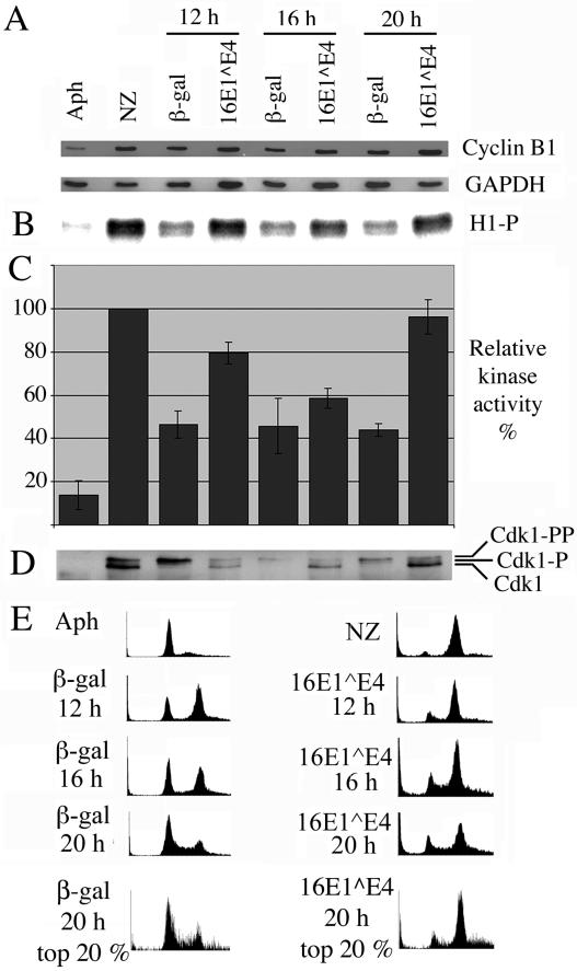
G1/S-synchronized cells were infected with recombinant adenoviruses expressing 16E1∧E4 or the control protein β-galactosidase and analyzed at 12, 16, and 20 h after release of the G1/S block. Cells reached the G2/M boundary between 12 and 20 h postrelease. Cells treated with aphidicolin, an inhibitor of DNA replication that arrests cells at G1/S before cyclin B1 has begun to accumulate, were used as a control. Cell populations arrested with aphidicolin contain only small numbers of cells in G2 and have low levels of Cdk1/cyclin B1 activity. Cells treated with nocodazole, an inhibitor of mitotic spindle formation that arrests cells in mitosis with fully active Cdk1/cyclin B1 complexes, were used as a control for high levels of Cdk1/cyclin B1 activity. Cell extracts were analyzed by Western blotting to determine their relative levels of cyclin B1 (Fig. 1A).
FIG. 1.
16E1∧E4 expression does not inhibit Cdk1/cyclin B kinase activity. Control G1/S-synchronized SiHa cells were either harvested without releasing the G1/S block (Aph) or released from the block for 6 h and then treated for 12 h with nocodazole prior to harvesting (NZ). G1/S-synchronized SiHa cells infected with recombinant adenoviruses expressing β-galactosidase or 16E1∧E4 were harvested at 12, 16, and 20 h post-block release. (A) Cell extracts were Western blotted for cyclin B1 and glyceraldehyde-3-phosphate dehydrogenase (GAPDH). (B) Cdk1/cyclin B1 was immunoprecipitated from cell extracts and used in an in vitro kinase assay to phosphorylate histone H1. Protein labeled with 32P was detected with a phosphorimager. (C) Levels of histone H1 phosphorylation were quantitated with ImageQuant software and expressed as a percentage of that obtained for the nocodazole-treated samples. The results show the means of four experiments ± standard error of the mean. (D) Immunoprecipitated complexes were Western blotted for Cdk1. (E) Cells were stained with propidium iodide, immunostained for β-galactosidase or 16E1∧E4, and analyzed by flow cytometry. Plots show cell number versus DNA content. The final row shows plots in which the top 20% of high-β-galactosidase- or 16E1∧E4-expressing cells were selected for analysis of DNA content.
As expected, cyclin B1 levels appeared low and high in cell populations treated with aphidicolin and nocodazole, respectively, since expression of cyclin B1 is cell cycle regulated, with expression peaking at the G2/M boundary. The levels of cyclin B1 in β-galactosidase-expressing cells were above the levels seen in aphidicolin-treated cells and below the levels seen in nocodazole-treated cells. The levels of cyclin B1 remained high throughout the 8-h period over which β-galactosidase-expressing cells were found to cross the G2/M boundary. The level of cyclin B1 in 16E1∧E4-expressing cells was not reduced compared to β-galactosidase-expressing cells, which suggests that the inhibitory effect of 16E1∧E4 on progression through G2 is not a result of a reduction in the level of cyclin B1.
The cell extracts used to analyze cyclin B1 levels were also used for immunoprecipitation of the Cdk1/cyclin B1 complex, which was then used in an in vitro kinase assay to phosphorylate histone H1 (Fig. 1B and C). As expected, kinase activity in the aphidicolin-treated cells was low, while in the nocodazole-treated cells it was high. Although there was not a single time point at which Cdk1/cyclin B1 activity was seen to peak in the β-galactosidase and 16E1∧E4 samples, we believe this is due to the asynchronicity of the population, with cells cycling quickly reaching mitosis at 12 h while others took 20 h. However, kinase activity in the 16E1∧E4 extracts was significantly higher than in the β-galactosidase extracts and approached the levels found in the nocodazole-treated extracts. It is therefore unlikely that 16E1∧E4-induced G2 arrest is mediated by inhibition of the enzyme activity of Cdk1/cyclin B1.
Interestingly, the Cdk1 immunoprecipitated from the 16E1∧E4-expressing cells was predominantly of the fastest-migrating, unphosphorylated form, while that derived from the β-galactosidase-expressing cells was generally of the slower-migrating, phosphorylated forms (Fig. 1D). These results suggest that the Cdk1 in 16E1∧E4-expressing cells is less subject to inhibitory phosphorylation, and this is compatible with the Cdk1/cyclin B1 complex's being active. As has been observed previously (42), the levels of unphosphorylated Cdk1 found in our samples did not correlate with kinase activity, suggesting that an additional level of regulation may be present.
The cells were also analyzed by flow cytometry to confirm their cell cycle distribution (Fig. 1E). β-Galactosidase-expressing cells proceed through the cell cycle after release from the G1/S block and eventually return to G1. 16E1∧E4-expressing cells, although able to exit the G1/S block and proceed through S phase, are unable to efficiently return to G1 and instead arrest with G2 DNA content. It is clear from the data presented here and in our previous paper (7) that not all the cells that express 16E1∧E4 arrest in G2. We hypothesized that the higher the level of 16E1∧E4 in a cell, the more likely it would be to arrest. Analysis of the top 20% of expressing cells (Fig. 1E) shows this to be true. The cells expressing high levels of 16E1∧E4 show a stronger tendency to arrest in G2 than cells expressing lower levels of 16E1∧E4. This is not similarly the case for cells expressing high levels of the control protein β-galactosidase, which have a cell cycle distribution very similar to that of the total population of β-galactosidase-expressing cells. That the 16E1∧E4-dependent cell cycle effect is not seen with only low levels of 16E1∧E4 makes sense, given the high levels of 16E1∧E4 expression found in vivo (12, 31).
Recombinant adenoviruses have been used throughout to direct expression of 16E1∧E4. Since the backbone of the recombinant viruses can have effects on the cell cycle, we have consistently used recombinant adenoviruses that express β-galactosidase with an identical backbone as controls for those that express 16E1∧E4 rather than using uninfected cells. Our experiments with uninfected cells have shown that cyclin B1 appears to behave in a similar manner in these cells as it does in our control cells expressing β-galactosidase, although the cell cycle progresses slightly faster in the adenovirus-infected cells (http://www.nimr.mrc.ac.uk/supplementary/doorbar, Fig. S1).
Cyclin B1 and Cdk1 colocalize with 16E1∧E4 on the cytokeratin filament network.
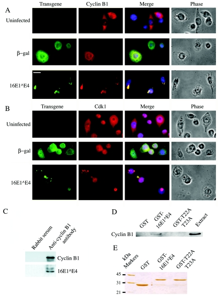
Since 16E1∧E4-induced G2 arrest is not due to inhibition of the enzyme activity of the Cdk1/cyclin B1 complex, the possibility that 16E1∧E4 could be affecting the complex in some other way was investigated. SiHa cells that expressed β-galactosidase or 16E1∧E4 were immunostained in order to reveal the intracellular distribution of cyclin B1 (Fig. 2A) and Cdk1 (Fig. 2B). In cells that expressed β-galactosidase, cyclin B1 appeared diffuse throughout the cytoplasm or had begun to accumulate in the nucleus prior to the onset of chromosome condensation (a similar distribution was observed in uninfected cells; Fig. 2A and Fig. S1 at http://www.nimr.mrc.ac.uk/supplementary/doorbar). In cells that expressed 16E1∧E4, cyclin B1 localized to discrete areas within the cytoplasm and colocalized with 16E1∧E4. This could be shown with several different antibodies to 16E1∧E4 (TVG402 and TVG405) and cyclin B1 (GNS1 and GNS11) and with multiple fixation and permeabilization protocols. Control antibodies did not give rise to similar staining patterns (http://www.nimr.mrc.ac.uk/supplementary/doorbar, Fig. S2), and as expected, cells in G0 showed no staining with the cyclin B1 antibody (http://www.nimr.mrc.ac.uk/supplementary/doorbar, Fig. S3).
FIG. 2.
16E1∧E4 associates with Cdk1/cyclin B1. G1/S-synchronized SiHa cells expressing β-galactosidase or 16E1∧E4 were fixed at 16 h post-block release. Cells were double stained for β-galactosidase or 16E1∧E4 (green) and cyclin B1 (A) or Cdk1 (B) (red). A DAPI nuclear stain was included (blue). Bar, 10 μm. (C) Cell extracts were immunoprecipitated with anti-cyclin B1 rabbit polyclonal antibody or normal rabbit serum and Western blotted for 16E1∧E4 and cyclin B1. (D) Cell extracts from nocodazole-treated SiHa cells were used in pull-down experiments with GST, GST-16E1∧E4, or GST-T22A,T23A-16E1∧E4 and Western blotted along with the cell extract for cyclin B1. The levels of cyclin B1 pulled down are compared to 20% of the input amount of cyclin B1. (E) The input of GST, GST-16E1∧E4, and GST-T22A,T23A-16E1∧E4 into the pull-down experiments is shown by silver staining.
The data shown in Fig. 2A were representative of the staining patterns observed in replicate experiments, and were obtained with monoclonal antibodies TVG 405 (16E1∧E4) and GNS1 (cyclin B1) on permeabilized cells fixed with formaldehyde. To confirm that the apparent colocalization of cyclin B1 with 16E1∧E4 was not limited to late time points, the cells were also stained at 8 h postrelease. At this time, the β-galactosidase and 16E1∧E4 populations are in the same cell cycle stage, and cyclin B1 colocalizes with 16E1∧E4 but not with β-galactosidase (http://www.nimr.mrc.ac.uk/supplementary/doorbar, Fig. S4). The 16E1∧E4 staining pattern at 16 h is typical of cells in which collapse of the cytokeratin network has occurred, and in these cells 16E1∧E4 and keratins form a perinuclear bundle, as described previously (11). Cdk1 was found diffusely in the cytoplasm and nucleus of uninfected cells and cells expressing β-galactosidase, but in cells that expressed 16E1∧E4, it was found predominantly in association with the 16E1∧E4 in the cytoplasm (Fig. 2B).
To further confirm the close physical association between the 16E1∧E4 protein and cyclin B1, a rabbit polyclonal antibody was used to immunoprecipitate cyclin B1. These experiments are not straightforward because 16E1∧E4 is predominantly insoluble in immunoprecipitation buffers and coimmunoprecipitation relies on the small proportion of the 16E1∧E4 protein that is soluble in NP-40-containing buffers (48). However, following Western blotting, 16E1∧E4 could be detected in the cyclin B1 immunoprecipitates from 16E1∧E4-expressing cells but was not apparent in immunoprecipitates carried out with the rabbit serum control (Fig. 2C). The slower-migrating 16E1∧E4 band represents a phosphorylated form that can be removed by treatment with λ phosphatase (data not shown).
We were unable to perform the reverse coimmunoprecipitation, i.e., precipitating cyclin B1 on 16E1∧E4 antibodies, which may suggest that the major epitope on 16E1∧E4 is masked as a result of cyclin B1 binding. To overcome this difficulty, N-terminal GST-tagged 16E1∧E4 expressed in Escherichia coli was used to pull down cyclin B1 extracted from nocodazole-treated SiHa cells on glutathione-coated Sepharose beads (Fig. 2D and E). Cyclin B1 was found to be pulled down only by GST-16E1∧E4 and not by GST alone, providing further evidence of the ability of cyclin B1 to associate specifically with 16E1∧E4. We have previously described a 16E1∧E4 mutant (T22A,T23A-16E1∧E4) that fails to induce G2 arrest (7). We made an N-terminal GST-tagged version of this mutant and used it in the same pull-down experiments. As expected, unlike wild-type 16E1∧E4, the T22A,T23A-16E1∧E4 mutant was unable to pull down cyclin B1 (Fig. 2D). This suggests that the interaction with cyclin B1 is significant for the G2 arrest phenotype.
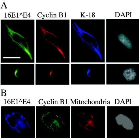
Since, when expressed in epithelial cells (in vivo and in vitro), 16E1∧E4 can associate with cytokeratin filaments (11, 48), triple staining was carried out with antibodies to 16E1∧E4, cyclin B1, and keratin 18. All three proteins had a similar staining pattern (Fig. 3A shows one cell with an intact keratin network prior to cytokeratin collapse and one cell postcollapse). These results suggest that cyclin B1 can be relocated to the cytokeratin network as a result of its association with 16E1∧E4 and that this phenomenon is independent of whether the cytokeratin network is intact or collapsed.
FIG. 3.
Cyclin B1 associated with 16E1∧E4 is found on the keratins or mitochondria. (A) G1/S-synchronized SiHa cells expressing 16E1∧E4 were fixed at 16 h post-block release. Cells were triple stained for 16E1∧E4 (green), cyclin B1 (red), and keratin 18 (K-18; blue). A DAPI nuclear stain was included (white). The upper panels show a cell prior to keratin collapse, while the lower panels show a cell with collapsed keratin. (B) Saos-2 cells infected with recombinant adenoviruses that express 16E1∧E4 were triple stained for 16E1∧E4 (blue), cyclin B1 (green), and mitochondria (red). A DAPI nuclear stain was included (white). Bar, 10 μm.
Cyclin B1 can interact with 16E1∧E4 independently of keratin binding.
We have previously shown that 16E1∧E4-induced G2 arrest can occur in cells (Saos-2) that lack keratins (7). To determine whether the colocalization of cyclin B1 with 16E1∧E4 is also independent of 16E1∧E4 binding to keratins, we examined the distribution of cyclin B1 in 16E1∧E4-expressing Saos-2 cells. In Saos-2 cells, 16E1∧E4 is found associated with the mitochondria (40), and we were interested to see whether this would result in cyclin B1 also becoming localized to the mitochondria. To examine this, 16E1∧E4-expressing Saos-2 cells were triple stained with reagents that detect 16E1∧E4 (TVG405) and cyclin B1 (GNS11) and mitochondria (MitoTracker) (Fig. 3B). The results show that cyclin B1 is found associated with 16E1∧E4 on the mitochondria. (In β-galactosidase-expressing Saos-2 cells, cyclin B1 cannot be detected on the mitochondria [data not shown]). It thus appears that the ability of 16E1∧E4 to colocalize with cyclin B1 is not dependent on keratins.
16E1∧E4 induces a reduction in solubility of active Cdk1/cyclin B1.
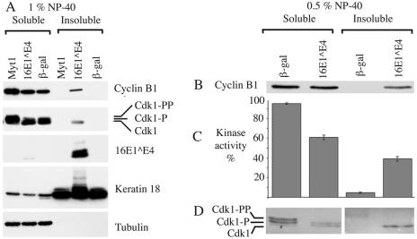
Cdk1/cyclin B1 is usually found as a soluble complex within the cytosol and as such is readily extracted in detergents such as NP-40. In contrast, 16E1∧E4 is predominantly insoluble in NP-40. Since cyclin B1 and Cdk1 appear to associate with 16E1∧E4, we examined whether this resulted in a change in the solubility of cyclin B1 or Cdk1. We expressed β-galactosidase, 16E1∧E4, or the control protein Myt1 in G1/S-synchronized SiHa cells. Myt1 induces G2 arrest by phosphorylating Cdk1 on Thr14 and Tyr15 (33) and/or by binding Cdk1 to hold it on the endoplasmic reticulum (28). The use of Myt1 provides a control to show that any 16E1∧E4-induced changes in the solubility of Cdk1/cyclin B1 are not simply a result of G2 arrest. The cells were harvested 20 h after release of the G1/S block and fractionated with buffers containing 1% NP-40 (extracts NP-40-soluble fraction) or 1% SDS (extracts NP-40-insoluble fraction) (Fig. 4A).
FIG. 4.
16E1∧E4 decreases the solubility of active Cdk1/cyclin B1. (A) G1/S-synchronized SiHa cells expressing Myt1, 16E1∧E4, or β-galactosidase were harvested at 20 h post-block release and fractionated with 1% NP-40 (NP-40-soluble fraction) and then with 1% SDS (NP-40-insoluble fraction). Fractions were Western blotted for 16E1∧E4, cyclin B1, Cdk1, keratin 18, and, as a control for fractionation, tubulin. (B) G1/S-synchronized SiHa cells expressing β-galactosidase or 16E1∧E4 were harvested at 20 h post-block release and fractionated first with 0.5% NP-40 (NP-40-soluble fraction) and then with 0.1% SDS (NP-40-insoluble fraction). The fractions were Western blotted for cyclin B1. (C) Cdk1/cyclin B1 was immunoprecipitated from fractions with anti-cyclin B1 monoclonal antibody. Immunoprecipitated complexes were used in an in vitro kinase assay to phosphorylate histone H1. 32P-labeled protein was detected with a phosphorimager and quantitated with ImageQuant. Kinase activity is expressed as a percentage of total activity in β-galactosidase- or 16E1∧E4-expressing cells. The results show the mean values of three experiments ± standard error of the mean. (D) Immunoprecipitated fractions were Western blotted for Cdk1.
In both β-galactosidase- and Myt1-expressing cells, Cdk1 and cyclin B1 localized exclusively to the NP-40-soluble fraction. However, in extracts containing 16E1∧E4, Cdk1 and cyclin B1 were additionally present in the NP-40-insoluble fraction, suggesting that they were no longer soluble in NP-40. In particular, only the fast-migrating form of Cdk1 (which lacks inhibitory phosphorylation on Thr14 or Tyr15) was found along with cyclin B1 in the NP-40-insoluble fraction, suggesting the presence of an active complex, in accordance with the data from Fig. 1. That the sequestration of Cdk1 and cyclin B1 into the insoluble fraction was specific was shown by the inability of 16E1∧E4 to sequester other proteins (Cdk4 and Cdc25C) into the insoluble fraction (http://www.nimr.mrc.ac.uk/supplementary/doorbar, Fig. S5).
To confirm that the Cdk1/cyclin B1 sequestered into the NP-40-insoluble fraction was active, the cells were fractionated with 0.5% NP-40 (extracts NP-40-soluble fraction) and 0.1% SDS (extracts NP-40-insoluble fraction). The fractions were Western blotted for cyclin B1 (Fig. 4B) and assayed for histone H1 kinase activity (Fig. 4C). Over one-third of the kinase activity that is present in 16E1∧E4-expressing cells is found in the NP-40-insoluble fraction of the cell, suggesting that it is 16E1∧E4 associated. In fact, the proportion of soluble Cdk1/cyclin B1 in 16E1∧E4-expressing cells may be lower than that shown in Fig. 4C, since the fractionation process itself leads to a loss of Cdk1/cyclin B1 from the insoluble fraction. Increased concentrations of NP-40 and increased incubation times decrease the amount of Cdk1/cyclin B1 in the insoluble fraction (compare the ratio of NP-40-soluble to -insoluble cyclin B1 in Fig. 4A, which was exposed to 1% NP-40 for 15 min, to the ratio in Fig. 4B, which was exposed to 0.5% NP-40 for only approximately 5 min). The immunofluorescence data (Fig. 2 and 3), which show the in situ distribution, reveal that the majority of the Cdk1 and cyclin B1 is associated with 16E1∧E4 on the keratin filament networks (Fig. 3A) or with 16E1∧E4 in collapsed cytokeratin bundles (Fig. 2A and B and Fig. 3A).
Successful entry into mitosis relies on a sensitive switch mechanism that requires the accumulation of a critical concentration of active Cdk1/cyclin B1 in the nucleus (14). Figure 4D shows that in 16E1∧E4-expressing cells, Cdk1 that did not have inhibitory phosphorylation on Thr14 or Tyr15 was apparent in the NP-40-insoluble fraction, where 16E1∧E4 and the keratins were predominantly found. These results suggest that the nuclear levels of active Cdk1/cyclin B1 may be lower in 16E1∧E4-expressing cells than in control cells. Attempts to verify this theory with biochemical assays are hindered by the insoluble nature of 16E1∧E4 (http://www.nimr.mrc.ac.uk/supplementary/doorbar, Fig. S7C) and its tendency to collapse with the cytokeratins into perinuclear bundles (Fig. 2 and 3A). This has prevented us from isolating nuclear fractions that are devoid of contaminating 16E1∧E4 and associated Cdk1/cyclin B1. In addition, previous attempts at biochemical fractionation of cyclin B1 have been unsuccessful (42), and similar difficulties were encountered by us when attempting to analyze cyclin B1 distribution (http://www.nimr.mrc.ac.uk/supplementary/doorbar, Fig. S7A and B).
Nuclear accumulation of Cdk1/cyclin B1 is inhibited in 16E1∧E4-expressing cells.
The data presented in Fig. 2 suggest that cyclin B1 does not accumulate in the nucleus of 16E1∧E4-expressing cells. To determine if this is because it is being prevented from entering the nucleus, cyclin B1 staining was carried out in the presence of leptomycin B, a drug that inhibits nuclear export of cyclin B1 (28). During G2, cyclin B1 is constantly shuttling in and out of the nucleus, but since its rate of export exceeds its rate of import, it is normally only observed in the cytoplasm. Leptomycin B treatment therefore causes cyclin B1 to accumulate in the nucleus of G2 cells, although this in itself is insufficient to trigger mitosis since the Cdk1/cyclin B1 complex remains inactive. The efficacy of leptomycin B treatment was determined (http://www.nimr.mrc.ac.uk/supplementary/doorbar, Fig. S6).
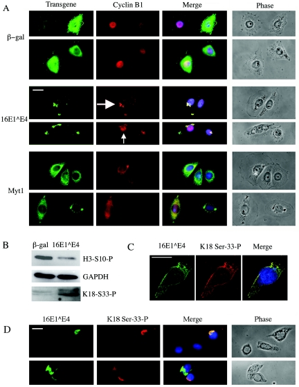
In β-galactosidase-expressing cells, cyclin B1 was predominantly nuclear following leptomycin B treatment, with virtually no cyclin B1 being found in the cytoplasm (Fig. 5A). This confirms that in cells that express β-galactosidase, cyclin B1 is free to traffic into the nucleus. However, in cells that express the G2 arrest protein Myt1, the pattern is different. Some cyclin B1 is found to colocalize with Myt1 in the cytoplasm, which is consistent with the inhibition of cyclin B1 nuclear import. Again, this is to be expected, since Myt1 binds Cdk1/cyclin B1 and localizes it to the endoplasmic reticulum (28). In the majority of 16E1∧E4-expressing cells, much of the cyclin B1 appears to be associated with 16E1∧E4 in the cytoplasm (large arrow in Fig. 5A), although in some cells cyclin B1 may also be found in the nucleus (small arrow in Fig. 5A).
FIG. 5.
16E1∧E4 expression reduces the ability of cyclin B1 to accumulate in the nucleus. G1/S-synchronized SiHa cells expressing β-galactosidase, 16E1∧E4, or Myt1 were treated with leptomycin B (LMB), beginning 14 h postrelease, and then harvested 2.5 h later. (A) The cells were double stained for β-galactosidase, 16E1∧E4, or Myt1 (green) and cyclin B1 (red). A DAPI nuclear stain was included (blue). The large white arrow indicates a cell with predominantly cytoplasmic cyclin B1, and the small white arrow indicates a cell in which some of the cyclin B1 has entered the nucleus. (B) Total cell extracts were Western blotted for phosphohistone H3 or phosphokeratin 18 serine 33 and compared to a glyceraldehyde-3-phosphate dehydrogenase (GAPDH) control. (C and D) G1/S-synchronized SiHa cells expressing 16E1∧E4 at 24 h post-block release were immunostained for 16E1∧E4 (green) and phosphokeratin 18 serine 33. A DAPI nuclear stain was included (blue). Bars, 10 μm.
Since nuclear-cytoplasmic fractionation of 16E1∧E4-expressing cells is unreliable, we used the alternative strategy of examining the phosphorylation status of nuclear and cytoplasmic proteins as a marker for the presence of active Cdk1/cyclin B1 in these cellular compartments. If the nuclear accumulation of active Cdk1/cyclin B1 is being inhibited in 16E1∧E4-expressing cells, then we would expect nuclear proteins normally phosphorylated as the cell crosses the G2/M boundary (e.g., histone H3) not to be phosphorylated (22). The phosphorylation status of serine 10 of histone H3 in β-galactosidase- and 16E1∧E4-expressing cells at 16 h post-block release was assessed by Western blotting with a phosphorylation-specific antibody (Fig. 5B). The results show that the levels of phosphorylated histone H3 in the 16E1∧E4 extracts were much lower than those found in the β-galactosidase extracts (while levels of the control protein glyceraldehyde-3-phosphate dehydrogenase are the same).
Although histone H3 serine 10 is phosphorylated by nuclear aurora B kinase, aurora B activity is regulated by Cdk1/cyclin B1 (34), providing further support to the hypothesis that 16E1∧E4 is preventing active Cdk1/cyclin B1 from reaching its nuclear targets. In contrast, we would expect that cytoplasmic proteins that are normally phosphorylated in G2/M, such as keratin 18 (26), to still be phosphorylated in 16E1∧E4-expressing cells. Analysis of keratin 18 serine 33 phosphorylation (Fig. 5B) showed it to be consistently higher in cells that express 16E1∧E4 than in those that express β-galactosidase. Interestingly, keratin 18 serine 33 is a known target for Cdk1/cyclin B1 phosphorylation (26), raising the possibility that the tethering of the Cdk1/cyclin B1 to the cytokeratin network by 16E1∧E4 may contribute to its phosphorylation. This is supported by immunostains of 16E1∧E4-expressing cells, where phosphorylation of keratin 18 serine 33 is found to colocalize with 16E1∧E4 in networks (Fig. 5C) and in collapsed bundles (Fig. 5D). In contrast, in control cells not expressing 16E1∧E4, keratin 18 phosphoserine 33 is absent (Fig. 5D) or found only in mitotic and apoptotic cells (data not shown) (26). Although indirect, these data strongly suggest that nuclear import of active Cdk1/cyclin B1 is inhibited following expression of high levels of 16E1∧E4, and instead the kinase remains in the cytoplasm.
A 16E1∧E4 mutant that fails to arrest in G2 also fails to disturb cyclin B1.
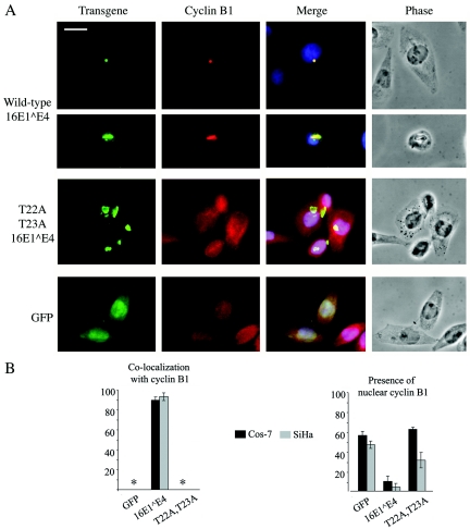
If the 16E1∧E4-induced G2 arrest is indeed mediated by it holding Cdk1/cyclin B1 in the cytoplasm, then we would expect to see a reduced association of the mutant T22A,T23A-16E1∧E4 with cyclin B1 and a greater propensity for cyclin B1 to enter the nucleus. To test this, we transfected SiHa and Cos-7 cells with plasmids that expressed wild-type 16E1∧E4, T22A,T23A-16E1∧E4, or the control protein GFP. The cells were stained for cyclin B1 and, when present, 16E1∧E4 (Fig. 6A shows SiHa cells, but the pattern was similar in Cos-7 cells; data not shown). In both SiHa and Cos-7 cells that were positive for cyclin B1, cyclin B1/16E1∧E4 colocalization was apparent in approximately 90% of cells (Fig. 6B).
FIG. 6.
Mutant unable to induce G2 arrest fails to colocalize with cyclin B1 and prevent its nuclear entry. Wild-type 16E1∧E4, mutant T22A,T23A-16E1∧E4, and GFP were expressed in SiHa and Cos-7 cells by transfection, and the localization of cyclin B1 was determined by immunofluorescence microscopy. (A) SiHa cells were double stained for 16E1∧E4 (green) and cyclin B1 (red). A DAPI nuclear stain was included (blue). Bar, 10 μm. (B) The percentages of cells showing cyclin B1 colocalized with 16E1∧E4 and showing some nuclear cyclin B1 were determined. The graphs show the mean values of three replicate experiments ± standard error of the mean.
As the data shown in Fig. 6 were obtained by transfection, this confirms that the association between 16E1∧E4 and cyclin B1 is independent of any other proteins that may have been expressed from the adenoviral vectors used to generate previous figures. That not all of the cyclin B1 is found colocalized with 16E1∧E4 is in agreement with the data presented in Fig. 4, which suggested that only Cdk1/cyclin B1 complexes that lack Thr14/Tyr15 phosphorylation were associated with 16E1∧E4. In cells showing evidence of colocalization, the two proteins were found in the perinuclear bundles that result from collapse of the cytokeratin network (Fig. 6A). Although T22A,T23A-16E1∧E4 retained the ability to associate with and collapse the cytokeratin network, cyclin B1 was never found to convincingly localize to these perinuclear bundles. This suggests that cyclin B1 fails to colocalize with the T22A,T23A-16E1∧E4 mutant and is in agreement with the data shown in Fig. 2D, in which the T22A,T23A-16E1∧E4 mutant is unable to pull down cyclin B1 in vitro.
As expected prior to mitosis, the majority of cyclin B1 was found in the cytoplasm of cells that express either wild-type 16E1∧E4,T22A, T23A-16E1∧E4, or GFP. However, in the cells that express GFP, some cyclin B1 could also be observed in the nucleus. Although cyclin B1 is considered predominantly cytoplasmic during interphase, it is actually trafficking in and out of the nucleus (44). In many cell types, the rate of export is so far in excess of the rate of import that cyclin B1 cannot be detected in the nucleus. However, in SiHa cells, small amounts of nuclear cyclin B1 can be detected prior to mitosis, presumably because of a decrease in the export rate or an increase in the import rate. This allows us to monitor the ability of 16E1∧E4 to directly inhibit the import of cyclin B1 without concerns that it is simply affecting the pathways that regulate the massive cyclin B1 import that occurs at the G2/M boundary.
In contrast to the results obtained with GFP, in cells that express 16E1∧E4, cyclin B1 was rarely found in the nucleus, even at low levels (Fig. 6A), supporting the leptomycin B data shown in Fig. 5A. Cyclin B1 was found in the nucleus of T22A,T23A-16E1∧E4-expressing cells as frequently as in cells that express GFP (Fig. 6B). These data further support our hypothesis that the 16E1∧E4-induced G2 arrest is mediated by the association of Cdk1/cyclin B1 with 16E1∧E4, which holds the complex out of the nucleus. The inability to sequester Cdk1/cyclin B1 out of the nucleus correlates with a loss in the ability of 16E1∧E4 to cause G2 arrest.
Coexpression of 16E1∧E4 and cyclin B1 in cells approaching the epithelial surface in vivo.
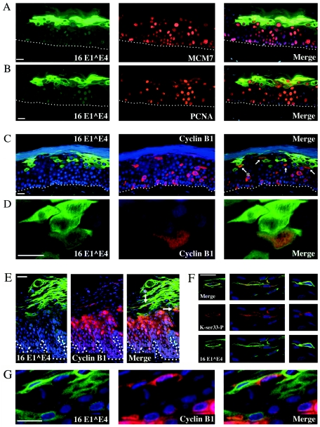
To examine the potential significance of these observations during productive infection, E1∧E4 and cyclin B1 were visualized in HPV16-positive rafts and HPV-induced lesions. High levels of 16E1∧E4 expression are found in a band in the upper layers of the differentiating epithelium (Fig. 7) (12). While in the top layers of this band, 16E1∧E4 is present in cells that have lost their nuclei and exited the cell cycle, in the lower layers of the band the 16E1∧E4 cells do contain nuclei and are positive for proteins such as MCM, PCNA, and cyclin A (Fig. 7A and B), suggesting that they are in the cell cycle. This is to be expected since previous work has shown that amplification of the papillomavirus genome correlates with expression of E1∧E4 (30, 38). Since replication of the papillomavirus genome requires the cells to contain S-phase proteins, such proteins must be found in E1∧E4-expressing cells. We show here that cyclin B1 is also present in some 16E1∧E4-expressing cells (Fig. 7C, D, E, and G). Such cells generally express lower levels of E1∧E4 than those in the upper epithelial layers, where nuclear degeneration occurs. Our data suggest that the ability of 16E1∧E4 to arrest cells in vitro may also occur in vivo following exit from S phase and entry into the G2 phase of the cell cycle.
FIG. 7.
Expression of 16E1∧E4 in cyclin B1-expressing cells during epithelial cell differentiation. HPV16-induced raft cultures were double stained for the presence of 16E1∧E4 (green) and (A) MCM7, (B) PCNA, or (C) cyclin B1 (red) to show that 16E1∧E4 levels begin to rise in cells that are expressing cell cycle markers. A subset of the cells that express moderate levels of 16E1∧E4 also express cyclin B1 and are arrowed in C. (D) An enlarged image of the cyclin B1/16 E1∧E4 double-positive cell that is marked with an asterisk in C. (E) HPV16-induced low-grade lesion double stained for the presence of 16E1∧E4 (green) and cyclin B1 (red). Coexpression of cyclin B1 and 16 E1∧E4 is limited to the region wherethe levels of 16E1∧E4 protein are beginning to rise during epithelial differentiation. Panel G shows a high-power image of cells from this region (indicated by an asterisk in E) that contain both cyclin B1 and 16 E1∧E4. Panel F shows that 16E1∧E4-expressing cells (green) in the upper layers of low-grade cervical lesions contain the keratin phosphoserine 33 epitope (red) that is also present following the expression of 16E1∧E4 in vitro (see Fig. 5). Merged images are shown in the uppermost panels within F. Where appropriate, the position of the basal layer is indicated by dotted white lines. A DAPI nuclear stain (blue) was included. Bars equal 20 μm except in panels D and G, where the bars equal 10 μm.
In an attempt to ascertain whether the cyclin B1 is active in these 16E1∧E4-expressing cells, the presence of phosphorylation on the Cdk1/cyclin B1 kinase site in keratin (serine 33) was investigated with phosphorylation-specific antibodies (Fig. 7F). We found abundant phosphoserine 33 colocalizing with 16E1∧E4. This supports the hypothesis that active Cdk1/cyclin B1 is associated with 16E1∧E4 on the keratins. This is in agreement with what was found following expression of 16E1∧E4 in SiHa cells (Fig. 5C and D).
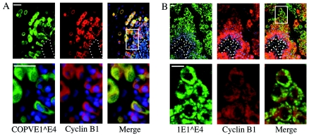
It is not yet clear whether G2 arrest is a general feature of all E1∧E4 proteins, but it is interesting that in lesions caused by viruses such as canine oral papillomavirus (Fig. 8A) and HPV1 (Fig. 8B), which begin their productive cycles in the basal and parabasal cell layers, that the vast majority of E1∧E4-expressing cells also express cyclin B1. Similarity in the distribution of cyclin B1 and E1∧E4 was most apparent in the cell layers where genome amplification is being supported (30, 36, 38), with cyclin B1 levels decreasing as the cells migrate towards the epithelial surface (Fig. 8A and B; compare distribution in the lesion caused by HPV16, Fig. 7E and G).
FIG. 8.
Coexpression of E4 and cyclin B1 in productive lesions caused by other papillomavirus types. (A) Canine oral papillomavirus (COPV)-induced lesion double stained for the presence of canine oral papillomavirus E1∧E4 (green) and cyclin B1 (red). The boxed region in the rightmost top panel is shown enlarged in the lower panels. Expression of E1∧E4 is apparent in a subset of cyclin B1-positive cells in the basal layer and in all the cyclin B1-expressing cells in the parabasal and superficial layers. (B) HPV1-induced lesion double stained for the presence of 1E1∧E4 (green) and cyclin B1 (red). The boxed region in the rightmost top panel is shown enlarged in the lower panels. HPV1 E1∧E4 expression begins in cyclin B1-positive cells in the parabasal layer. The intensity of cyclin B1 staining decreases as the cells migrate towards the epithelial surface. Bars, 20 μm.
DISCUSSION
We have previously shown that the HPV 16E1∧E4 protein induces G2 arrest (7), an observation that was subsequently confirmed and extended to 18E1∧E4 (35). We now present the observation that arrest does not result from inhibition of the kinase activity of the Cdk1/cyclin B1 complex but instead appears to result from the retention of active Cdk1/cyclin B1 complexes in the cytoplasm away from their nuclear substrates.
Our data show that the levels of Cdk1/cyclin B1 kinase activity in 16E1∧E4-expressing cells are either similar to or higher than those found in β-galactosidase-expressing cells. This implies that 16E1∧E4-induced G2 arrest is not mediated by inhibition of the enzyme activity of Cdk1/cyclin B1. Since the Cdk1/cyclin B1 enzyme complex in 16E1∧E4-expressing cells appears to be active, why is entry into mitosis prevented? Analysis by immunofluorescence microscopy showed that Cdk1 and cyclin B1 colocalized with 16E1∧E4, suggesting that although 16E1∧E4 may not inhibit the enzyme activity of the complex, it may be having another type of direct effect. This was supported by coimmunoprecipitation and GST pull-down experiments, in which 16E1∧E4 was found to associate with cyclin B1, although it is not as yet clear whether this is a direct interaction. One consequence of this association appears to be that Cdk1/cyclin B1 can become associated with the NP-40-insoluble fraction, probably as a result of linkage to the relatively insoluble cytokeratin filament network. Also apparent is a reduced ability of cyclin B1 to enter the nucleus. Since we know that there were plenty of active Cdk1/cyclin B1 complexes in 16E1∧E4-expressing cells, we hypothesize that insufficient active Cdk1/cyclin B1 complexes accumulate in the nucleus to trigger mitosis. This is supported by data from the mutant T22A,T23A-16E1∧E4, which showed failure to pull down cyclin B1 in vitro, lack of colocalization with cyclin B1 in cells, an increased propensity for cyclin B1 to enter the nucleus, and an increased ability to enter mitosis.
Sequestration of Cdk1/cyclin B1 by 16E1∧E4 may also explain the observations in Fig. 1D and Fig. 4D, which show that the Cdk1 coimmunoprecipitated with cyclin B1 from 16E1∧E4-expressing cells is less phosphorylated than that from β-galactosidase-expressing cells. The two kinases responsible for the inhibitory phosphorylation of Cdk1 are Wee1, which is found in the nucleus (21), and Myt1, which is found membrane bound on the endoplasmic reticulum (33). The Cdk1 that is localized to the cytokeratin networks may therefore not be readily accessible to either Wee1 or Myt1. In contrast, Cdc25, the phosphatase that removes the inhibitory phosphorylation, is mobile in the cytoplasm (19) and would therefore still be likely to reach the cytokeratin-associated Cdk1. As with human cytomegalovirus-infected cells (42), as yet unknown changes to the activity of these cell cycle regulatory proteins may also contribute to the hypophosphorylation of Cdk1.
Several other examples of virus-induced G2 arrest have been characterized recently, and it seems that G2 arrest may be a common strategy employed by viruses to optimize virion production. Contrary to the arrest observed with 16E1∧E4, G2 arrest induced by other viral proteins is often associated with inhibition of the kinase activity of the Cdk1/cyclin B1 complex. Other viral proteins capable of inhibiting Cdk1/cyclin B1 include the human immunodeficiency virus Vpr protein (20), reovirus σ1s protein (39), and HPV E2 protein (15). In contrast, activation of the Cdk1/cyclin B1 kinase complex is observed in human cytomegalovirus-infected G2-arrested cells, but the situation is complex, with several cyclins (E, A, and B) being upregulated and cell cycle arrest additionally occurring in G1 and mitosis (24, 41). In fact, only the cell cycle arrest induced by the human parvovirus B19 NS1 protein appears similar to that induced by 16E1∧E4. Cells infected with B19 virus arrest in G2 with high Cdk1/cyclin B1 kinase activity levels and cytoplasmic cyclin B1 (32), although the mechanism for this has not been elucidated. Despite the similarities between 16E1∧E4-induced and NS1-induced G2 arrest, there appear to be no obvious similarities between these two proteins at the level of amino acid sequence.
A number of cellular proteins are also able to induce G2 arrest in the presence of active Cdk1/cyclin B1. These include Myt1 (28), used here as a control, Patched 1 (3), and 14-3-3σ (5), which sequester the Cdk1/cyclin B1 complex onto the endoplasmic reticulum membrane, onto the cell membrane, and in the cytosol, respectively. 16E1∧E4 appears to be unique in its use of the cytokeratin filament networks for sequestration of Cdk1/cyclin B1. However, binding to the cytokeratin network is not crucial for 16E1∧E4-induced G2 arrest. Even in the absence of cytokeratin expression in Saos-2 cells, 16E1∧E4 induced G2 arrest. The reason for this appears to be that cytokeratin binding is not essential for confining Cdk1/cyclinB1/16E1∧E4 complexes to the cytoplasm. In the absence of keratin binding, 16E1∧E4 remains insoluble in NP-40 (presumably as result of the formation of 16E1∧E4 hexamers) (48) and is maintained in the cytoplasm due to association with other cytoplasmic structures, such as mitochondria (Fig. 3B) (40).
Why does 16E1∧E4 induce G2 arrest? Human immunodeficiency virus Vpr-associated G2 arrest has been shown to be involved in upregulating virus production (17), and the requirement for E1∧E4 for the onset of viral DNA amplification (37) makes it tempting to speculate that G2 arrest may in some way create an intracellular environment that is optimized for viral DNA replication or other late events. The consequences of 16E1∧E4-induced G2 arrest may include changes in the phosphorylation status of viral and cellular proteins. It has been shown for herpes simplex virus type 1 that expression of some of its late genes is dependent on stabilization of Cdk1 (1). The persistence of active Cdk1/cyclin B1 in 16E1∧E4-expressing cells may have a similar significance in addition to cell cycle arrest.
It is interesting that in HPV 16-induced lesions (and in cervical lesions caused by other supergroup A HPV types [data not shown]), the number of cells expressing both cyclin B1 and E1∧E4 is relatively small, whereas in canine oral papillomavirus- and HPV1-induced lesions (where the productive cycle begins in the basal and parabasal cell layers), the number of double-positive cells is much more extensive (36, 38). It appears from these results that E4 may be able to accumulate in cells that already express cyclin B during productive infection. As the ability to detect E4 correlates closely with the onset of vegetative viral genome amplification (38), such cells are unlikely to be in a true G2 phase and must be competent for viral DNA replication. Lesions induced by canine oral papillomavirus or HPV1 are usually highly productive, while those induced by HPV16 (which contain relatively few cyclin B/E1∧E4 double-positive cells) contain relatively few cells that support genome amplification and capsid synthesis (31, 38). In HPV16-induced lesions as well as in lesions caused by HPV1 and by canine oral papillomavirus, the E4 present in the highest layers of the epithelium does not appear to be associated with cyclin B1, which is not surprising, as by this stage the cells have exited the cell cycle and lost their nuclei. We suspect that the highly abundant 16E1∧E4 at this location may performs a different function, e.g., aiding viral egress, perhaps related to its ability to bind and disturb the keratin networks (11).
The expression of 16E1∧E4 is lost during progression from early-stage premalignant lesions (cervical intraepithelial neoplasia 1) to late stage premalignant lesions (cervical intraepithelial neoplasia 3) (31). In cervical intraepithelial neoplasia 1 lesions, cell proliferation and/or the expression of proliferation markers is confined to the epithelial layers that extend from the basal layer to the region where 16E1∧E4 is expressed. In contrast, in cervical intraepithelial neoplasia 3 lesions, cell proliferation extends from the basal layer to the surface of the lesion and 16E1∧E4 expression is absent or restricted to pockets of cells close to the epithelial surface (31). It seems plausible that the loss of G2 arrest as a consequence of lack of 16E1∧E4 expression may contribute to the increase in proliferation in the upper epithelial layers. Loss of E2, which negatively regulates E7 expression and also causes G2 arrest (15), has been regarded as an important event in cancer progression (46). It seems likely that E1∧E4 and E2 may act synergistically to achieve cell cycle arrest in vivo, and, as with E2, the loss of E1∧E4 may be a predisposing factor in the development of cervical cancer. Since reintroduction of E1∧E4 into cervical cancer cells, e.g., SiHa and HeLa cell lines, causes their arrest, E1∧E4 may be regarded as a viral tumor suppressor.
Acknowledgments
We thank Sir John Skehel and Jonathan Stoye for support during the course of this work. We thank H. Piwnica-Worms (Howard Hughes Medical Institute, St. Louis, Mo.) for the gift of the β-galactosidase and Myt1 recombinant adenoviruses and M. B. Omary (Veterans Affairs Palo Alto Health Care System, Palo Alto, Calif.) for the gift of antikeratin antibodies 8592 and 8250.
C.D., D.J., P.D., R.S., K.R., K.M., J.B.A.M., P.J.M., W.P., P. L., and Q.W. were supported by the United Kingdom Medical Research Council. S.A.S was supported by an Association for International Cancer Research joint award to J.D. and J.B.A.M. J.D was supported by the Royal Society and the United Kingdom Medical Research Council.
REFERENCES
- 1.Advani, S. J., R. R. Weichselbaum, and B. Roizman. 2000. The role of cdc2 in the expression of herpes simplex virus genes. Proc. Natl. Acad. Sci. USA 97:10996-11001. [DOI] [PMC free article] [PubMed] [Google Scholar]
- 2.Baker, C. C., W. C. Phelps, V. Lindgren, M. J. Braun, M. A. Gonda, and P. M. Howley. 1987. Structural and transcriptional analysis of human papillomavirus type 16 sequences in cervical carcinoma cell lines. J. Virol. 61:962-971. [DOI] [PMC free article] [PubMed] [Google Scholar]
- 3.Barnes, E. A., M. Kong, V. Ollendorff, and D. J. Donoghue. 2001. Patched1 interacts with cyclin B1 to regulate cell cycle progression. EMBO J. 20:2214-2223. [DOI] [PMC free article] [PubMed] [Google Scholar]
- 4.Bryan, J. T., and D. R. Brown. 2000. Association of the human papillomavirus type 11 E1()E4 protein with cornified cell envelopes derived from infected genital epithelium. Virology 277:262-269. [DOI] [PubMed] [Google Scholar]
- 5.Chan, T. A., H. Hermeking, C. Lengauer, K. W. Kinzler, and B. Vogelstein. 1999. 14-3-3Sigma is required to prevent mitotic catastrophe after DNA damage. Nature 401:616-620. [DOI] [PubMed] [Google Scholar]
- 6.Crum, C. P., S. Barber, M. Symbula, K. Snyder, A. M. Saleh, and J. K. Roche. 1990. Coexpression of the human papillomavirus type 16 E4 and L1 open reading frames in early cervical neoplasia. Virology 178:238-246. [DOI] [PubMed] [Google Scholar]
- 7.Davy, C. E., D. J. Jackson, Q. Wang, K. Raj, P. J. Masterson, N. F. Fenner, S. Southern, S. Cuthill, J. B. Millar, and J. Doorbar. 2002. Identification of a G2 arrest domain in the E1 wedge E4 protein of human papillomavirus type 16. J. Virol. 76:9806-9818. [DOI] [PMC free article] [PubMed] [Google Scholar]
- 8.Doorbar, J., D. Campbell, R. J. Grand, and P. H. Gallimore. 1986. Identification of the human papilloma virus-1a E4 gene products. EMBO J. 5:355-362. [DOI] [PMC free article] [PubMed] [Google Scholar]
- 9.Doorbar, J., R. C. Elston, S. Napthine, K. Raj, E. Medcalf, D. Jackson, N. Coleman, H. M. Griffin, P. Masterson, S. Stacey, Y. Mengistu, and J. Dunlop. 2000. The E1E4 protein of human papillomavirus type 16 associates with a putative RNA helicase through sequences in its C terminus. J. Virol. 74:10081-10095. [DOI] [PMC free article] [PubMed] [Google Scholar]
- 10.Doorbar, J., S. Ely, N. Coleman, M. Hibma, D. H. Davies, and L. Crawford. 1992. Epitope-mapped monoclonal antibodies against the HPV16E1-E4 protein. Virology 187:353-359. [DOI] [PubMed] [Google Scholar]
- 11.Doorbar, J., S. Ely, J. Sterling, C. McLean, and L. Crawford. 1991. Specific interaction between HPV-16 E1-E4 and cytokeratins results in collapse of the epithelial cell intermediate filament network. Nature 352:824-827. [DOI] [PubMed] [Google Scholar]
- 12.Doorbar, J., C. Foo, N. Coleman, L. Medcalf, O. Hartley, T. Prospero, S. Napthine, J. Sterling, G. Winter, and H. Griffin. 1997. Characterization of events during the late stages of HPV16 infection in vivo using high-affinity synthetic Fabs to E4. Virology 238:40-52. [DOI] [PubMed] [Google Scholar]
- 13.Doorbar, J., A. Parton, K. Hartley, L. Banks, T. Crook, M. Stanley, and L. Crawford. 1990. Detection of novel splicing patterns in a HPV 16-containing keratinocyte cell line. Virology 178:254-262. [DOI] [PubMed] [Google Scholar]
- 14.Ferrell, J. E., Jr. 1998. How regulated protein translocation can produce switch-like responses. Trends Biochem. Sci. 23:461-465. [DOI] [PubMed] [Google Scholar]
- 15.Fournier, N., K. Raj, P. Saudan, S. Utzig, R. Sahli, V. Simanis, and P. Beard. 1999. Expression of human papillomavirus 16 E2 protein in Schizosaccharomyces pombe delays the initiation of mitosis. Oncogene 18:4015-4021. [DOI] [PubMed] [Google Scholar]
- 16.Genther, S. M., S. Sterling, S. Duensing, K. Munger, C. Sattler, and P. F. Lambert. 2003. Quantitative role of the human papillomavirus type 16 E5 gene during the productive stage of the viral life cycle. J. Virol. 77:2832-2842. [DOI] [PMC free article] [PubMed] [Google Scholar]
- 17.Goh, W. C., M. E. Rogel, C. M. Kinsey, S. F. Michael, P. N. Fultz, M. A. Nowak, B. H. Hahn, and M. Emerman. 1998. HIV-1 Vpr increases viral expression by manipulation of the cell cycle: a mechanism for selection of Vpr in vivo. Nat. Med. 4:65-71. [DOI] [PubMed] [Google Scholar]
- 18.Goodwin, E. C., E. Yang, C. J. Lee, H. W. Lee, D. DiMaio, and E. S. Hwang. 2000. Rapid induction of senescence in human cervical carcinoma cells. Proc. Natl. Acad. Sci. USA 97:10978-10983. [DOI] [PMC free article] [PubMed] [Google Scholar]
- 19.Graves, P. R., C. M. Lovly, G. L. Uy, and H. Piwnica-Worms. 2001. Localization of human Cdc25C is regulated both by nuclear export and 14-3-3 protein binding. Oncogene 20:1839-1851. [DOI] [PubMed] [Google Scholar]
- 20.He, J., S. Choe, R. Walker, P. Di Marzio, D. O. Morgan, and N. R. Landau. 1995. Human immunodeficiency virus type 1 viral protein R (Vpr) arrests cells in the G2 phase of the cell cycle by inhibiting p34cdc2 activity. J. Virol. 69:6705-6711. [DOI] [PMC free article] [PubMed] [Google Scholar]
- 21.Heald, R., M. McLoughlin, and F. McKeon. 1993. Human wee1 maintains mitotic timing by protecting the nucleus from cytoplasmically activated Cdc2 kinase. Cell 74:463-474. [DOI] [PubMed] [Google Scholar]
- 22.Hendzel, M. J., Y. Wei, M. A. Mancini, A. Van Hooser, T. Ranalli, B. R. Brinkley, D. P. Bazett-Jones, and C. D. Allis. 1997. Mitosis-specific phosphorylation of histone H3 initiates primarily within pericentromeric heterochromatin during G2 and spreads in an ordered fashion coincident with mitotic chromosome condensation. Chromosoma 106:348-360. [DOI] [PubMed] [Google Scholar]
- 23.Howley, P. M., and D. R. Lowy. 2001. Papillomaviruses and their replication, p. 2197-2229. In B. N. Fields, D. M. Knipe, and P. M. Howley (ed.), Virology, vol. 2. Lippincott Williams and Wilkins, Philadelphia, Pa. [Google Scholar]
- 24.Jault, F. M., J. M. Jault, F. Ruchti, E. A. Fortunato, C. Clark, J. Corbeil, D. D. Richman, and D. H. Spector. 1995. Cytomegalovirus infection induces high levels of cyclins, phosphorylated Rb, and p53, leading to cell cycle arrest. J. Virol. 69:6697-6704. [DOI] [PMC free article] [PubMed] [Google Scholar]
- 25.Keen, N., R. Elston, and L. Crawford. 1994. Interaction of the E6 protein of human papillomavirus with cellular proteins. Oncogene 9:1493-1499. [PubMed] [Google Scholar]
- 26.Ku, N. O., J. Liao, and M. B. Omary. 1998. Phosphorylation of human keratin 18 serine 33 regulates binding to 14-3-3 proteins. EMBO J. 17:1892-1906. [DOI] [PMC free article] [PubMed] [Google Scholar]
- 27.Ku, N. O., and M. B. Omary. 1997. Phosphorylation of human keratin 8 in vivo at conserved head domain serine 23 and at epidermal growth factor-stimulated tail domain serine 431. J. Biol. Chem. 272:7556-7564. [DOI] [PubMed] [Google Scholar]
- 28.Liu, F., C. Rothblum-Oviatt, C. E. Ryan, and H. Piwnica-Worms. 1999. Overproduction of human Myt1 kinase induces a G2 cell cycle delay by interfering with the intracellular trafficking of Cdc2-cyclin B1 complexes. Mol. Cell. Biol. 19:5113-5123. [DOI] [PMC free article] [PubMed] [Google Scholar]
- 29.Lowy, D. R., and P. M. Howley. 2001. Papillomaviruses, p. 2231-2264. In B. N. Fields, D. M. Knipe, and P. M. Howley (ed.), Virology, vol. 2. Lippincott Williams and Wilkins, Philadelphia, Pa. [Google Scholar]
- 30.Middleton, K., W. Peh, S. Southern, H. Griffin, K. Sotlar, T. Nakahara, A. El-Sherif, L. Morris, R. Seth, M. Hibma, D. Jenkins, P. Lambert, N. Coleman, and J. Doorbar. 2003. Organization of human papillomavirus productive cycle during neoplastic progression provides a basis for selection of diagnostic markers. J. Virol. 77:10186-10201. [DOI] [PMC free article] [PubMed] [Google Scholar]
- 31.Middleton, K., W. Peh, S. A. Southern, H. M. Griffin, K. Sotlar, T. Nakahara, A. El-Sherif, L. Morris, R. Seth, M. Hibma, D. Jenkins, P. F. Lambert, N. Coleman, and J. Doorbar. Organization of human papillomavirus productive cycle during neoplastic progression provides a basis for selection of diagnostic markers. J. Virol. 77:10186-10201. [DOI] [PMC free article] [PubMed]
- 32.Morita, E., K. Tada, H. Chisaka, H. Asao, H. Sato, N. Yaegashi, and K. Sugamura. 2001. Human parvovirus B19 induces cell cycle arrest at G2 phase with accumulation of mitotic cyclins. J. Virol. 75:7555-7563. [DOI] [PMC free article] [PubMed] [Google Scholar]
- 33.Mueller, P. R., T. R. Coleman, A. Kumagai, and W. G. Dunphy. 1995. Myt1: a membrane-associated inhibitory kinase that phosphorylates Cdc2 on both threonine-14 and tyrosine-15. Science 270:86-90. [DOI] [PubMed] [Google Scholar]
- 34.Murata-Hori, M., M. Tatsuka, and Y. L. Wang. 2002. Probing the dynamics and functions of aurora B kinase in living cells during mitosis and cytokinesis. Mol. Biol. Cell 13:1099-1108. [DOI] [PMC free article] [PubMed] [Google Scholar]
- 35.Nakahara, T., A. Nishimura, M. Tanaka, T. Ueno, A. Ishimoto, and H. Sakai. 2002. Modulation of the cell division cycle by human papillomavirus type 18 E4. J. Virol. 76:10914-10920. [DOI] [PMC free article] [PubMed] [Google Scholar]
- 36.Nicholls, P. K., J. Doorbar, R. A. Moore, W. Peh, D. M. Anderson, and M. A. Stanley. 2001. Detection of viral DNA and E4 protein in basal keratinocytes of experimental canine oral papillomavirus lesions. Virology 284:82-98. [DOI] [PubMed] [Google Scholar]
- 37.Peh, W. L., J. L. Brandsma, N. D. Christensen, N. M. Cladel, X. Wu, and J. Doorbar. 2004. The viral E4 protein is required for the completion of the cottontail rabbit papillomavirus productive cycle in vivo. J. Virol. 78:2142-2151. [DOI] [PMC free article] [PubMed] [Google Scholar]
- 38.Peh, W. L., K. Middleton, N. Christensen, P. Nicholls, K. Egawa, K. Sotlar, J. Brandsma, A. Percival, J. Lewis, W. J. Liu, and J. Doorbar. 2002. Life cycle heterogeneity in animal models of human papillomavirus-associated disease. J. Virol. 76:10401-10416. [DOI] [PMC free article] [PubMed] [Google Scholar]
- 39.Poggioli, G. J., T. S. Dermody, and K. L. Tyler. 2001. Reovirus-induced sigma1-dependent G2/M phase cell cycle arrest is associated with inhibition of p34cdc2. J. Virol. 75:7429-7434. [DOI] [PMC free article] [PubMed] [Google Scholar]
- 40.Raj, K., S. Berguerand, S. Southern, J. Doorbar, and P. Beard. 2004. E1∧E4 protein of human papillomavirus type 16 associates with mitochondria. J. Virol. 78:7199-7207. [DOI] [PMC free article] [PubMed] [Google Scholar]
- 41.Salvant, B. S., E. A. Fortunato, and D. H. Spector. 1998. Cell cycle dysregulation by human cytomegalovirus: influence of the cell cycle phase at the time of infection and effects on cyclin transcription. J. Virol. 72:3729-3741. [DOI] [PMC free article] [PubMed] [Google Scholar]
- 42.Sanchez, V., A. K. McElroy, and D. H. Spector. 2003. Mechanisms governing maintenance of Cdk1/cyclin B1 kinase activity in cells infected with human cytomegalovirus. J. Virol. 77:13214-13224. [DOI] [PMC free article] [PubMed] [Google Scholar]
- 43.Southern, S. A., I. W. McDicken, and C. S. Herrington. 2000. Evidence for keratinocyte immortalization in high-grade squamous intraepithelial lesions of the cervix infected with high-risk human papillomaviruses. Lab. Investig. 80:539-544. [DOI] [PubMed] [Google Scholar]
- 44.Takizawa, C. G., and D. O. Morgan. 2000. Control of mitosis by changes in the subcellular location of cyclin-B1-Cdk1 and Cdc25C. Curr. Opin. Cell Biol. 12:658-665. [DOI] [PubMed] [Google Scholar]
- 45.Taylor, W. R., and G. R. Stark. 2001. Regulation of the G2/M transition by p53. Oncogene 20:1803-1815. [DOI] [PubMed] [Google Scholar]
- 46.Tonon, S. A., M. A. Picconi, P. D. Bos, J. B. Zinovich, J. Galuppo, L. V. Alonio, and A. R. Teyssie. 2001. Physical status of the E2 human papilloma virus 16 viral gene in cervical preneoplastic and neoplastic lesions. J. Clin. Virol. 21:129-134. [DOI] [PubMed] [Google Scholar]
- 47.Walboomers, J., M. Jacobs, Manos, M. M., F. Bosch, J. Kummer, K. Shah, P. Snijders, J. Peto, C. Meijer, and N. Munoz. 1999. Human papillomavirus is a necessary cause of invasive cervical cancer worldwide. J. Pathol. 189:12-19. [DOI] [PubMed] [Google Scholar]
- 48.Wang, Q., H. Griffin, S. Southern, D. Jackson, A. Martin, P. McIntosh, C. Davy, P. J. Masterson, P. A. Walker, P. Laskey, M. B. Omary, and J. Doorbar. 2004. Functional analysis of the human papillomavirus type 16 E1∧E4 protein provides a mechanism for in vivo and in vitro keratin filament reorganization. J. Virol. 78:821-833. [DOI] [PMC free article] [PubMed] [Google Scholar]


