Abstract
Nitrogen-vacancy (NV) centers in nanodiamond hold great promise for creating superior biological labels and quantum sensing methods. Yet, inefficient photon generation and extraction from excited NV centers restricts the achievable sensitivity and temporal resolution. Herein, we report an entirely complementary route featuring pyramidal hyperbolic metasurface to modify the spontaneous emission of NV centers. Fabricated using nanosphere lithography, the metasurface consists of alternatively stacked silica-silver thin films configured in a pyramidal fashion, and supports both spectrally broadband Purcell enhancement and spatially extended intense local fields owing to the hyperbolic dispersion and plasmonic coupling. The enhanced photophysical properties are manifested as a simultaneous amplification to the spontaneous decay rate and emission intensity of NV centers. We envision the reported pyramidal metasurface could serve as a versatile platform for creating chip-based ultrafast single-photon sources and spin-enhanced quantum biosensing strategies, as well as aiding in further fundamental understanding of photoexcited species in condensed phases.
Keywords: Hyperbolic Metasurfaces, Plasmonics, Pyramid, Nitrogen-Vacancy Centers, Nanodiamonds
Graphical Abstract
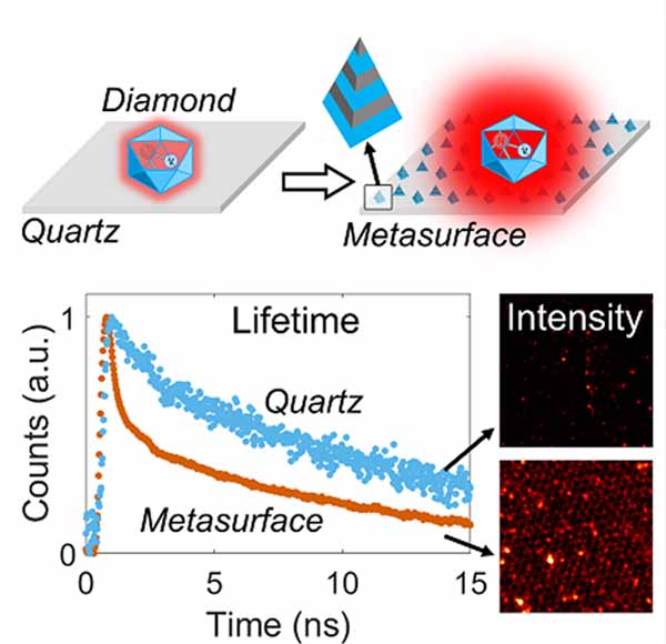
A plasmonic hyperbolic metasurface consisting of alternatively stacked silica-silver thin films configured in a pyramidal fashion is developed and found to support both spectrally broadband Purcell enhancement and spatially extended intense local fields. The metasurface allows a simultaneous amplification to the spontaneous decay rate and emission intensity of nitrogen-vacancy centers in nanodiamond.

Introduction
Nitrogen-vacancy (NV) centers in nanodiamond represent a unique type of solid-state quantum emitter owing to their outstanding optical and spin properties.[1] Exhibiting a broadband and photostable fluorescence, coupled with the chemical stability and low cytotoxicity of nanodiamonds, NV centers can serve as a robust anti-bunched single-photon source and an excellent biological imaging contrast agent.[2] The distinct spin-preserving and spin-selective optical transitions of NV centers form the basis of optically detected magnetic resonance and have fostered a plethora of interest in developing quantum biosensing methods, as exemplified by the nanoscale magnetometry[1e, 2a] and spin-enhanced ultrasensitive diagnostics[1a-c]. Yet, the achievable sensitivity and temporal resolution are compromised by inefficient photon generation and extraction from excited NV centers owing to their long radiative lifetime, which ranges from 10 to 30 ns depending on the size and refractive index of nanodiamonds.
Boosting the efficiency of photon generation hinges on accelerating the spontaneous emission of NV centers.[2c, 3] For conventional quantum emitters such as semiconductor quantum dots and organic dyes, their transition dynamics can be modified by the Purcell effect, which is linearly dependent on the quality factor but inversely scales with the mode volume of an optical cavity.[3b, 3c, 4] Owing to the high-Q cavity mode of a dielectric resonator and the subwavelength mode volume of a plasmonic nanocavity, they are both well suited to provide a significant Purcell enhancement to accelerate the transition dynamics of quantum emitters, provided that the optical cavity is tuned to be in resonance with the emission spectrum of quantum emitters. However, as an optical cavity usually supports a much narrower spectral linewidth than the broadband emission spectrum of NV centers, it proves less effective at providing a broadband enhancement mechanism. The lack of a broadband enhancement strategy underscores the challenges in accelerating the spontaneous emission of NV centers by merely using conventional methods.
Recently, hybrid dielectric-metallic nanostructures judiciously structured at the subwavelength scale, known as metamaterials, have unleashed new possibilities for engineering spontaneous emission in nanophotonics.[2c, 5] For example, metamaterials made of alternatively stacked dielectric-metallic thin films have shown great promise for broadband amplification of the local density of optical states (LDOS) and extraordinary light confinement at the deep subwavelength for enhanced light-matter interactions.[3a, 5a] Underpinning such exceptional properties is the highly directional response of free electrons as their motion is spatially constrained to the plane of each constituent thin film, which gives rise to the ultra-anisotropic dielectric tensors.[5a] When one of the principal components of the dielectric tensors is opposite in sign to the other two, the isofrequency surface (IFS) of TM polarized waves exhibits a hyperbolic dispersion with respect to the corresponding components of the k-vector. One important consequence is the activation of metamaterial modes with a high momentum, or high-k modes, which allows extraordinary light-matter interactions and has formed the basis for realizing quantum nanophotonic functionalities on hyperbolic metamaterials.[6] Indeed, high-k modes have been found to facilitate lasing action for quantum emitters[7] and can significantly enhance the transition dynamics of quantum emitters over an extended wavelength range[3a, 5b-e]. Nevertheless, the evanescent nature makes it difficult for high-k modes to couple to the free space, and therefore, the enhanced transition dynamics is mostly manifested as the accelerated non-radiative decay rates, without pronounced fluorescence intensity enhancement.
To address such challenges, we propose to develop a pyramidal hyperbolic metasurface, which can potentially provide desired broadband optical enhancement and effective coupling to the far field. Important clues from previous studies suggest that the pyramidal geometry serves as an effective scatterer[8] and contributes to strong enhancement for both surface-enhanced Raman scattering[9] and plasmon-enhanced fluorescence[10]. We hypothesize that the pyramidal geometry would similarly facilitate an accelerated radiative decay rate of NV centers with enhanced fluorescence brightness. To test the hypothesis, a comprehensive study is launched to first investigate its optical properties and the capability to modify the photophysical properties of NV centers. Nanosphere lithography is then employed to fabricate (3-cycle, 6-layer) pyramidal metasurfaces. Finally, the spontaneous emission of NV centers in nanodiamond is measured on the fabricated metasurface using fluorescence lifetime imaging microscopy (FLIM) to provide an experimental validation to the hypothesis.
Results and Discussion
Broadband amplification of Purcell effect by hyperbolic dispersion
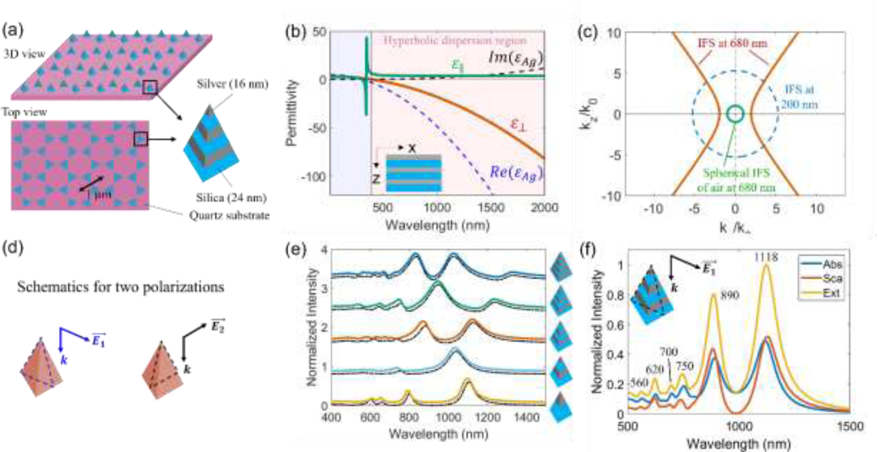
The conceptualized metasurface is a nanopyramid array sitting on a quartz slide with each individual nanopyramid made of three cycles of alternatively stacked silica-silver thin films, as schematically depicted in Fig. 1a. A cycle is defined to consist of one silica-silver layer. The silica and silver layer each has a thicknesses of 24 nm and 16 nm. The nanopyramid array is hexagonally patterned with a periodicity of 1 µm. Each nanopyramid has a height of 120 nm with a base edge length of about 310 nm. Selection of the parameters is made based on the requirement of exhibiting hyperbolic dispersion, and by considering the ease of nanofabrication. Particularly, despite the low filling density of the nanostructured elements on the quartz substrate, previous research has shown that the hyperbolic dispersion for such metasurfaces consisting of layered dielectric-metallic thin films is mostly preserved, whereas the effective permittivity can be approximated by the effective medium theory (EMT) in the long-wavelength limit.[5e, 11]. Following the convention defined previously,[5a] the components and of the effective permittivity for the metasurface are defined as parallel and perpendicular to the anisotropy axis, i.e. the z-axis, as shown in Fig. 1b. Based on EMT, as analytically laid out in the supporting information, the orthogonal components are opposite in sign with and for a wavelength larger than 390 nm. The metasurface thus displays a hyperbolic dispersion for a given isofrequency surface (IFS) if its frequency falls into the hyperbolic dispersion region as highlighted in light red color in Fig. 1b. One important consequence is the broadband amplification of the Purcell effect. Based on Fermi’s Golden Rule,[4e] the spontaneous emission rate for a two-level quantum system is given by
| (1a) |
Figure 1.

(a) Schematic for the conceptualized nanopyramid array-based hyperbolic metasurface with each individual nanopyramid made of three cycles of alternatively stacked silica (24 nm) and silver (16 nm) thin films on the quartz substrate. The hexagonally patterned nanopyramid array has a periodicity of 1 µm as indicated in the figure. Each nanopyramid has a height of 120 nm and a base edge length of about 310 nm; (b) calculated permittivity parallel ( in the x-y plane) and perpendicular ( in the x-z plane) to the metasurface, i.e., the inset schematic: alternatively stacked silica (24 nm) and silver (16 nm) thin films, as compared to the real and imaginary part of the permittivity of silver based on Drude model; (c) IFS at 680 nm for the metasurface in (b) displaying a hyperbolic shape as compared to spherical IFS of air at 680 nm and spherical IFS at 200 nm. (d) Definition of the two polarizations, (along with k in the cross-sectional plane as depicted by the dashed triangle) and which are orthogonal to each other; (e) Extinction spectral tunability produced by varying the number of cycles (schematics not to scale) from one to five for the alternatively stacked silica (24 nm)-Ag (16 nm) pyramidal metasurface. The base edge length was fixed at about 310 nm. The colored curves were under polarization while the black dashed curves were under polarization (curves are arbitrarily offset vertically for clarity of presentation. (f) The absorption (Abs), scattering (Sca), and extinction (Ext) spectra for the pyramidal metasurface consisting of three cycles of alternatively stacked silica (24 nm)-Ag (16 nm) layers under polarization, whereas the spectra under polarization was shown in Fig. S1.
where is the transition matrix from the initial state to the final state with as the interaction Hamiltonian. As an approximation, scales with , which demands summing over all the available wavevectors k with polarization σ that have a frequency equal to the transition energy . By combining the IFS in Eq. (S3), we have
| (1b) |
The right term in Eq. (1b) is the area of the IFS with a frequency as defined in Eq. (S3). Therefore, scales with the area enclosed by the IFS at the transition frequency . From Fig. 1c, for a hyperbolic dispersion, Eq. (1b) diverges and practically, it leads to a strong and broadband amplification of the spontaneous emission rate , which is drastically different from a spherical dispersion. Such unique broadband optical amplification has important applications in enhancing spontaneous emission of NV centers in nanodiamond, given the associated broadband emission spectrum. In what follows, a systematic study is launched using finite-different time-domain (FDTD) simulations to first elucidate the optical and photophysical properties of the pyramidal metasurface, followed by an experimental demonstration of an enhanced spontaneous emission of NV centers on the fabricated pyramidal metasurface.
Spectral tunability and Fabry-Pérot-like cavity mode profiles
The spectral tunability of the layered pyramidal metasurface, studied by calculating the extinction spectra of an individual layer pyramid under two orthogonal polarizations as defined in Fig. 1d, was manifested as the rich spectral variations across the visible to the near-infrared wavelength range that are dependent on the number of silica-silver cycles, as shown in Fig. 1e. A closer study of the optical properties and the electric field mode profiles of a pyramidal metasurface consisting of three silica-silver cycles reveals Fabry-Pérot (FP) cavity-like resonances, as shown in Fig. 1f, S2-S3. On the long-wavelength side, tends to be confined in the silica layer near the vertices and edges; on the short-wavelength side, spreads to the facets. Such observations are consistent with previous findings in metallic nanocubes, tetrahedronal and pyramidal nanoparticles.[8, 10, 12], which suggest that vertex/corner and edge modes occur at a lower frequency while higher-frequency modes are featured by spreading to the facets. Following previous study on multilayer metamaterials at the nanoscale,[11] the mode at 1118 nm can be assigned as (1,1,3), while the modes at 890 nm and 750 nm can be assigned as (1,1,2) with reversed parity, as shown in Fig. S2. Likewise, in Fig. S3, the mode at 1113 nm can be assigned as (1,1,3), whereas the modes at 890 nm, 705 nm, 686 nm, and 633 nm can all be assigned as (1,1,2) with different densities of surface polarization charges.
Excitation rate enhancement
To understand the photophysical properties enabled by the pyramidal metasurfaces, following our previous treatment,[10] the NV center in nanodiamond was modelled as a point dipole-like two-level quantum system represented by , with the excitation rate given by
| (2) |
From Eq. (2), the excitation rate enhancement scales with the local electric field enhancement . Using FDTD simulations, the calculated local field enhancement for the pyramidal metasurface with three cycles of silica-silver layers is shown in Fig. 2a, c under two orthogonal polarizations. As a control, the local field enhancement for a solid silver nanopyramid with the same dimension is shown in Fig. 2b, d. While the solid silver nanopyramid displayed an intense field enhancement at the vertices and edges, the pyramidal metasurface exhibits an electric field that spreads across the vertices, edges, and facets. Such a spatially extended local field distribution with a consistently high magnitude underscores the unique capability of the pyramidal metasurface in enhancing the excitation rates of solid-state quantum emitters including but not limited to the studied NV centers.
Figure 2. Electric field enhancement at an excitation wavelength of 532 nm.

(a) and (c) Electric field enhancement under two orthogonal polarizations with different views for the pyramidal metasurface consisting of three cycles of alternatively stacked silica (24 nm)-Ag (16 nm) layers, as compared to (b) and (d) for solid silver nanopyramid arrays with the same dimension. The excitation rate is proportional to the square of the electric field enhancement.
Spontaneous emission rate enhancement
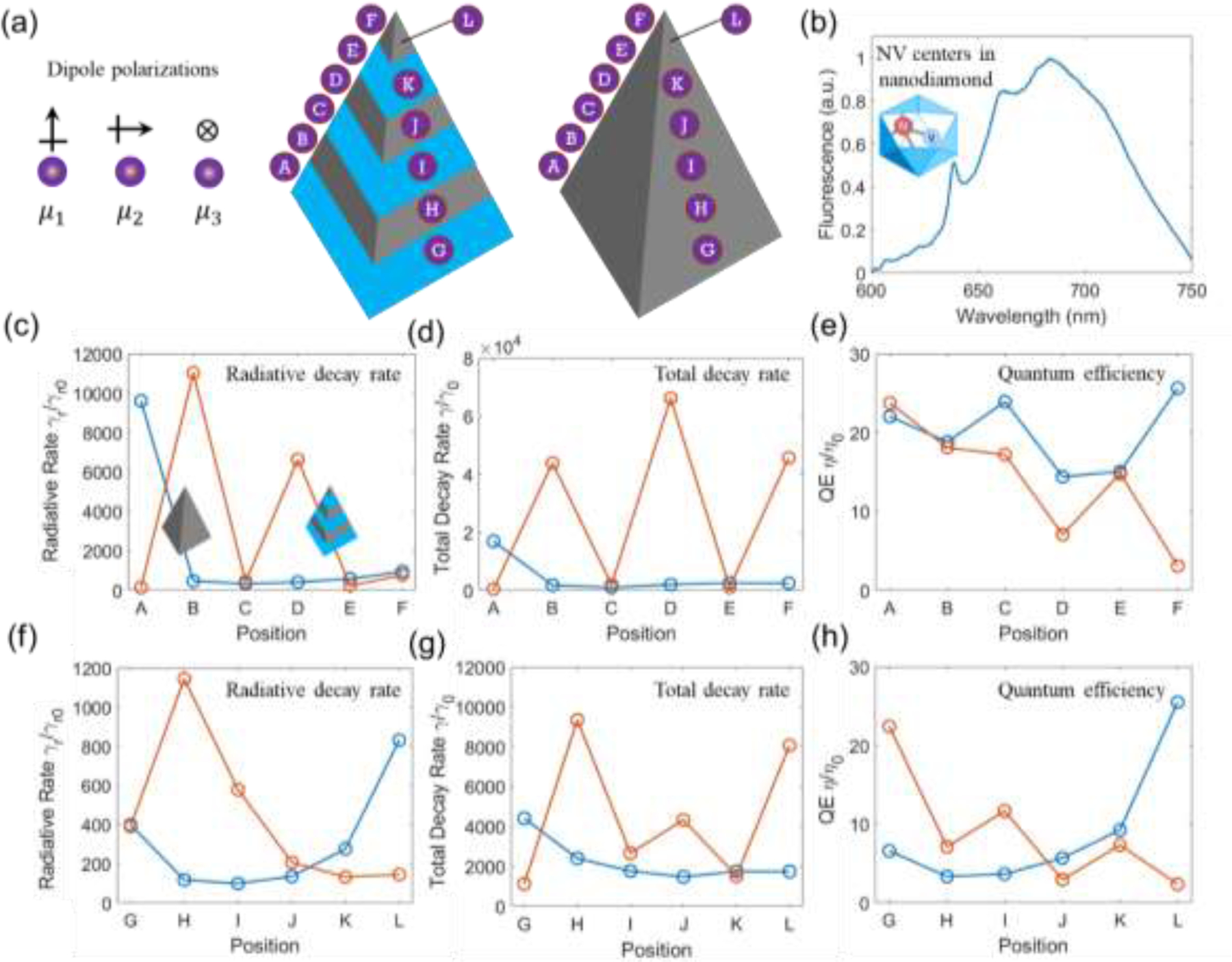
To understand how the spontaneous emission of NV centers in nanodiamond was modified by the metasurface, the spontaneous emission was calculated following our previous method.[2b, 10] The intrinsic quantum efficiency (QE) of NV centers in nanodiamond with a mean diameter of 40 nm was assumed to be 0.7. The NV centers displayed a broadband fluorescence emission from 600 nm to 750 nm as measured on the quartz slide (Fig. 3b), whereas the zero-phonon line is clearly discernible at around 638 nm. To test the broadband optical effects enabled by the pyramidal metasurface, the calculated radiative rate, total decay rate, and QE enhancements were integrated over the wavelength range from 650 nm to 720 nm. The results in Fig. 3c-h were averaged from the three orthogonal polarizations as depicted in Fig. 3a to account for any arbitrary direction in which the NV centers could orient. The position-dependent modifications to the spontaneous emission were further obtained by moving the point dipole, which modelled the NV centers, along the edge (point A to F in Fig. 3a) and along the facet (point G to L in Fig. 3a). It is noted that NV centers created by nitrogen ion implantation technique[13] are shallow and can be found near the surface of a nanodiamond. Also, there are multiple NV centers randomly distributed in a nanodiamond. It is possible that when one NV center is positioned at the far side of a nanodiamond away from the metasurface, the rest of NV centers could be located near the metasurface. These NV centers close to the metasurface will experience enhancement to the emission dynamics while those farther away likely contribute to the background emission. To give a qualitative picture, in this study, we modelled each NV center to be located at about 6 nm away from the nearest point of the metasurface and horizontally aligned to the center of each layer. As a control, a solid silver nanopyramid with the same dimension was also studied following the same method, where the NV center was similarly modelled to be 6 nm away from the nanopyramid. The results were presented in Fig. 3c-h for comparison.
Figure 3. Spontaneous emission rate enhancement.

(a) Left: NV centers in nanodiamond are modelled as an electric dipole with three orthogonal polarizations (, , ); right: schematic for spatial variation of the electric dipole along the edge (A to F) and facet (G to L) of the metasurface. (b) Fluorescence spectrum from NV centers in nanodiamond under 532 nm excitation. Results shown in (c-e) and (f-h) correspond to results from the edge and facet, respectively. (c) and (f) Radiative decay rate enhancement , (d) and (g) total decay rate enhancement , (e) and (h) quantum efficiency (QE) enhancement for an electric dipole spatially varied on the metasurface as compared to on the silver nanopyramid arrays with the same dimension. For (c) to (h), all the enhancement factors were obtained by integrating the enhancement factor for each studied dipole in the wavelength range from 650 nm to 720 nm and averaged over the three orthogonal polarizations; red curves are for data obtained on the layered pyramidal metasurface while blue curves are for the silver nanopyramid arrays.
It was observed that, for the point dipole positioned along either the edge or the facet, the pyramidal metasurface enabled an overall larger radiative and total decay rate. As a comparison, the solid silver nanopyramid allowed a larger radiative and total decay rate only when the dipole was located near either the top vertex or the bottom vertices/edges. If considering the dipole positioned along both the edge and the facet of the pyramid, the metasurface and the solid silver nanopyramid enabled a largely similar QE enhancement, as shown in Fig. 3e, h. Observations made in Fig. 2–3 suggest that, despite the considerable optical enhancement at the vertices and edges for a solid silver nanopyramid, the pyramidal metasurface enabled a spatially extended and broadband amplification to the transition dynamics including both the excitation and spontaneous emission processes. Further reinforcing such observations are the calculated radiative rate, total decay rate, and QE enhancements integrated over an extended wavelength range from 550 nm to 1500 nm, as depicted in Fig. S4, which consistently showed superior photophysical properties enabled by the pyramidal metasurface. Additionally, the pyramidal metasurface was also found to enable a larger non-radiative decay rate, as shown in Fig. S5, for the calculated non-radiative rate integrated over 650 nm to 720 nm and 550 nm to 1500 nm, respectively.
Fabrication and characterization of the layered pyramidal metasurface
To experimentally test the capability of the pyramidal metasurface in accelerating the transition dynamics of NV centers, we fabricated layered pyramidal metasurfaces by leveraging nanosphere lithography[8-9, 10, 14], as schematically shown in Fig. 4a, and performed FLIM studies of fluorescent nanodiamonds on the metasurface. Scanning electron microscope (SEM) images (Fig. 4b-d) confirmed the sharp pyramidal geometry with layered structures. Optical characterization unveiled two prominent resonances in the near-infrared wavelength range as shown in Fig. 4e, which is largely consistent with the calculated optical spectra where two strong resonances were similarly observed in Fig. 1f, S1. The fine spectral features in the visible wavelength range seen in Fig. 1f, S1 are obscured in the experimental data in Fig. 4e, likely due to the rounded geometry and the less than perfect thin film deposited by e-beam evaporator. The experimentally observed broadened peaks are likely caused by inhomogeneous size distribution of the constituent layered nanopyramids rather than energy losses, as silver becomes less lossy in the near-infrared wavelength range.
Figure 4. Fabrication and characterization of the pyramidal metasurface.

(a) Nanosphere lithography for fabricating the pyramidal hyperbolic metasurface consisting of 3 cycles of alternatively stacked silica (24 nm) and silver (16 nm) thin films; (b-d) SEM images of the metasurface with different magnificationI(e) UV-Vis spectra for the fabricated metasurface (red curve) whereas the calculated extinction spectrum (black dashed curve, which was also shown in Fig. 1f) was shown for comparison.
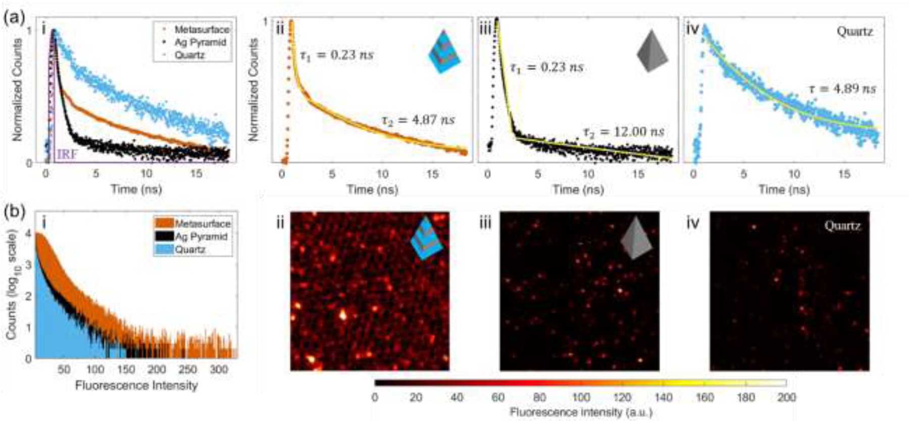
For FLIM measurements, an incident wavelength of 532 nm was used to excite the NV centers in nanodiamonds that were drop-coated on the pyramidal metasurface. Two controls, i.e., a solid silver nanopyramid with the same dimension and a quartz substrate, were similarly studied. The fluorescence signal was collected through a bandpass filter in the wavelength range of 650 nm to 720 nm. By integrating the collected signals over an area of about 14 µm × 14 µm on each studied substrate, both the excited state decay profiles and the fluorescence intensity were obtained and are shown in Fig. 5. The pyramidal metasurface and the solid silver nanopyramid were found to enable a largely similar relaxation dynamics for the faster component with a lifetime of about 0.23 ns, versus 4.89 ns on the quartz substrate. As a large population of nanodiamonds were likely distributed near the vertices and bottom edges rather than on the facets of the pyramidal geometry, NV centers were likely to have benefited from the vertex- and bottom edge-induced plasmonic effects of the solid silver nanopyramid, which is the primary optical enhancement mechanism to enhance the spontaneous decay dynamics as numerically shown in Fig. 3, S4-S5. The accelerated relaxation dynamics for the faster component of NV centers on the pyramidal metasurface was most likely enabled by a combination of plasmonic effects and the hyperbolic dispersion-activated broadband enhancement mechanism, as analytically shown in Eq. (1) and numerically shown in Fig. 3, S4-S5. It is noted that the relaxation dynamics of NV centers could be made even faster if they were optimally positioned near/on the facets of the pyramidal metasurface where a much larger decay rates can be activated.
Figure 5. Characterization of photophysical properties for the fabricated pyramidal metasurface.

(a) Lifetime (normalized counts in log10 scale) and (b) intensity measured for nanodiamonds (~ 40 nm in diameter) on the metasurface consisting of 3 cycles of alternatively stacked silica (24 nm) – Ag (16 nm) layers as compared to on Ag nanopyramid and quartz substrates. The instrument response function (IRF) is shown as a purple curve in (a-i). (a-ii) and (a-iii) are bi-exponential fittings for the decay curves of NV centers measured on the pyramidal metasurface and the Ag nanopyramid, respectively. (a-iv) is the single exponential fitting for the decay curve of NV centers measured on the quartz substrate. Yellow lines in (a) are the fitted curves. (b-ii) to (b-iv) are the fluorescence intensity images of NV centers measured on the pyramidal metasurface, the Ag nanopyramid, and the quartz substrate, respectively. The fluorescence images in (b) have a dimension of about 14 µm × 14 µm. An incident wavelength of 532 nm was used to excite NV centers with the fluorescence signal collected in the wavelength range of 650 nm to 720 nm.
The measured fluorescence intensity histogram in Fig. 5b suggests that NV centers on the pyramidal metasurface displayed an enhanced fluorescence intensity with respect to these on the quartz slide and on the solid silver nanopyramid. It is noteworthy that, despite the similarly enhanced relaxation dynamics for the faster component of NV centers as observed in Fig. 5a, NV centers benefited from a higher fluorescence enhancement on the pyramidal metasurface than on the solid silver nanopyramid, likely contributed by the spatially extended excitation enhancement on the pyramidal metasurface. A comparison of the fluorescence images in Fig. 5b-ii to 5b-iv further establishes that, regardless of the population of coated NV centers on each substrate, the pyramidal metasurface qualitatively exhibited a superior fluorescence enhancement for NV centers as compared to the solid silver nanopyramid and the quartz slide. Collectively, the simultaneous enhancement of the transition dynamics, including both the excitation and spontaneous emission rates and the fluorescence intensity of NV centers, corroborate the distinct capabilities and strong potentials of the pyramidal metasurface in modifying the photophysical properties of broadband-emission quantum emitters including but beyond NV centers in nanodiamond.
Conclusion
In this study, a pyramidal hyperbolic metasurface is reported to enhance the spontaneous emission of NV centers in nanodiamond. Through systematic FDTD-based numerical studies, the pyramidal metasurface is found to display highly desired optical and photophysical properties. The uncovered mode profiles suggest that the electric field becomes spread towards the facets from the vertices and edges at the shorter wavelength range which overlaps with the emission spectrum of NV centers in nanodiamond. Interrogation of the photophysical properties confirms that in addition to the vertices and edges, the facets of the pyramidal metasurface also provide significant enhancement to both the excitation and radiative decay rate enhancement. The spatially extended enhancement of the local electric fields and the transition dynamics highlight the distinct capability of the pyramidal metasurface in providing a broadband enhancement mechanism in both the spectral and spatial domains to amplify the spontaneous emission of NV centers in nanodiamond. These numerically obtained results have been validated by performing FLIM measurements of NV centers in nanodiamond on the fabricated pyramidal metasurface. Collectively, the reported pyramidal metasurface provides an attractive platform for efficient photon generation and outcoupling to the far field and pave the way for realizing chip-based ultrafast photonic devices and developing spin-enhanced quantum optical sensing strategies.
Supplementary Material
Acknowledgement
This work was supported by a grant from the National Institute of Standards and Technology (NIST) Professional Research Experience Program (PREP). I.B. acknowledges support from the National Institute of General Medical Sciences (Grant No. DP2GM128198). The authors also acknowledge the use of facilities and technical support made possible by the Center for Nanoscale Science and Technology’s NanoFab at NIST.
Footnotes
Associated Contents
Supporting Information: Materials and Methods; Figures S1-S5.
Conflict of Interest
The authors declare no competing financial interest.
NIST equipment/supplies disclaimer:
Commercial equipment and materials are identified in order to adequately specify certain procedures. In no case does such identification imply recommendation or endorsement by the National Institute of Standards and Technology, nor does it imply that the materials or equipment identified are necessarily the best available for the purpose.
REFERENCE
- [1].a)Miller BS, Bezinge L, Gliddon HD, Huang D, Dold G, Gray ER, Heaney J, Dobson PJ, Nastouli E, Morton JJL, McKendry RA, Nature 2020, 587, 588; [DOI] [PubMed] [Google Scholar]; b) Li C, Soleyman R, Kohandel M, Cappellaro P, Nano Letters 2022, 22, 43; [DOI] [PubMed] [Google Scholar]; c) Barton J, Gulka M, Tarabek J, Mindarava Y, Wang Z, Schimer J, Raabova H, Bednar J, Plenio MB, Jelezko F, Nesladek M, Cigler P, ACS Nano 2020, 14, 12938; [DOI] [PubMed] [Google Scholar]; d) Tetienne JP, Hingant T, Rondin L, Cavaillès A, Mayer L, Dantelle G, Gacoin T, Wrachtrup J, Roch JF, Jacques V, Physical Review B 2013, 87, 235436; [Google Scholar]; e) Maze JR, Stanwix PL, Hodges JS, Hong S, Taylor JM, Cappellaro P, Jiang L, Dutt MVG, Togan E, Zibrov AS, Yacoby A, Walsworth RL, Lukin MD, Nature 2008, 455, 644. [DOI] [PubMed] [Google Scholar]
- [2].a) Schirhagl R, Chang K, Loretz M, Degen CL, Annual Review of Physical Chemistry 2014, 65, 83; [DOI] [PubMed] [Google Scholar]; b) Liang L, Zheng P, Jia S, Ray K, Chen Y, Barman I, bioRxiv 2021, 2021.11.09.467982;; c) Choy JT, Hausmann BJM, Babinec TM, Bulu I, Khan M, Maletinsky P, Yacoby A, Lončar M, Nature Photonics 2011, 5, 738. [Google Scholar]
- [3].a) Galfsky T, Krishnamoorthy HNS, Newman W, Narimanov EE, Jacob Z, Menon VM, Optica 2015, 2, 62; [Google Scholar]; b) Andersen SKH, Kumar S, Bozhevolnyi SI, Nano Letters 2017, 17, 3889; [DOI] [PubMed] [Google Scholar]; c) Bogdanov SI, Shalaginov MY, Lagutchev AS, Chiang C-C, Shah D, Baburin AS, Ryzhikov IA, Rodionov IA, Kildishev AV, Boltasseva A, Shalaev VM, Nano Letters 2018, 18, 4837. [DOI] [PubMed] [Google Scholar]
- [4].a) Kolchin P, Pholchai N, Mikkelsen MH, Oh J, Ota S, Islam MS, Yin X, Zhang X, Nano Letters 2015, 15, 464; [DOI] [PubMed] [Google Scholar]; b) Yuan S, Qiu X, Cui C, Zhu L, Wang Y, Li Y, Song J, Huang Q, Xia J, ACS Nano 2017, 11, 10704; [DOI] [PubMed] [Google Scholar]; c) Zhao D, Silva REF, Climent C, Feist J, Fernández-Domínguez AI, García-Vidal FJ, ACS Photonics 2020, 7, 3369; [DOI] [PMC free article] [PubMed] [Google Scholar]; d) Krivenkov V, Samokhvalov P, Nabiev I, Rakovich YP, The Journal of Physical Chemistry Letters 2020, 11, 8018; [DOI] [PubMed] [Google Scholar]; e) Novotny L, Hecht B, Principles of nano-optics, Cambridge university press, 2012; [Google Scholar]; f) Zhao Y, Mukherjee K, Benkstein KD, Sun L, Steffens KL, Montgomery CB, Guo S, Semancik S, Zaghloul ME, Nanoscale 2019, 11, 11922. [DOI] [PubMed] [Google Scholar]
- [5].a)Poddubny A, Iorsh I, Belov P, Kivshar Y, Nature Photonics 2013, 7, 948; [Google Scholar]; b) Lu D, Kan JJ, Fullerton EE, Liu Z, Nature Nanotechnology 2014, 9, 48; [DOI] [PubMed] [Google Scholar]; c) Li L, Wang W, Luk TS, Yang X, Gao J, ACS Photonics 2017, 4, 501; [Google Scholar]; d) Roth DJ, Krasavin AV, Wade A, Dickson W, Murphy A, Kéna-Cohen S, Pollard R, Wurtz GA, Richards D, Maier SA, Zayats AV, ACS Photonics 2017, 4, 2513; [Google Scholar]; e) Indukuri SRKC, Bar-David J, Mazurski N, Levy U, ACS Nano 2019, 13, 11770. [DOI] [PubMed] [Google Scholar]
- [6].a)Mahmoodi M, Tavassoli SH, Takayama O, Sukham J, Malureanu R, Lavrinenko AV, Laser & Photonics Reviews 2019, 13, 1800253; [Google Scholar]; b) Cortes CL, Newman W, Molesky S, Jacob Z, Journal of Optics 2012, 14, 063001; [Google Scholar]; c) Solntsev AS, Agarwal GS, Kivshar YS, Nature Photonics 2021, 15, 327. [Google Scholar]
- [7].a)Lin H-I, Tan H-Y, Liao Y-M, Shen K-C, Shalaginov MY, Kataria M, Chen C-T, Chang J-W, Chen Y-F, ACS Applied Materials & Interfaces 2021, 13, 49224; [DOI] [PubMed] [Google Scholar]; b) Shen K-C, Ku C-T, Hsieh C, Kuo H-C, Cheng Y-J, Tsai DP, Advanced Materials 2018, 30, 1706918; [DOI] [PubMed] [Google Scholar]; c) Lin H-I, Shen K-C, Liao Y-M, Li Y-H, Perumal P, Haider G, Cheng BH, Liao W-C, Lin S-Y, Lin W-J, Lin T-Y, Chen Y-F, ACS Photonics 2018, 5, 718; [Google Scholar]; d) Lin H-I, Wang C-C, Shen K-C, Shalaginov MY, Roy PK, Bera KP, Kataria M, Paul Inbaraj CR, Chen Y-F, npj Flexible Electronics 2020, 4, 20. [Google Scholar]
- [8].Zheng P, Kasani S, Wu N, Nanoscale Horizons 2019, 4, 516. [DOI] [PMC free article] [PubMed] [Google Scholar]
- [9].a)Zheng P, Kasani S, Shi X, Boryczka AE, Yang F, Tang H, Li M, Zheng W, Elswick DE, Wu N, Analytica Chimica Acta 2018, 1040, 158; [DOI] [PMC free article] [PubMed] [Google Scholar]; b) Zheng P, Wu L, Raj P, Mizutani T, Szabo M, Hanson WA, Barman I, Small 2022, n/a, 2200090; [DOI] [PMC free article] [PubMed]; c) Tabatabaei M, Sangar A, Kazemi-Zanjani N, Torchio P, Merlen A, Lagugné-Labarthet F, The Journal of Physical Chemistry C 2013, 117, 14778. [Google Scholar]
- [10].Zheng P, Kasani S, Tan W, Boryczka J, Gao X, Yang F, Wu N, Analytica Chimica Acta 2022, 1203, 339721. [DOI] [PubMed] [Google Scholar]
- [11].Yang X, Yao J, Rho J, Yin X, Zhang X, Nature Photonics 2012, 6, 450. [Google Scholar]
- [12].Zheng P, Paria D, Wang H, Li M, Barman I, Nanoscale 2020, 12, 832. [DOI] [PMC free article] [PubMed] [Google Scholar]
- [13].Ofori-Okai BK, Pezzagna S, Chang K, Loretz M, Schirhagl R, Tao Y, Moores BA, Groot-Berning K, Meijer J, Degen CL, Physical Review B 2012, 86, 081406. [Google Scholar]
- [14].a)Zheng P, Cushing SK, Suri S, Wu N, Physical Chemistry Chemical Physics 2015, 17, 21211; [DOI] [PMC free article] [PubMed] [Google Scholar]; b) Zheng P, Li M, Jurevic R, Cushing SK, Liu Y, Wu N, Nanoscale 2015, 7, 11005; [DOI] [PMC free article] [PubMed] [Google Scholar]; c) Kasani S, Zheng P, Wu N, The Journal of Physical Chemistry C 2018, 122, 13443. [DOI] [PMC free article] [PubMed] [Google Scholar]
Associated Data
This section collects any data citations, data availability statements, or supplementary materials included in this article.


