Abstract
Chronic lymphocytic leukemia (CLL) cells are highly dependent on interactions with the immunosuppressive tumor micro-environment (TME) for survival and proliferation. In the search for novel treatments, pro-inflammatory cytokines have emerged as candidates to reactivate the immune system. Among those, interleukin 27 (IL-27) has recently gained attention, but its effects differ among malignancies. Here, we utilized the Eμ-TCL1 and EBI3 knock-out mouse models as well as clinical samples from patients to investigate the role of IL-27 in CLL. Characterization of murine leukemic spleens revealed that the absence of IL-27 leads to enhanced CLL development and a more immunosuppressive TME in transgenic mice. Gene-profiling of T-cell subsets from EBI3 knock-out highlighted transcriptional changes in the CD8+ T-cell population associated with T-cell activation, proliferation, and cytotoxicity. We also observed an increased anti-tumor activity of CD8+ T cells in the presence of IL-27 ex vivo with murine and clinical samples. Notably, IL-27 treatment led to the reactivation of autologous T cells from CLL patients. Finally, we detected a decrease in IL-27 serum levels during CLL development in both pre-clinical and patient samples. Altogether, we demonstrated that IL-27 has a strong anti-tumorigenic role in CLL and postulate this cytokine as a promising treatment or adjuvant for this malignancy.
Introduction
Chronic lymphocytic leukemia (CLL) is the most frequent type of adult leukemia in the USA and Europe, affecting mainly older adults.1 Clinically, CLL is defined as a B-cell hematological malignancy characterized by accumulation of abnormal, monoclonal, B lymphocytes in the peripheral blood (PB) and secondary lymphoid organs.2
Current treatments against CLL do not have a curative potential, and a significant percentage of patients do not respond or become resistant.3 Consequently, there is a pressing need for the development of novel therapies for advanced and aggressive CLL, which unfortunately remains an incurable disease. CLL cells are highly dependent on interactions with surrounding non-malignant cells for survival and proliferation.4,5 In fact, they spontaneously undergo apoptosis in monoculture, but co-culture with accessory cells significantly extends their survival.6-8 Given the pivotal role of the tumor microenvironment (TME) in CLL, an increasing number of studies are focusing on the identification of micro-environmental signals that could play a role in the pathogenesis of this disease.
Interleukin 27 (IL-27) is a heterodimeric cytokine composed of two non-covalently linked subunits: IL-27p28 (P28) and Epstein-Barr-virus-induced molecule 3 (EBI3).9 IL-27 is produced by activated antigen presenting cells (APC) and it signals through a heterodimeric receptor (IL-27R) that comprises the gp130 and WSX-1 subunits, both essential for efficient signaling.10 IL-27 was initially characterized as being pro-inflammatory, given its ability to promote Th1 immunity. Nevertheless, it was later reported that IL-27 exerts a potent inhibitory role during Th2, Th17 and regulatory T-cell (Treg) differentiation. Hence, IL-27 mediates a wide range of functions involved in T-cell-mediated immunity.11
Not surprisingly, IL-27 is known to have pleotropic functions in the setting of cancer development in relationship to the biological context and experimental models considered. Most existing evidence refers to the anti-tumor activities of this cytokine.12 IL-27 has been reported to hinder tumor development and progression mainly by modulating the immune landscape surrounding the malignant cells.13 While most evidence points towards the upregulation of Th1 and Cytotoxic T lymphocyte (CTL) responses as the main anti-tumor contribution of IL-27 to the TME,14-16 other reports suggest that IL-27 can also mediate natural killer (NK) cell responses and inhibit M2 macrophage polarization.17,18 On the other hand, a few studies indicate that IL-27 might also contribute to tumorigenesis in specific settings. For example, elevated IL-27 serum levels are associated with poor prognosis and disease progression in some malignancies such as gastroesophageal cancer, melanoma and adult acute myeloid leukemia (AML).19-21 Additionally, IL-27 was found to modulate transcriptional programs and induce the expression of immune-regulatory molecules such as PD-L1 and IDO in human ovarian cancer cells in vitro.22,23
Given the dynamic nature of cytokine biology, and consistent with a rather variable role in cancer biology, the role of IL-27 and its mechanism of action must be carefully investigated in each malignancy. With this background, we asked whether IL-27 has an effect in the development and progression of CLL. Here, we describe a strong anti-tumorigenic role of IL-27 in CLL in distinct preclinical mouse models and patient samples. First, we show that the genetic depletion of the IL-27 subunit Ebi3 leads to a strongly enhanced CLL development and a more immunosuppressive TME using two in vivo approaches. Secondly, we elucidate a mechanism by which IL-27 enhances CD8+ T-cell anti-tumor immunity in CLL. Moreover, we measured lower plasma levels of IL-27 concomitant with CLL development in mice and patients. We functionally validate the enhanced anti-tumor ability of CD8+ T cells in the presence of IL-27 both in murine and human samples. Finally, we show that IL-27 neutralization recapitulates the enhanced leukemic progression observed in the transgenic mouse models.
Methods
Additional information regarding the materials and methods used in this publication can be found in the Online Supplementary Appendix.
Patient samples
All experiments involving the use of human samples were conducted in accordance with the declaration of Helsinki and approved by the appropriate local Ethics Committee: the Jules Bordet Institute Ethics Committee (Belgium), The Comité National d'Ethique de Recherche (Luxembourg), and the University of Saarland (Germany).
PB samples were obtained from CLL patients and from age-matched healthy donors following informed consent. PB mononuclear cells (PBMC) were isolated from whole blood by density gradient centrifugation using the Leuco-Sep™ Separation Medium (Greiner Bio-One) as described in the manufacturer’s protocol.
Animal experiments
All animal experiments were performed under specific pathogen-free conditions with the approval of the Luxembourg Ministry of Agriculture in accordance with the guidelines from the European Union. Ebi3-/- (RRID: IMSR_JAX:008701) and FoxP3YFP/Cre (RRID: IMSR_JAX:016959) mice were purchased from the Jackson Laboratory (Bar Harbour, ME); and C57BL/6 (MGI: 3028467, RRID: IMSR_JAX:000664) from Janvier Labs (France). Eµ-TCL1 mice (on C57BL/6 background; MGI: 3527221) were kindly provided by Pr. Carlo Croce and Pr. John Byrd (OSU, OH). The Eμ-TC L1 Ebi3-/- strain was generated in-house by crossing Ebi3-/- mice with Eμ-T C L1, together with FoxP3YFP/Cre Ebi3-/- mice, obtained by breeding FoxP3YFP/Cre and Ebi3-/-mice for several generations. Rag2-/- mice were held at specific pathogen-free conditions at the central animal facility of the German Cancer Research Centre (DKFZ).
Flow cytometry
Single-cell suspensions were stained with cell-surface antibodies (30 minutes [min], 4 °C) and washed twice with FACS buffer. Dead cell discrimination was performed using Zombie dye (Biolegend), resuspended in PBS (20 min, 4 °C). For the staining of intracellular proteins, surface-stained cells were fixed (30 min, room temperature) with eBio-scienceTM Foxp3/Transcription Factor Staining Buffer Set (ThermoFisher Scientific). After additional washing steps, cells were permeabilized using eBioscienceTM Permeabilization Buffer (ThermoFisher Scientific) and stained with the intracellular antibody mix (30 min, 4°C). Samples were stored at 4°C in dark conditions until acquisition. Antibodies used for flow cytometry are listed in the Online Supplementary Table S1.
Statistical analyses
Sample size was determined based on expected variance of read-out. No samples or animals were excluded from the analyses. Statistical analysis was performed using GraphPad Prism software (version 9.1.2; RRID: SCR_002798). Data are displayed as mean ± standard error of mean (SEM). For the percentage of CLL cells in PB over time, we performed two-way ANOVA followed by multiple comparison test. The unpaired t test was used for the rest of the figures. A P value lower than 0.05 was considered statistically significant. Significance displayed in each figure is explained in figure legends.
Results
EBI3 depletion promotes leukemia development and induces an enhanced immunosuppressive tumor microenvironment.
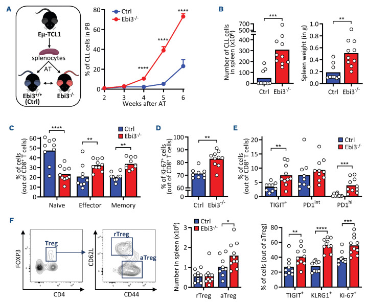
In order to investigate the role of IL-27 in CLL development, we used Ebi3-/- mice, defective in the production of the heterodimeric IL-27. First, we validated the specific deletion of the Ebi3 subunit in murine splenocytes via quantitative polymerase chain reaction (qPCR), and confirmed the suitability of the models for further analyses (Online Supplementary Figure S1A). We adoptively transferred (AT) Ebi3-/-mice and Ebi3+/+ mice (wild-type [WT], used as controls) with two independent clones of CLL cells obtained from the spleen of leukemic Eμ-TCL1 mice. We then followed up leukemia development in the PB of recipient mice using flow cytometry (FC), which led to the observation of a strikingly enhanced tumor development in the absence of Ebi3 (Figure 1A; Online Supplementary Figure S1B). Consistently, we observed an increased number of splenocytes and spleen weight in mice lacking Ebi3 (Figure 1B). Then, we sorted multiple immune cell subsets from leukemic mice and we identified dendritic cells and monocytes as the main producers of IL-27 in leukemic mice, as they expressed high levels of both subunits Ebi3EBI3 and p28 (Online Supplementary Figure S1C). After, we characterized the immune landscape in the splenic CLL TME, focusing on T cells as main mediators of the anti-tumor immune response in vivo.24 We found an increased number of CD8+ effector T cells and CD4+ conventional T cells (Tconv) in mice lacking EBI3, while the Treg number remained stable (Online Supplementary Figure S1D). Moreover, Ebi3-/- mice presented more effector (CD62L- CD44+) and memory (CD62L+ CD44+) CD8+ T cells, concomitant to a decrease in the frequency of naive cells (CD62L+ CD44-) (Figure 1C). In addition, we observed an increased frequency in Ki-67+ CD8+ T cells in Ebi3-/- mice (Figure 1D). It is known that some immune checkpoints (IC) are expressed by activated CD8+ T cells, but only terminally exhausted cells co-express several IC.25 Here, we observed an increased percentage of TIGIT+ and PD1hi in CD8+ T cells lacking EBI3, while no differences were observed in the frequency of PD1int cells (Figure 1E). In order to analyze the functional status of CD8+ T cells, we performed hierarchical clustering of CD8+ T cells based on marker expression. Seven CD8+ T-cell clusters (C) were identified based on CD44, Ki-67, PD1, IFN-γ, TIGIT and CD62L (Online Supplementary Figure S1E). In the absence of Ebi3, there was a decrease in naïve CD8+ T cells (C1) together with an increase in memory T cells (Tmem, C3) and effector T cells (Te ff) showing high IC and Ki-67 expression (Online Supplementary Figure S1F), suggesting an exhausted phenotype. Moreover, activated Tregs (aTregs, CD62L- CD44+) are more abundant in leukemic Ebi3-/- mice (Figure 1F, left panel) and display higher levels of TIGIT and KLRG1 along with increased Ki-67 positivity (Figure 1F, right panel), thus exhibiting enhanced immunosuppression and proliferation. As observed for CD8+ T cells, the proportion of antigen-experienced CD4+ Tconv was also increased in Ebi3-/- mice (Online Supplementary Figure S1G). Clustering of CD4+ T cells revealed the presence of nine clusters (Online Supplementary Figure S1H, I), showing that activated and immunosuppressive Tregs were expanded in absence of Ebi3. Altogether, these findings suggest an anti-tumor role of EBI3 during CLL development, as its depletion enhances tumor growth, promotes CD8+ T-cell exhaustion and increases Treg immunosuppressive phenotype.
Figure 1.
EBI3 depletion promotes leukemia development and induces an enhanced immunosuppressive tumor microenvironment. (A) Two cohorts of recipient Ebi3-/- and control (Ctrl) mice were injected with splenocytes isolated from leukemic Eμ-TCL1 mice. Two weeks after adoptive transfer (AT), mice were bled weekly to monitor chronic lymphocytic leukemia (CLL) development in peripheral blood (PB). Percentages of neoplastic CD5+ CD19+ cells were detected in PB by flow cytometry (FC); (N=18 for control [Ctrl] and N=19 for Ebi3-/-, two-way ANOVA). (B-F). For the second cohort, splenic tumor microenvironment (TME) was characterized by FC (N=9 for Ctrl and N=10 for Ebi3-/-). (B) Number of CD5+ CD19+ CLL cells in the spleen (left), and spleen weight (right) in Ebi3-/- compared to Ctrl mice. (C) Frequency of naive (CD62L+ CD44-), effector (CD62L- CD44+) or memory (CD62L+ CD44+) CD8+ T cells. (D) Percentage of proliferative Ki-67+ cells among CD8+ T cells in the spleen of Ctrl and Ebi3-/- mice. (E) Frequency of the indicated populations in Ctrl and Ebi3-/- CD8+ T cells. (F) Representative flow cytometry plot (left) depicting the gating strategy used to discriminate activated regulatory T cells (aTregs, CD62Lhi CD44lo) from resting regulatory T cells (rTregs, CD62Llo CD44hi). Number of rTregs and aTregs in spleens (middle). Frequency of the indicated populations in Ctrl and Ebi3-/- Tregs (right). Unpaired t test, *P<0.05, **P<0.01, ***P<0.001, ****P<0.0001.
In parallel, we deeply characterized the immune landscape in Ebi3+/+ and Ebi3-/- mice to confirm that the aforementioned differences in CLL growth were not due to major intrinsic differences between the transgenic strains. We used Foxp3YFP/Cre mice to analyze T cells and Tregs. As expected, immunophenotyping of Foxp3YFP/Cre and Foxp3YFP/Cre Ebi3-/-splenocytes did not reveal any major difference between Ebi3+/+ and Ebi3-/- mice. We found no statistical differences in the frequency of T cells, NK cells, NK-T cells or myeloid cells between groups (Online Supplementary Figure S2A).
Moreover, the analysis of the myeloid cell compartment showed no differences in the frequency of neutrophils, dendritic cells, monocytes nor macrophages (Online Supplementary Figure S2B). Only a slight increase/decrease in the frequencies of CD8+ T cells and CD4+ Tconv cells, respectively, was observed, with no differences for Tregs (Online Supplementary Figure S2C). In order to evaluate T-cell functionality between phenotypes, we isolated splenic CD4+ and CD8+ T cells and analyzed IC and cytokines expression following ex vivo stimulation for 4 hours (h). We neither observed any difference in the frequency of indicated IC and cytokines in CD8+ T cells, CD4+ Tconv cells, nor in Tregs (Online Supplementary Figure S2D-F). These results indicate that Ebi3 depletion does not drastically impact immune cells distribution, effector cytokine secretion and IC expression in T cells, pointing towards a fundamental role of IL-27 in the tumor context rather than in physiological conditions.
EBI3 depletion in transgenic Eµ-TCL1 mice affects mice survival
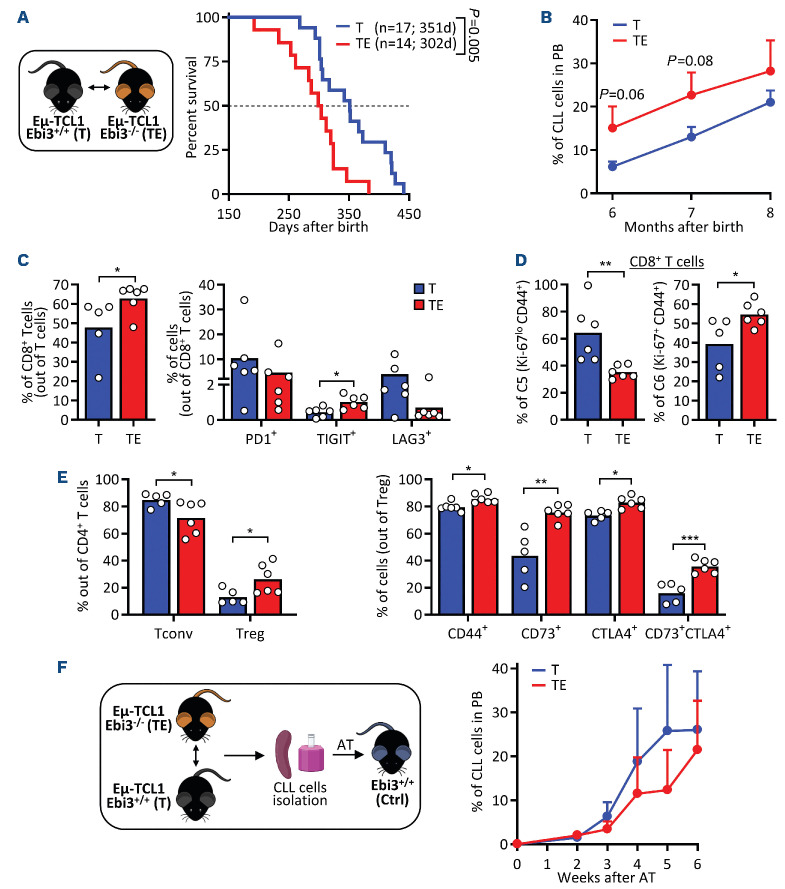
In order to better understand the role of EBI3 during CLL development, we crossed the E/i-TCL1 mouse model (referred as T mice) with Ebi3-/- mice to generate the E^i-TCL1 Ebi3-/- mouse model (referred as TE mice) that spontaneously develop CLL in the absence IL-27 production. As opposed to the TCL1 AT model, TE mice lack Ebi3 expression in all cell types, including CLL cells. We observed a shorter survival for TE mice (median survival of 302 vs. 351 days; Figure 2A) and an increased percentage and number of CLL cells in the PB at an earlier time point, although not statistically significant (Figure 2B; Online Supplementary Figure S3A). The analysis of the splenic TME following euthanasia indicated, despite a similar tumor load (Online Supplementary Figure S3B, C), an increased frequency of CD8+ T cells among total T cells in leukemic TE mice (Figure 2C left panel), as well as an increase in TIGIT+ CD8+ T cells (Figure 2C right panel). Clustering analysis identified six clusters based on CD44, Ki-67, PD1, KLRG1, TIGIT and CD62L expression in CD8+ T cells, as well as an enrichment in proliferative Ki67+ CD8+ T cells (C6) (Figure 2D; Online Supplementary Figure S3D, E). In addition, a general increase in CD8+ T-cell subpopulations expressing several IC was observed in TE mice compared with their Ebi3+/+ counterparts (clusters C1-C4) (Online Supplementary Figure S3E), pointing towards a more exhausted phenotype of CD8+ T cells in TE mice. Additionally, we observed a decrease in CD4+ T-cell percentage in leukemic mice lacking Ebi3 compared to Ebi3+/+ controls (Online Supplementary
Figure 2.
EBI3 depletion in transgenic Eµ-TCL1 mice affects mice survival. (A) Transgenic Eμ-T C L1 mice were crossed with Ebi3-/-mice to obtain leukemic mice deficient in interleukin 27 (IL-27). Starting at 6 months after birth, mice were bled every month to evaluate peripheral disease development (T=Eμ-TC L1 mice; TE=Eμ-TCL1 Ebi3-/- mice). Mouse survival was compared between groups (survival curve analysis). (B) Percentages of neoplastic CD5+ CD19+ cells were detected by flow cytometry (FC) in peripheral blood (PB) (N=17 for T mice and N=14 for TE mice, two-way ANOVA test). (C-E) Leukemic T and TE mice were euthanized and their splenocytes were analyzed using FC. (C) Percentage of CD8+ T cells in control (Ctrl) and Ebi3-/- CD3+ T cells and frequency of indicated populations among T and TE CD8+ T cells (N=6 for T group and N=6 for TE group). (D) Percentages of CD8+ T-cell clusters distribution in T and TE mice. (E) Percentage of conventional T-cell (Tconv) and regulatory T-cell (Tregs) populations among CD4+ T cells (left) and frequency of indicated cell populations among Tregs (right). (F) Leukemic Eμ-T C L1 mice and Eμ-TCL1 Ebi3-/- mice were euthanized, and chronic lymphocytic leukemia (CLL) cells were isolated from splenocytes. Isolated CLL cells were adoptively transferred into wild-type (WT) mice. Recipient mice were bled weekly to evaluate peripheral disease development. Percentages of neoplastic CD5+ CD19+ cells were detected in PB by FC (N=12 mice divided in 6 groups. Each group of 2 animals received 1 independent CLL clone coming from diseased T or TE mice). Unpaired t test, *P<0.05, **P<0.01.
Figure S3F). More precisely, TE mice exhibited a decreased proportion of CD4+ Tconv together with increased frequency of Tregs (Figure 2E left panel), which were more immunosuppressive, as highlighted by higher expression of CD44, CD73 and CTLA4 in the absence of Ebi3 (Figure 2E right panel). Unsupervised clustering revealed eight CD4+ T-cell clusters (Online Supplementary Figure S3G, I). Of particular relevance, a cluster representing highly immunosuppressive Tregs (C1) was enriched in Ebi3-deficient leukemic mice. Immunophenotyping of T and TE CLL cells showed that the expression of immunosuppressive cytokines, major histocompatibility complex molecules, activation markers, and IC remained unchanged; suggesting that EBI3 depletion in CLL cells does not play a role in the observed phenotype (Online Supplementary Figure S3J).
In order to investigate whether the effects of Ebi3 depletion observed in vivo were mediated by cells of the TME or by CLL cells, we depleted Ebi3 exclusively in CLL cells. For this purpose, we isolated CLL cells from spleens of diseased T and TE mice and injected them in recipient control Ebi3+/+ mice. In this experimental setting, we could not observe any difference in tumor growth in mice injected with Ebi3-/- leukemic cells (Figure 2F). Altogether our results indicate that EBI3 depletion in cells of the TME and not in CLL cells favors CLL progression by impacting T-cell-mediated immunity.
Specific T-cell-EBI3 depletion promotes leukemia development
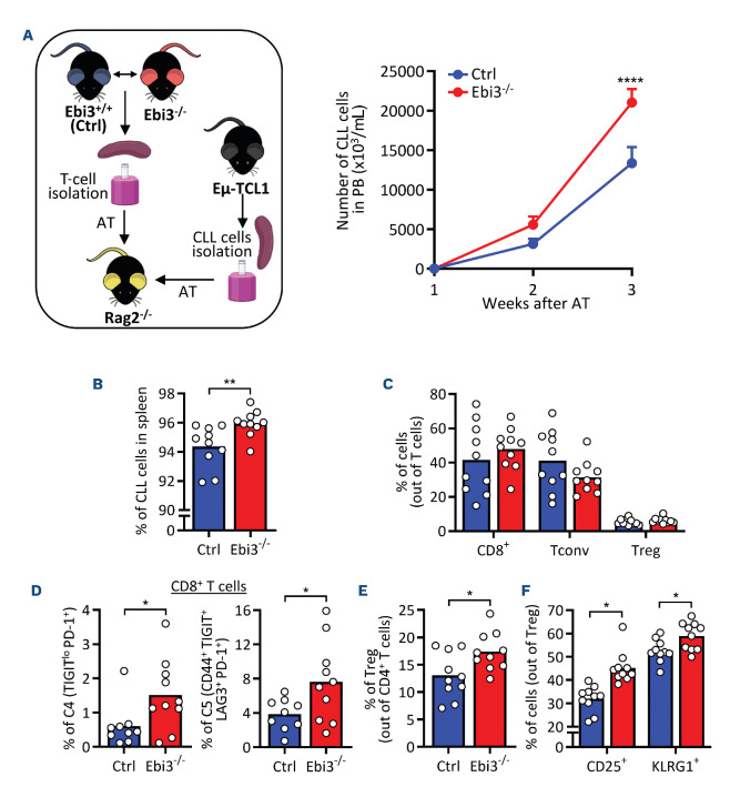
As the AT of EBI3-depleted CLL cells did not impact tumor growth (Figure 2F) while substantial changes in T cells were observed upon EBI3 depletion in the AT model and Eμ-TCL1 model (Figures 1C, D and 2C), we proceeded to specifically investigate the impact of Ebi3-/- CD3+ T cells on CLL development. We used Rag2-/- mice, deficient in producing mature T and B cells. CD3+ T cells were isolated from the spleen of Ebi3-/- and Ebi3+/+ mice and intravenously injected in recipient Rag2-/- mice. Recipient mice were subsequently adoptively transferred with Eμ-T C L1 leukemic cells (Figure 3A). Monitoring of leukemic growth in the PB revealed that Rag2-/- mice injected with Ebi3-/- T cells showed an enhanced CLL development compared to mice injected with the WT counterpart (Figure 3A). Importantly, the differences observed between the two groups were not due to a variation in the T-cell number throughout the experiment (Online Supplementary Figure S4A). Consistently, post-euthanasia analysis of the leukemic spleens showed an increase in the percentage of CLL cells in the Ebi3-/-group (Figure 3B). Regarding T cells, we did not observe any difference in the frequency of subpopulations (Figure 3C). Nonetheless, unsupervised analysis identified nine clusters of CD8+ T cells (Online Supplementary Figure S4B), and revealed the expansion of activated/exhausted CD8+ T cells in the mice injected with Ebi3-/- CD3+ T cells (Figure 3D; Online Supplementary Figure S4C). In addition, we identified an increased frequency of Tregs within the CD4+ T-cell population (Figure 3E) and a higher percentage of CD25+ and KLRG1+ Tregs (Figure 3F), indicating an enhanced immuno-suppressive ability of Tregs in absence of EBI3. Moreover, unsupervised clustering analysis of analyzing CD4+ T cells identified an accumulation of activated and immunosuppressive Tregs (C2) in recipient mice injected with Ebi3-/- T cells (Online Supplementary Figure S4D, E).
CD8+ T cells from Ebi3-/- mice have altered expression of genes involved in activation and functionality
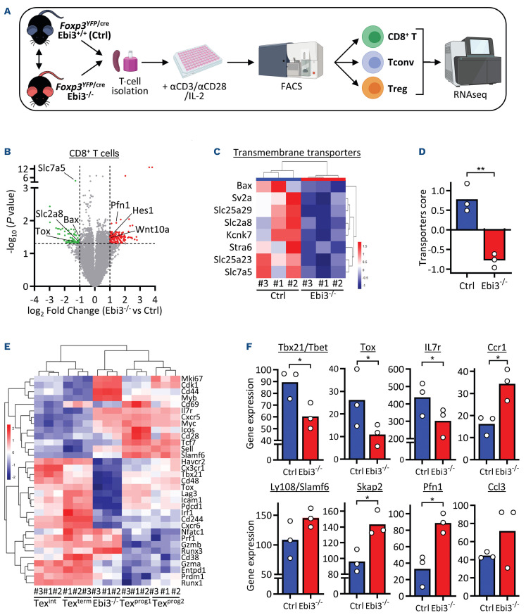
As our data demonstrates that IL-27 controls CLL development in a T-cell-mediated mechanism in vivo, we proceeded to investigate the transcriptional differences between T cells from Ebi3+/+ and Ebi3-/- mice after ex vivo activation (Figure 4A; Online Supplementary Figure S5A). Among the upregulated genes in CD8+ T cells, we identified Profilin-1 (Pfn1) (Figure 4B), an actin-binding protein and a negative regulator of effector T-cell-mediated cytotoxicity.26 Amidst the downregulated genes, we found several transmembrane transporters known to mediate T-cell activation27 (Slc2a8 and Slc7a5; Figure 4C, D). An ontology analysis indicated a reprograming of crucial functions as translation initiation and rRNA processing (Online Supplementary Figure S5B). We also compared the gene expression profile of Ebi3-/- CD8+ T cells with transcriptional signatures of four CD8+ T-cell subsets showing different degrees of exhaustion and dysfunctionality previously published.28 We observed that Ebi3-/- CD8+ T cells present similarities with progenitor CD8+ exhausted T cells (TexProg) but also with more terminally exhausted cells (Texterm; Online Supplementary Figure S5C, D). Indeed, looking at the third component of the PCA, explaining 13% of the variability, indicated that Ebi3-/- T cells and Texterm present similar gene expression profiles (Online Supplementary Figure S5D right panel). Therefore, we selected genes described to mirror T-cell activation and exhaustion and we observed that Ebi3-/- T cells clustered between progenitor and terminally exhausted T cells (Figure 4E). The expression of these marker genes also showed differences between Ebi3+/+ and Ebi3-/- T cells, mostly for Tox, Tbet, Il7r, Ccr1 and Pfn1 (Figure 4F). Regarding CD4+ Tconv and Treg compartments, gene expression analysis revealed no differences between groups (Online Supplementary Figure S5E-F), suggesting that, in absence of Ebi3, effector CD8+ T cells are the major T-cell population affected and appear less functional when activated in vitro.
Figure 3.
Specific T-cell-EBI3 depletion promotes leukemia development. (A) Control (Ctrl) and Ebi3-/- mice were euthanized, and CD3+ T cells were isolated from the splenocytes. Recipient Rag2-/- mice were injected with either Ctrl (N=10) or Ebi3-/- CD3+ T cells (N=10) and subsequently injected with splenocytes derived from leukemic Eμ-TCL1 mice. Experimental mice were bled weekly to evaluate peripheral disease development. Number of circulating neoplastic CD5+ CD19+ cells is shown in the 2 groups (two-way ANOVA test). (B) Percentage of chronic lymphocytic leukemia (CLL) cells in the spleen of recipient Rag2-/- mice. (C) Percentages of CD8+, conventional T cells (Tconv), and regulatory T cells (Tregs) among the injected Ctrl and Ebi3-/- CD3+ T cells. (D) Percentages of CD8+ T cells in clusters identified in spleens of recipient Rag2-/- mice. (E, F) Frequency of Tregs and of CD25+ and KLRG1+ Tregs in the spleen of recipient Rag2-/- mice. Unpaired t test, *P<0.05, ****P<0.0001.
Ebi3-/- CD8+ T cells present impacted cytotoxic activity while IL-27 enhances their potential both in murine and human setings
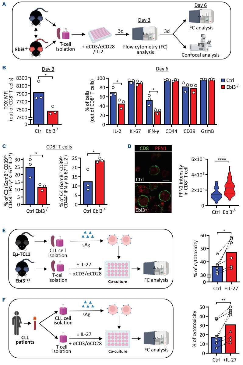
In order to functionally characterize Ebi3-/- and Ebi3+/+ CD8+ T cells and validate the gene expression data, we performed ex vivo activation of CD3+ T cells for 3 days and analyzed CD8+ T cells by FC and confocal microscopy (Figure 5A). In line with previous findings, after 3 days, we could observe a decrease in TOX abundance in Ebi3-/- CD8+ T cells (Figure 5B left panel). Very interestingly, we could not observe any difference in cytokine production between the two conditions after 3 days of activation (Online Supplementary Figure S6A), whereas after 6 days, Ebi3-/- CD8+ T cells presented decreased production of IL-2 and IFN-γ compared to WT cells (Figure 5B right panel). Clustering analysis of CD8+ T cells co-expressing different cytokines (C3) appeared significantly reduced (Figure 5C; Online Supplementary Figure S6B-D), confirming that in the absence of Ebi3, CD8+ T cells are less polyfunctional. After 6 days of activation, we quantified Profilin1 protein level in CD8+ T cells by immunofluorescence. As expected, the analysis revealed an increase in Profilin1 in Ebi3-/- CD8+ T cells (Figure 5D). In order to assess if IL-27 could indeed affect the cytotoxic capabilities of CD8+ T cells, we performed a cytotoxic assay with WT or Ebi3-/- CD8+ T cells treated with IL-27 for 48 h (Figure 5E), and super antigen (sAg)-loaded Eμ-TCL1 CLL cells as target cells. We noted an increased cytotoxic ability of CD8+ T cells in the presence of IL-27 (Figure 5E, Online Supplementary Figure S6E). In order to inspect whether the effect of IL-27 on CD8+ T cells was conserved in human cells, first, we purified and activated T cells from PBMC of four healthy donors, and then cultured them in the presence or absence of IL-27 for 6 days (Online Supplementary Figure S6F). T cells were co-cultured with target cells for 30 h and the killing efficiency of CD8+ T cells during co-culture was quantified. In line with previous results, we observed a decrease in the percentage of live target cells, translated into an increased killing efficiency of human CD8+ T cells in the presence of IL-27 for all donors (Online Supplementary Figure S6G, H). Finally, we isolated T cells and CLL cells from CLL patients, and performed a killing assay in the absence or presence of IL-27. Again, we confirmed the positive effect of IL-27 on the cytotoxic activity of CLL patients’ T cells (Figure 5F).
Figure 4.
CD8+ T cells from Ebi3-/- mice have altered expression of genes involved in activation and functionality. (A) Foxp3YFP/Cre and Foxp3YFP/Cre Ebi3-/- mice (N=3 per group) were euthanized at 8 weeks of age and CD3+ T cells were isolated from the spleno-cytes. T cells were activated for 72 hours, sorted into CD8+ T cells, CD4+ conventional T cells (Tconv), and regulatory T cells (Tregs) (YFP+) by flow cytometry (FC), and subjected to RNA sequencing (RNAseq). (B) Volcano plot depicting differentially expressed genes (DEG) between control (Ctrl) and Ebi3-/- CD8+ T cells. (C, D) Heatmap and score of transmembrane transporters in Ctrl and Ebi3-/- CD8+ T cells. (E) Heatmap depicting the similarity of Ebi3-/- CD8+ T cells with the subset of proliferating progenitor exhausted T cells described in 43. (F) Expression profiles of single genes in CD8+ T cells from Ctrl and Ebi3-/- mice. Unpaired t test, *P<0.05, **P<0.01.
IL-27 level is reduced in the blood of leukemic mice and chronic lymphocytic leukemia patients and IL-27 neutralization enhances chronic lymphocytic leukemia development
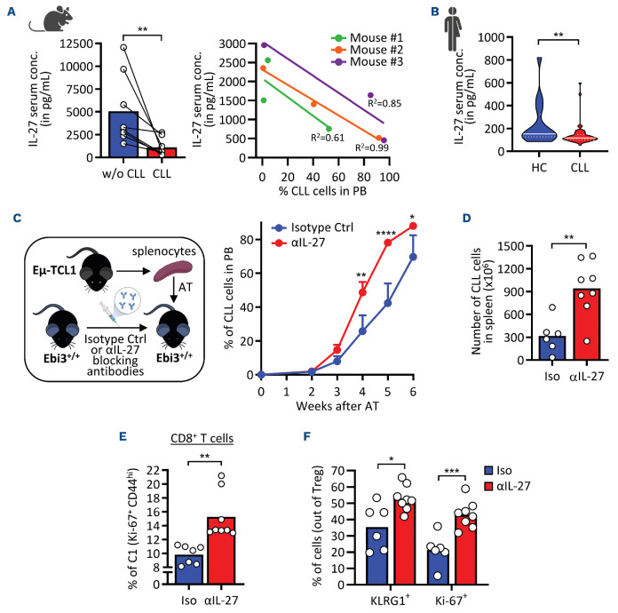
In order to better understand the role of IL-27 in CLL, we quantified IL-27 in the serum of leukemic mice and of CLL patients by ezyme-linked immunosorbant assay (Figure 6A, B). Leukemic mice presented a decreased level of IL-27 in serum during leukemia progression (Figure 6A). Similar findings were made in CLL patients, where IL-27 concentration was decreased compared to healthy control individuals (HC) (Figure 6B), in line with the anti-tumor role of IL-27 in CLL development. In addition, the analysis of several publicly available gene expression datasets revealed a lower EBI3 expression in different immune populations between CLL patients and HC (Online Supplementary Figure S7A) and between leukemic Eμ-TCL1 and WT mice (Online Supplementary Figure S7B). Since a number of studies indicate that IL-27 exerts a direct inhibitory effect on malignant cells,29 we assessed whether IL-27 can also have a direct anti-tumor effect on CLL cells. Thus, we treated both patient and mouse CLL samples ex vivo for 48 h with different concentrations of human and murine IL-27 respectively (25 and 50 ng/mL) (Online Supplementary Figure S7C, F). Assessment of viability and expression of key immunosuppressive markers indicated that neither the apoptosis rate nor the expression of markers were significantly altered, suggesting that Il-27 treatment does not impact the viability and the phenotype of murine and human CLL cells.
In order to support our results on Ebi3 genetic deletion and confirm the implication of IL-27 in CLL progression, we depleted IL-27 in vivo in C57BL/6 mice before injecting TCL1 cells (Figure 6C). Monitoring leukemic growth in the PB revealed an increase in CLL in mice depleted for IL-27, consistent with a higher number of CLL cells in the spleen following euthanasia (Figure 6D). Despite not observing differences in the frequency of CD8+ T cells between groups (Online Supplementary Figure S8A), conventional and clustering analyses confirmed the enrichment of proliferative and activated CD8+ T cells (Ki-67+ CD44hi) (Figure 6E; Online Supplementary Figure S8D-E), similarly to the adoptive transfer in Ebi3-/- mice (Figure 1D). Moreover, we observed a significant decrease in the percentage of CD4+ T cells (Online Supplementary Figure S8E), even though the frequencies of Tconv and Tregs within the CD4+ T-cell compartment were unaltered between the two groups (Online Supplementary Figure S8F). Analysis of CD4+ T cells confirmed the increased immunosuppressive and activated phenotype of Tregs in the absence of IL-27 in recipient mice (C2) (Online Supplementary Figure S8G, H). Importantly, Tregs appeared more suppressive and proliferative in IL-27-depleted mice, as demonstrated by the increased percentage of KLRG1 and Ki 67-expressing Tregs (Figure 6F).
Discussion
Given the pleiotropic function of IL-27 in the tumorigenesis of multiple solid and hematologic malignancies, we investigated the role of this cytokine in the development and progression of CLL. Using a wide range of mouse models and patient samples, we demonstrated that IL-27 has an anti-tumoral role in CLL pathobiology. Moreover, we explored the microenvironment, cellular, and transcriptional mediators responsible for this observation.
A number of studies have previously assessed the role of IL-27 in CLL using a variety of cell lines and clinical samples.30-32 Nevertheless, the existing research remains contradictory and inconclusive. Here, using adoptive transfer of TCL1 leukemia cells in C57BL/6 as well as transgenic mouse models, we demonstrate for the first time that the lack of IL-27 results in a much faster CLL development and earlier death. Additionally, CD8+ T cells are known to be dysfunctional in CLL.33,34 Phenotypic characterization of the splenic immune subsets revealed an increasingly immunosuppressive TME in the absence of IL-27, suggesting that this cytokine has a role in modulating the anti-tumor response in CLL.
In line with the varying effects of IL-27 reported in the lit erature, the serum levels of this cytokine have been found to be either significantly elevated or decreased in a wide range of malignancies,19,35,36 contributing to disease progression or control respectively. In consonance with our previous findings, our data revealed a significant and consistent decrease of IL-27 serum levels as CLL progresses in both murine and patient samples. This observation highlights both the involvement of this cytokine in CLL development as well as further supports its antitumor role in this malignancy.
Figure 5.
Ebi3-/- CD8+ T cells present impacted cytotoxic activity while IL-27 enhances their potential both in murine and human. (A) Control (Ctrl) and Ebi3-/- mice (N=3 per group) were euthanized at 8 weeks and CD3+ T cells were isolated from splenocytes. T cells were activated in vitro for 6 days and analyzed after 3 days by flow cytometry (FC) and after 6 days by FC and confocal microscopy. (B) Mean fluorescence intensity (MFI) of TOX after 3 days of activation and frequency of cells expressing the selected markers after 6 days of activation in Ctrl vs. Ebi3-/- CD8+ T cells. (C) Percentages of CD8+ T cells in clusters of wild-type (WT) vs. Ebi3-/- CD8+ T cells. (D) Detection and quantification of Profilin1 (red) between Ctrl vs. Ebi3-/- activated CD8+ T cells (green) after 6 days of activation (scale bar, 5μm). (E) In vitro cytotoxic assay. CD3+ T cells were isolated from Ctrl mice (N=6) and incubated with or without interleukin-27 (IL-27) for 48 hours (h) (left). TCL1-derived chronic lymphocytic leukemia (CLL) cells loaded with the super-antigen (sAg) were co-cultured with T cells for 4 h and stained to investigate cytotoxicity efficiency by FC (right). (F) Patient-derived CLL cells cytotoxicity assay (left panel). Peripheral blood mononucelar cells (PBMC) were isolated from CLL patient PB. One fraction was enriched in CD3+ T cells, and these cells were then activated in the presence or absence of IL-27. Antigen-pulsed CLL cells were co-cultured for 4 h with previously activated autologous CD3+ T cells. The cytotoxicity efficiency was measured by FC (right panel). Unpaired t test, *P<0.05, **P<0.01, ****P<0.0001.
Even though the EBI3 subunit is shared by IL-35, EBI3-deficient C57BL/6 mice are often used to investigate the role of IL-27 as they reportedly result in a dominant IL-27-deficient phenotype due to the higher expression of IL-27 and the wider range of cells producing the cytokine. This is evidenced by the presence of functional Tregs expressing high levels of IL-10.11 Nevertheless, the interpretation of data obtained from knock-out mouse models of cytokine subunits or their receptors is often complicated due to the promiscuous usage of chains among the different members of each family. In order to overcome this source of variability, we further validated the role of IL-27 by neutralizing this cytokine in an in vivo leukemia model.
Previous studies reported a direct anti-tumor role of IL-27 in several tumors, including hematological malignancies such as pediatric AML and CLL.30,37 Nonetheless, our data indicated that there are no significant changes in the viability or phenotype of murine and patient CLL cells, both in vivo and following in vitro treatment with IL-27. Alternatively, we provided evidence that T cells are the main mediators in the observed enhanced CLL development in the absence of IL-27, as CD3+ T cells controlled CLL development more efficiently in the presence of this cytokine.
The T-cell compartment has a critical function in antitumor immunity, and has been widely reported to show a significant functional impairment in CLL.38 Using gene expression analysis, we identified transcriptional changes in CD8+ T cells in the presence or absence of IL-27. Interestingly, we detected a decreased expression of transporters key in T-cell activation, proliferation, and cytotoxicity, most notably SLC7a5, which could potentially contribute to the altered T-cell activity described in the IL-27-depleted environment. Moreover, the expression of profilin-1 was dramatically increased in this setting. This actin binding protein plays a pivotal role in cytoskeleton remodeling, and has been shown to severely hinder CTL-mediated cell killing by disrupting the formation of the T-cell cytolytic synapse.38 Altogether, these results suggest that the progressively decreasing levels of IL-27 in CLL patients leads to dysfunctional T-cell dynamics through different mechanisms, including the downregulation of important metabolic pathways and the inability to mount an appropriate immune response due to the lack of a functional lytic immune synapse. Further research must investigate the mechanisms behind the suggested mediators of T-cell dysfunction in the absence of IL-27.
A number of studies suggest that the immune enhancing properties of IL-27 lead to a potent anti-tumor response through the upregulation of Th1 and CTL responses.39,40 Here, we show that IL-27 enhances the cytotoxicity of CD8+ T cells in mice and in humans. These observations are consistent with previous reports in the literature suggesting that IL-27 acts on CD8+ T cells through the activation of STAT1 and induces the T-bet and EOMES transcription factors, which are in turn critical for induction of effector molecules, and hence, the anti-tumor immune response.15,41,42 Of particular relevance, we demonstrate that IL-27 is able to partially restore the anti-tumor ability of CLL patient-derived T cells against autologous leukemic cells, emphasizing the potential clinical prospects of this approach in the treatment of CLL.
Here we identified IL-27 as a relevant mediator in CLL pathogenesis, highlighting its correlation with disease progression and suggesting a potential role as disease biomarker. Collectively, our data unveiled a novel mechanism by which IL-27 promotes anti-tumor immunity in CLL by enhancing CD8+ T-cell cytotoxicity, and established this cytokine as a promising immunotherapeutic agent in CLL. Further research should assess its efficacy in a pre-clinical setting in combination with other existing treatments, such as chemotherapy and immunotherapy agents, paving the path towards an affordable and effective personalized medicine regimen for the treatment of CLL.
Figure 6.
IL-27 level is reduced in the blood of leukemic mice and chronic lymphocytic leukemia patients and IL-27 neutralization enhanced chronic lymphocytic leukemia development as observed in Ebi3-/- mice. (A) (Left) Serum levels of interleukin 27 (IL-27) in mice before and after chronic lymphocytic leukemia (CLL) development. (Right) Correlation between IL-27 serum levels and CLL development in mice measured by enzyme-linked immosorbant assay (ELISA). (B) Serum levels of IL-27 in CLL patients (N=53) and healthy controls (N=16) measured by ELISA. (C-F) (Right) A cohort of recipient Ctrl mice was intraperitoneally (i.p.) injected with α-IL-27 or isotype control antibodies and subsequently adoptively transferred with splenocytes from leukemic Eμ-TCL1 mice. Mice were bled weekly to evaluate peripheral disease development. The injection of antibodies was maintained once per week until euthanasia. (C) (Left) Percentages of circulating leukemic CD5+ CD19+ cells in the 2 groups (N=7 for isotype group and N=8 for α-IL-27 group, two-way ANOVA test. (D-F) Mice were euthanized and their splenocytes were analyzed by flow cytometry. (D) Number of CLL cells in the spleen of both experimental conditions. (E) Distribution of cells in cluster C1 among CD8+ T cells from isotype- and α-IL-27-treated mice. (F) Percentage of KLRG1+ or Ki67+ Treg from isotype- and α-IL-27-treated mice. Unpaired t test, *P<0.05, **P<0.01, ****P<0.0001.
Supplementary Material
Acknowledgments
We thank Pr. Carlo Croce and Pr. John Byrd (Ohio State University, US) for the kind gift of Eµ-TCL1 mouse. We thank the National Cytometry Platform (LIH; Dr Antonio Cosma, Dr Céline Hoffmann, Thomas Cerutti, Fanny Hedin) for assistance in flow cytometry and confocal microscopy, and all the Animal Facility (LIH) staff, particularly Anais Oudin and Coralie Pulido. We thank Pr. Markus Hoth for inspiring discussion and continuous support, as well as for NALM6-pCasper cells (with Eva C. Schwarz). We thank the LUXGEN platform LIH/LNS; Nathalie Nicot, Pol Hoffmann, Arnaud Muller and Dr Daniel Stieber) for RNA sequencing. We thank Dr Guy Berchem, Dr Jean-Hugues François, Dr Susan Cortez Clemente, Dr Vincent Schlesser, Dr Sigrid De Wilde and Dr Laurent Plawny from the Centre Hospitalier du Luxembourg for their help in sample collection.
Funding Statement
Funding: This work was supported by grants from FNRS-Télévie to GP (7.4501.18, 7.6518.20), IFB (7.4529.19, 7.6603.21), MW (7.4508.16, 7.6504.18), SG (7.4502.19, 7.6604.21), CB (7.4577.22), and AL (7.4502.17, 7.4503.19), and from the Luxembourg National Research Fund (FNR) and Fondation Cancer to EG, EM and JP (PRIDE15/10675146/CANBIO, C20/BM/14582635, and C20/BM/14592342).
References
- 1.Swerdlow SH, Campo E, Pileri SA, et al. The 2016 revision of the World Health Organization classification of lymphoid neoplasms. Blood. 2016;127(20):2375-2390. [DOI] [PMC free article] [PubMed] [Google Scholar]
- 2.Cree IA. The WHO classification of haematolymphoid tumours. Leukemia. 2022;36(7):1701-1702. [DOI] [PMC free article] [PubMed] [Google Scholar]
- 3.Burger JA. Treatment of chronic lymphocytic leukemia. N Engl J Med. 2020;383(5):460-473. [DOI] [PubMed] [Google Scholar]
- 4.Svanberg R, Janum S, Patten PEM, Ramsay AG, Niemann CU. Targeting the tumor microenvironment in chronic lymphocytic leukemia. Haematologica. 2021;106(9):2312-2324. [DOI] [PMC free article] [PubMed] [Google Scholar]
- 5.Largeot A, Klapp V, Viry E, et al. Inhibition of MYC translation through targeting of the newly identified PHB-eIF4F complex as therapeutic strategy in CLL. Blood. 2023;141(26):3166-3183. [DOI] [PubMed] [Google Scholar]
- 6.Collins RJ, Verschuer LA, Harmon BV, Prentice RL, Pope JH, Kerr JF. Spontaneous programmed death (apoptosis) of B-chronic lymphocytic leukaemia cells following their culture in vitro. Br J Haematol. 1989;71(3):343-350. [DOI] [PubMed] [Google Scholar]
- 7.Burger JA, Tsukada N, Burger M, Zvaifler NJ, Dell'Aquila M, Kipps TJ. Blood-derived nurse-like cells protect chronic lymphocytic leukemia B cells from spontaneous apoptosis through stromal cell-derived factor-1. Blood. 2000;96(8):2655-2663. [PubMed] [Google Scholar]
- 8.Asslaber D, Grössinger EM, Girbl T, et al. Mimicking the microenvironment in chronic lymphocytic leukaemia - where does the journey go? Br J Haematol. 2013;160(5):711-714. [DOI] [PubMed] [Google Scholar]
- 9.Yoshida H, Hunter CA. The immunobiology of interleukin-27. Annu Rev Immunol. 2015;33.417-443. [DOI] [PubMed] [Google Scholar]
- 10.Molle C, Nguyen M, Flamand V, et al. IL-27 Synthesis induced by TLR ligation critically depends on IFN regulatory factor 3. J Immunol. 2007;178(12):7607-7615. [DOI] [PubMed] [Google Scholar]
- 11.Fabbi M, Carbotti G, Ferrini S. Dual roles of IL-27 in cancer biology and immunotherapy. Mediators Inflamm. 2017;2017:3958069. [DOI] [PMC free article] [PubMed] [Google Scholar]
- 12.Kourko O, Seaver K, Odoardi N, Basta S, Gee K. IL-27, IL-30, and IL-35: a cytokine triumvirate in cancer. Front Oncol. 2019;9:969. [DOI] [PMC free article] [PubMed] [Google Scholar]
- 13.Hisada M, Kamiya S, Fujita K, et al. Potent antitumor activity of interleukin-27. Cancer Res. 2004;64(3):1152-1156. [DOI] [PubMed] [Google Scholar]
- 14.Liu Z, Yu J, Carson WE, 3rd, Bai XF. The role of IL-27 in the induction of anti-tumor cytotoxic T lymphocyte response. Am J Transl Res. 2013;5(5):470-480. [PMC free article] [PubMed] [Google Scholar]
- 15.Morishima N, Owaki T, Asakawa M, Kamiya S, Mizuguchi J, Yoshimoto T. Augmentation of effector CD8+ T cell generation with enhanced granzyme B expression by IL-27. J Immunol. 2005;175(3):1686-1693. [DOI] [PubMed] [Google Scholar]
- 16.Salcedo R, Stauffer JK, Lincoln E, et al. IL-27 mediates complete regression of orthotopic primary and metastatic murine neuroblastoma tumors: role for CD8+ T cells. J Immunol. 2004;173(12):7170-7182. [DOI] [PubMed] [Google Scholar]
- 17.Matsui M, Kishida T, Nakano H, et al. Interleukin-27 activates natural killer cells and suppresses NK-resistant head and neck squamous cell carcinoma through inducing antibody-dependent cellular cytotoxicity. Cancer Res. 2009;69(6):2523-2530. [DOI] [PubMed] [Google Scholar]
- 18.Yao L, Wang M, Niu Z, et al. Interleukin-27 inhibits malignant behaviors of pancreatic cancer cells by targeting M2 polarized tumor associated macrophages. Cytokine. 2017;89:194-200. [DOI] [PubMed] [Google Scholar]
- 19.Diakowska D, Lewandowski A, Markocka-Mączka K, Grabowski K. Concentration of serum interleukin-27 increase in patients with lymph node metastatic gastroesophageal cancer. Adv Clin Exp Med. 2013;22(5):683-691. [PubMed] [Google Scholar]
- 20.Gonin J, Carlotti A, Dietrich C, et al. Expression of IL-27 by tumor cells in invasive cutaneous and metastatic melanomas [corrected]. PLoS One. 2013;8(10):e75694. [DOI] [PMC free article] [PubMed] [Google Scholar]
- 21.Ahmed HA, Maklad AM, Khaled SA, Elyamany A. Interleukin-27 and interleukin-35 in de novo acute myeloid leukemia: expression and significance as biological markers. J Blood Med. 2019;10:341-349. [DOI] [PMC free article] [PubMed] [Google Scholar]
- 22.Carbotti G, Barisione G, Airoldi I, et al. IL-27 induces the expression of IDO and PD-L1 in human cancer cells. Oncotarget. 2015;6(41):43267-43280. [DOI] [PMC free article] [PubMed] [Google Scholar]
- 23.Chihara N, Madi A, Kondo T, et al. Induction and transcriptional regulation of the co-inhibitory gene module in T cells. Nature. 2018;558(7710):454-459. [DOI] [PMC free article] [PubMed] [Google Scholar]
- 24.Sawant DV, Yano H, Chikina M, et al. Adaptive plasticity of IL-10(+) and IL-35(+) Treg cells cooperatively promotes tumor T cell exhaustion. Nat Immunol. 2019;20(6):724-735. [DOI] [PMC free article] [PubMed] [Google Scholar]
- 25.Palma M, Gentilcore G, Heimersson K, et al. T cells in chronic lymphocytic leukemia display dysregulated expression of immune checkpoints and activation markers. Haematologica. 2017;102(3):562-572. [DOI] [PMC free article] [PubMed] [Google Scholar]
- 26.Schoppmeyer R, Zhao R, Cheng H, et al. Human profilin 1 is a negative regulator of CTL mediated cell-killing and migration. Eur J Immunol. 2017;47(9):1562-1572. [DOI] [PubMed] [Google Scholar]
- 27.Ren W, Liu G, Yin J, et al. Amino-acid transporters in T-cell activation and differentiation. Cell Death Dis. 2017;8(3):e2655. [DOI] [PMC free article] [PubMed] [Google Scholar]
- 28.Beltra JC, Manne S, Abdel-Hakeem MS, et al. Developmental relationships of four exhausted CD8(+) T cell subsets reveals underlying transcriptional and epigenetic landscape control mechanisms. Immunity. 2020;52(5):825-841. [DOI] [PMC free article] [PubMed] [Google Scholar]
- 29.Kachroo P, Lee MH, Zhang L, et al. IL-27 inhibits epithelial-mesenchymal transition and angiogenic factor production in a STAT1-dominant pathway in human non-small cell lung cancer. J Exp Clin Cancer Res. 2013;32(1):97. [DOI] [PMC free article] [PubMed] [Google Scholar]
- 30.Manouchehri-Doulabi E, Abbaspour S, Rostami S, et al. Evaluating the mechanism underlying antitumor effect of interleukin 27 on B cells of chronic lymphocytic leukemia patients. J Cell Physiol. 2020;235(12):9424-9431. [DOI] [PubMed] [Google Scholar]
- 31.Hemati M, Rasouli Nejad Z, Shokri MR, Ghahremanfard F, Mir Mohammadkhani M, Kokhaei P. IL-27 impact on NK cells activity: Implication for a robust anti-tumor response in chronic lymphocytic leukemia. Int Immunopharmacol. 2020;82:106350. [DOI] [PubMed] [Google Scholar]
- 32.Jia H, Dilger P, Bird C, Wadhwa M. IL-27 promotes proliferation of human leukemic cell lines through the MAPK/ERK signaling pathway and suppresses sensitivity to chemotherapeutic drugs. J Interferon Cytokine Res. 2016;36(5):302-316. [DOI] [PMC free article] [PubMed] [Google Scholar]
- 33.Gargiulo E, Viry E, Morande PE, et al. Extracellular vesicle secretion by leukemia cells in vivo promotes CLL progression by hampering antitumor T-cell responses. Blood Cancer Discov. 2023;4(1):54-77. [DOI] [PMC free article] [PubMed] [Google Scholar]
- 34.Wierz M, Pierson S, Guyonnet L, et al. Dual PD1/LAG3 immune checkpoint blockade limits tumor development in a murine model of chronic lymphocytic leukemia. Blood. 2018;131(14):1617-1621. [DOI] [PMC free article] [PubMed] [Google Scholar]
- 35.Lu D, Zhou X, Yao L, Liu C, Jin F, Wu Y. Clinical Implications of the interleukin 27 serum level in breast cancer. J InvestMed. 2014;62(3):627-631. [DOI] [PubMed] [Google Scholar]
- 36.Duan M, Ning Z, Fu Z, et al. Decreased IL-27 negatively correlated with Th17 cells in non-small-cell lung cancer patients. Mediators Inflamm. 2015;2015:802939. [DOI] [PMC free article] [PubMed] [Google Scholar]
- 37.Zorzoli A, Di Carlo E, Cocco C, et al. Interleukin-27 inhibits the growth of pediatric acute myeloid leukemia in NOD/SCID/Il2rg-/- mice. Clin Cancer Res. 2012;18(6):1630-1640. [DOI] [PubMed] [Google Scholar]
- 38.Roessner PM, Seiffert M. T-cells in chronic lymphocytic leukemia: guardians or drivers of disease? Leukemia. 2020;34(8):2012-2024. [DOI] [PMC free article] [PubMed] [Google Scholar]
- 39.Takeda A, Hamano S, Yamanaka A, et al. Cutting edge: role of IL-27/WSX-1 signaling for induction of T-bet through activation of STAT1 during initial Th1 commitment. J Immunol. 2003;170(10):4886-4890. [DOI] [PubMed] [Google Scholar]
- 40.Salcedo R, Hixon JA, Stauffer JK, et al. Immunologic and therapeutic synergy of IL-27 and IL-2: enhancement of T cell sensitization, tumor-specific CTL reactivity and complete regression of disseminated neuroblastoma metastases in the liver and bone marrow. J Immunol. 2009;182(7):4328-4338. [DOI] [PMC free article] [PubMed] [Google Scholar]
- 41.Mayer KD, Mohrs K, Reiley W, et al. Cutting Edge: T-bet and IL-27R are critical for in vivo IFN-γ production by CD8 T cells during infection. J Immunol. 2008;180(2):693-697. [DOI] [PubMed] [Google Scholar]
- 42.Schneider R, Yaneva T, Beauseigle D, El-Khoury L, Arbour N. IL-27 increases the proliferation and effector functions of human naïve CD8+ T lymphocytes and promotes their development into Tc1 cells. Eur J Immunol. 2011;41(1):47-59. [DOI] [PubMed] [Google Scholar]
- 43.Beltra JC, Manne S, Abdel-Hakeem MS, et al. Developmental relationships of four exhausted CD8(+) T cell subsets reveals underlying transcriptional and epigenetic landscape control mechanisms. Immunity. 2020;52(5):825-841.e8. [DOI] [PMC free article] [PubMed] [Google Scholar]
Associated Data
This section collects any data citations, data availability statements, or supplementary materials included in this article.


