Abstract
Spores of Bacillus thuringiensis subsp. israelensis and their toxic crystals are bioencapsulated in the protozoan Tetrahymena pyriformis, in which the toxin remains stable. Each T. pyriformis cell concentrates the spores and crystals in its food vacuoles, thus delivering them to mosquito larvae, which rapidly die. Vacuoles containing undigested material are later excreted from the cells. The fate of spores and toxin inside the food vacuoles was determined at various times after excretion by phase-contrast and electron microscopy as well as by viable-cell counting. Excreted food vacuoles gradually aggregated, and vegetative growth of B. thuringiensis subsp. israelensis was observed after 7 h as filaments that stemmed from the aggregates. The outgrown cells sporulated between 27 and 42 h. The spore multiplication values in this system are low compared to those obtained in carcasses of B. thuringiensis subsp. israelensis-killed larvae and pupae, but this bioencapsulation represents a new possible mode of B. thuringiensis subsp. israelensis recycling in nontarget organisms.
The bacterium Bacillus thuringiensis subsp. israelensis (12, 15, 27) is used worldwide to control mosquitoes and blackflies, vectors of many human infectious diseases (for example, see reference 29). Its larvicidal activity is caused by insecticidal crystal proteins (ICP) that are produced during sporulation (31). Following ingestion, the crystal dissolves in the alkaline pH prevailing in the larval midgut, releasing protoxin polypeptides which are then activated by proteolytic enzymes and act as a stomach poison (14).
Although B. thuringiensis subsp. israelensis was originally isolated from a temporary mosquito-breeding site, treatment of larval populations does not result in epizootic outbreaks of disease, in contrast to the situation with classical biocontrol agents (23). No recycling or amplification of B. thuringiensis subsp. israelensis has been observed under field conditions (4, 22), and its use as a biological control agent is therefore restricted by its limited efficacy in the field.
Despite its similarity to B. thuringiensis subsp. israelensis, the mosquitocidal activity of Bacillus sphaericus persists longer in the field (2), due to several possible factors: protection of the toxic crystal within the exosporium (7) and failure to attach to sedimenting organic particulates in water (32), reproduction of B. sphaericus within the guts of nontarget arthropods (17), and recycling (germination, vegetative growth, and sporulation) in the carcasses of toxin-killed larvae (9–11). Recycling of ingested spores in the carcasses of mosquito larvae (1, 3, 33) and pupae (19) was also demonstrated for B. thuringiensis subsp. israelensis in the laboratory, but the linkage between its larvicidal activity and proliferation does not necessarily hold in nature.
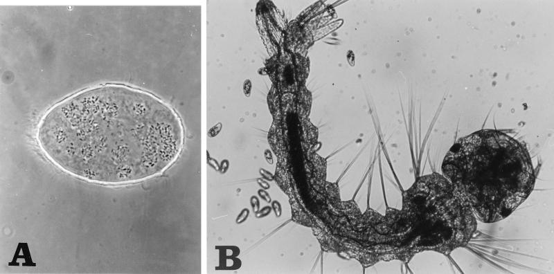
We have previously shown that toxicity of B. thuringiensis subsp. israelensis to larvae of Aedes aegypti and Anopheles stephensi was enhanced three- and eightfold, respectively, by bioencapsulation in food vacuoles of the protozoan Tetrahymena pyriformis, in which the toxins remain stable; each T. pyriformis cell concentrates in its food vacuoles between 180 and 240 spores and their associated ICP (5, 20, 21, 34) and delivers them to the target organisms (Fig. 1). Larvae of mosquitoes fed on B. thuringiensis subsp. israelensis-loaded T. pyriformis ingest large, lethal quantities of toxin and rapidly die (20, 21).
FIG. 1.
B. thuringiensis subsp. israelensis spores in food vacuoles of T. pyriformis (A) and second-instar Aedes aegypti larvae (B). Note the relative sizes of the three component organisms involved. Magnifications, ca. ×1,500 (A) and ×200 (B).
It is predicted that the interaction between ingested spores and T. pyriformis yields a natural site for recycling of B. thuringiensis subsp. israelensis. Here we show that spores indeed germinated, grew, and sporulated in excreted food vacuoles of T. pyriformis, forming new active ICP during this cycle.
MATERIALS AND METHODS
B. thuringiensis subsp. israelensis.
Isolated B. thuringiensis subsp. israelensis colonies (on Luria-Bertani [LB] plates) from a commercial powder (R-153-78, 1,000 IU mg−1 [13]; Roger Bellon Laboratories, Neuilly-sur-Seine, Belgium) were used for inoculation. Cells were grown (30°C) with shaking (260 rpm) in 20 ml of LB medium and harvested after 4 days, when sporulation and crystallization (observed by phase-contrast microscopy) were complete. The cells were washed thrice with sterile distilled water before each experiment to remove traces of nutrients.
T. pyriformis.
The protozoan was maintained and grown axenically as previously reported (20). Experiments were performed with 1 × 10⁵ to 3 × 10⁵ cells per ml of exponentially growing cultures (28°C, 60 strokes min−1). Cells were counted microscopically in a Sedgwick chamber after fixation in 1% formaldehyde. The cells were washed thrice with sterile distilled water by centrifugation (90 s at 1,500 × g) to remove carried-over nutrients.
Bioencapsulation.
Washed T. pyriformis cells (104 ml−1) were incubated (in 150-ml Erlenmeyer flasks) in a water bath (28°C, 60 strokes min−1) with washed spores (7 × 10⁶ ml−1) and crystals of B. thuringiensis subsp. israelensis in 40 ml of sterile distilled water (suspension 1). B. thuringiensis subsp. israelensis cells similarly treated without or with lysed (incubated at 35°C for 20 min) T. pyriformis cells (suspensions 2 and 3, respectively) were used as controls.
Subsequent recycling.
Recycling (in duplicates) was detected by viable-cell counting (for CFU) and by phase-contrast microscopy (for vegetative cells, spores, and crystals). Aliquots from the bioencapsulation suspensions were appropriately diluted in sterile distilled water and evenly spread on LB plates. The number of colonies was determined, as an average for a duplicate in three different dilutions, after 24 h of incubation at 37°C. For spore count, each aliquot (1 ml) was first heated (70°C for 10 min) and then sonicated with 1% Tween 80 (MSE Sonifier, 4× 30 s each with 30-s intervals, 0°C) before further dilutions. Total count (vegetative cells and spores) was determined without heat shock and sonication treatments.
Electron microscopy.
Bioencapsulated B. thuringiensis subsp. israelensis in T. pyriformis was fixed in 2% (vol/vol) glutaraldehyde in 0.1 M cacodylate buffer (pH 7.4) for 30 min. Samples were incubated with osmium tetroxide for 1 h. After a brief washing, the cells were dehydrated in a graded series of ethanol and finally in propylene oxide before being embedded in Epon. The sectioned material was stained with uranyl acetate and lead citrate. The sections were examined in a JEOL 100B electron microscope at magnifications between 9,500 and 45,000.
Bioassays.
Dry strips of paper bearing eggs of Aedes aegypti were submerged, and larvae were grown in 1 liter of sterile tap water supplemented with 1.5 g of Pharmamedia (Traders Protein, Memphis, Tenn.) at 30°C as described before (18). Larvae of the same age and size were selected and washed before every experiment. Twenty third-instar Aedes aegypti larvae, in duplicates, were incubated (28°C) in 100 ml of sterile tap water with the appropriate dilutions of suspensions (suspension 1, 2, or 3) as necessary. Larval mortality was scored after 24 h.
RESULTS
We have previously shown that under the bioencapsulation conditions specified here, each T. pyriformis cell forms up to 30 food vacuoles with an average of 8 B. thuringiensis subsp. israelensis spores per vacuole (5, 21). Vacuoles containing undigested material are excreted from T. pyriformis cells through the cytoproct at the end of their digestion process (26). Vacuoles excreted from B. thuringiensis subsp. israelensis-loaded cells should contain intact spores and crystals (34). Their fate inside the vacuoles was determined at various times after excretion by phase-contrast and electron microscopy as well as by viable-cell counting.
Phase-contrast microscopy.
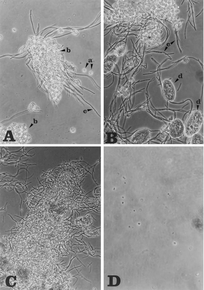
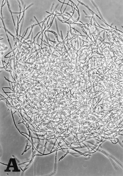
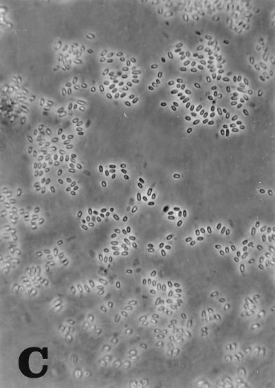
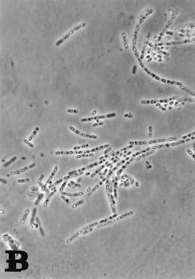
Excreted food vacuoles under our laboratory conditions joined together and were found in aggregates that gradually grew during the incubation of the bioencapsulation suspension. Few free spores were detected in the medium after 5 h. At 7 h, vegetative growth of B. thuringiensis subsp. israelensis was observed as chains of rods that stem from the aggregates (Fig. 2A), but most spores inside the excreted food vacuoles had not germinated. Vegetative growth continued: chains elongated, and new ones emerged as the result of additional spore germination (Fig. 2B and C). The outgrown cells sporulated after about 27 h (Fig. 3A), and sporulation seemed to be complete at 42 h, when most of the new B. thuringiensis subsp. israelensis cells contained spores and crystals (panel B). Sporangia lysed at 60 h, when spores and crystals were separated (Fig. 3C). No change in original spores was observed in a control water suspension (suspension 2) with the same concentration of B. thuringiensis subsp. israelensis during the same incubation period (Fig. 2D). When spores were incubated with heat-killed, lysed T. pyriformis cells (suspension 3), on the other hand, they germinated after 5 h of incubation and formed small chains of evenly suspended rods (two to four bacteria) which completed sporulation around 27 h (data not shown).
FIG. 2.
(A–C) Germination and outgrowth of B. thuringiensis subsp. israelensis spores and vegetative bacteria in excreted T. pyriformis food vacuoles (suspension 1) after 7 h (A), 15 h (B), and 20 h (C) of incubation. a, single food vacuoles; b, aggregates of excreted vacuoles; c, chains of vegetative bacteria breaking out of the aggregates; d, live T. pyriformis cells loaded with spores. Magnification, ×400. (D) Single spores in water (control suspension 2) after 20 h of incubation. Magnification, ×1,000.
FIG. 3.
Sporulation of newly formed B. thuringiensis subsp. israelensis in suspension 1 at 27 h (A), 42 h (B), and 60 h (C) of incubation. Magnifications, ×400, ×1,000, and ×1,000, respectively.
The T. pyriformis cells in the bioencapsulation system (suspension 1) were seen to run energetically, occasionally gathering around the food vacuole aggregates, seemingly trying to feed on the vegetative cells (Fig. 2B). At this time (15 h), their food vacuoles were all still loaded with B. thuringiensis subsp. israelensis spores and ICP. Around 27 h, cell size and the number of food vacuoles started to decline gradually, followed by excretion of white mucus, indicating starvation (28). At the end of the incubation period (60 h), the cells were very small, probably as a result of continued divisions without mass growth (6).
Electron microscopy observation.
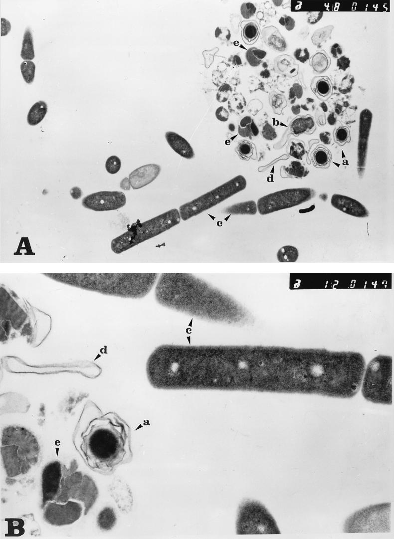
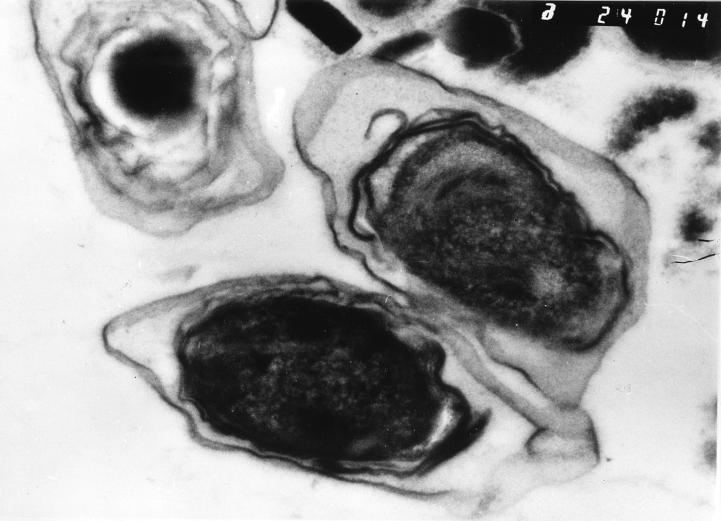
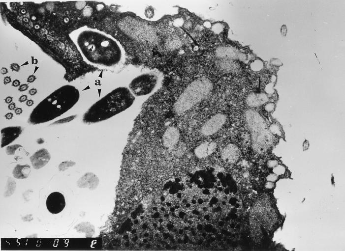
To investigate further the fate of B. thuringiensis subsp. israelensis spores and their ICP inside the excreted food vacuoles, we zoomed in on one of these vacuoles at 11 h of bioencapsulation with an electron microscope: spores and ICP were not damaged during passage through T. pyriformis. Typical intact spores of B. thuringiensis subsp. israelensis with their envelopes were clearly observed (Fig. 4). As expected from the phase-contrast micrographs (Fig. 2), some of these spores were in the process of germination (Fig. 4A and 5), and empty envelopes, probably left after the vegetative cells had emerged from them, were also detected inside the vacuole (Fig. 4). A number of vegetative cells, organized in chains, were seen emerging intact from vacuoles; ICP also retained their typical form, with three distinct inclusions of different densities (Fig. 4) (16). The numbers of crystals and spores (intact, germinating, and empty envelopes) seen inside this excreted food vacuole were 10 and 8, respectively. At this stage, T. pyriformis cells were viable and attempted to ingest chains of vegetative B. thuringiensis subsp. israelensis cells. A cross section through the oral apparatus of a cell in the process of such an attempt is shown in Fig. 6.
FIG. 4.
Electron micrographs of sections through a single excreted food vacuole loaded with spores, after 11 h of incubation. a, single spores; b, a germinating spore; c, chains of vegetative bacteria breaking out of the food vacuoles’ envelope; d, spore coat; e, intact ICP. (Note the inclusions of different densities in the crystal.) Magnifications, ×9,000 (A) and ×22,500 (B).
FIG. 5.
Electron micrograph of germinating B. thuringiensis subsp. israelensis spores inside an excreted food vacuole at 11 h of incubation. Magnification, ×45,000.
FIG. 6.
Electron micrograph of the oral region of T. pyriformis during ingestion of newly formed vegetative B. thuringiensis subsp. israelensis cells (a). Note the peripheral microtubules of kinetosomes of the oral apparatus (b). Magnification, ×11,500.
Viable-cell counting.
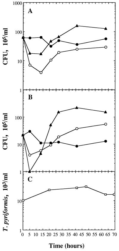
Recycling of ingested B. thuringiensis subsp. israelensis spores inside the excreted vacuoles was also demonstrated by counting the concentration of total CFU (including vegetative cells and spores), as well as spores alone during the period of incubation of the bioencapsulation suspension (suspension 1). The data presented here (Fig. 7) are for a typical representative experiment, which was followed simultaneously by electron microscopy and phase-contrast microscopy.
FIG. 7.
Recycling of B. thuringiensis subsp. israelensis spores. (A and B) Total cell count (spores, germinated spores, and vegetative cells) (A) and spore counts (B) in the bioencapsulation system (suspension 1) (○), in water (suspension 2) (•), and in lysed, heat-killed T. pyriformis cells (suspension 3) (▴). (C) Concentrations of T. pyriformis cells in suspension 1.
Total CFU decreased by approximately an order of magnitude within the first 5 h (Fig. 7A). The low CFU value was maintained for at least 15 h; later, it increased and reached a maximum of 3 × 10⁶ ml−1 at 27 h. This final CFU level was about one-half that at the beginning of the experiment.
When B. thuringiensis subsp. israelensis-T. pyriformis suspension was scored for spores, after treatment with detergent, sonication, and heat shock, the initial CFU concentration dropped from 6 × 10⁶ ml−1 to 2 × 10⁶ ml−1 (Fig. 7B). This initial low value further decreased by almost an order of magnitude after 5 h of incubation. At 15 h, spore count started to increase, and it reached a level threefold higher than the initial value at the end of the experiment.
Practically no changes in either total CFU or spore count were observed in the B. thuringiensis subsp. israelensis control suspension (suspension 2) during the experiment (Fig. 7A and B).
Fast germination of B. thuringiensis subsp. israelensis spores in the heat-killed lysate of T. pyriformis (suspension 3) was demonstrated as a sharp drop in spore count during the first 5 h of incubation (Fig. 7B). Resporulation was initiated after 15 h, and a maximal spore concentration of 2 × 10⁷ ml−1 was reached at 27 h. Here, there was no difference in the maximal CFU between the total and the spore counts.
T. pyriformis.
Live T. pyriformis cells were detected by phase-contrast microscopy and counting during the entire experiment. Their number increased 2.5-fold during the first 25 h of incubation. The high concentration remained constant until 47 h (Fig. 7C). No apparent change in cell volume was observed during the first 25 h (microscopical observations not shown). A small decline in T. pyriformis concentration was detected at 65 h, when cell volume decreased markedly and some of the cells started to die, as noted by paralysis and lysis.
Some of our relevant microscopical observations are displayed in color on a Web Page, at http://www.bgu.ac.il/life/zaritsky.html.
DISCUSSION
Lack of reproduction of B. thuringiensis subsp. israelensis in natural mosquito breeding sites after application is its major disadvantage as a biological control agent (4, 22–24). Originally isolated from a temporary pond with Culex pipiens larvae (15), it seems able to reproduce and survive under natural conditions, but the actual reproduction niche is still a mystery. Previous results demonstrated that B. thuringiensis subsp. israelensis spores and crystals maintained their viability and larvicidal activity, respectively, when bioencapsulated in T. pyriformis (5, 20, 21). The discovery that the spores can germinate and multiply in excreted food vacuoles of the protozoan, which shares the mosquito larval habitat, is of special importance; this complements the already-known recycling processes in carcasses of mosquito larvae (1, 3, 33) and pupae (19). The recycling mode described here was demonstrated in the same experiment qualitatively by microscopic observations (Fig. 2–5) and quantitatively by viable-cell counting (Fig. 7).
An exponentially growing T. pyriformis cell can form up to 30 food vacuoles, with an average of 3 every 10 min of growth at 28°C (8, 25). Each cell concentrates between 180 and 240 B. thuringiensis subsp. israelensis spores and their associated ICP (5, 21) and is fully loaded at spore/T. pyriformis ratios above 240:1. The high ratio used in this study (700:1) guarantees that all potential food vacuoles were formed during the first 90 min of incubation and were full of spores and crystals. The loaded vacuoles are excreted through the cytoproct at the end of the digestion process. The cells require at least an additional 60 min to recover their potential for a new cycle of vacuole formation (25). This analysis explains why free spores could hardly be detected in the medium after 5 h of incubation of the bioencapsulation system (data not shown).
The excreted food vacuoles can be visualized as small capsules each containing between six and eight spores and their associated crystals (Fig. 2A and 4). As the spores germinated and emerged, live T. pyriformis cells ingested them (Fig. 2B and 6), but with minor success because their oral apparatus is unable to ingest long filaments, formed by outgrowth. This caused starvation of the cells, which resulted in excretion of white mucus that contributed to the aggregation process (28).
Germination of B. thuringiensis subsp. israelensis spores on T. pyriformis lysate (suspension 3) started earlier and was dispersed in the medium, in contrast to the focal germination in the excreted vacuole aggregates of suspension 1. This difference derives from the germination conditions: the lysate provides the spores with a rich homogeneous nutrient source, while the spores in suspension are locked up in the vacuoles and can use their undigested content only. This difference explains the differences in germination and growth characteristics of B. thuringiensis subsp. israelensis organisms in the two suspensions, as can be seen from viable-cell counts as well (Fig. 7). As expected, the spores in water without T. pyriformis (live or dead) did not germinate during the whole experiment because they had no nutrient source (Fig. 2D).
In the bioencapsulation suspension (suspension 1), total CFU declined 20-fold during the first 15 h of incubation (Fig. 7A), after which it started to rise but did not reach the original bacterial concentration (6 × 10⁶ ml−1). The decline stems from the large number of spore clusters formed by immediate encapsulation of the spores in the food vacuoles and following aggregation after excretion (Fig. 2). Detection of newly formed B. thuringiensis subsp. israelensis in suspension 1 is thus masked by the aggregates, now in addition containing filamentous vegetative cells: a single CFU on an LB plate is formed by all cells that compose an aggregate, and the number of CFU (Fig. 7A) may represent the total number of aggregates. The moderate decline in the extract of lysed T. pyriformis (Fig. 7A, suspension 3) may have resulted from small aggregates of vegetative cells sporadically found in the suspension (data not shown).
To estimate the true concentration of spores, samples were counted after heat shock and sonication. This treatment dispersed the aggregates and destroyed vegetative B. thuringiensis subsp. israelensis cells. The initial concentration at the beginning of the experiment was 2 × 10⁶ ml−1 (Fig. 7B), about threefold lower than the total cell count (Fig. 7A). The treatment itself thus damaged a high proportion of the spores.
The decrease in the spore count observed (Fig. 7B) in suspension 3 (lysed T. pyriformis cells) during the first 5 h was sharper than in suspension 1 because a high proportion of the spores in it were sensitized to the treatment by germination (as was also seen by phase-contrast microscopy). Suspension 3 supported sporulation of 1.7 × 10⁷ cells ml−1, while the final concentration in suspension 1 after 65 h was lower (6 × 10⁶ ml−1) because the original spores multiplied in the content of excreted food vacuoles with limited nutritional value. In addition, newly formed spores are more sensitive to heat and sonication treatments than fully mature spores (4 days old) used in the bioencapsulation experiments (unpublished results). Another factor that might have influenced the final yield is digestion of newly formed vegetative bacteria by T. pyriformis (Fig. 6). The higher final yield of spores relative to the initial bacterial concentration in suspension 1 confirms that better estimates of spore concentration can be achieved by performing the treatment before counting, since treatment disperses the aggregates. As expected, the protozoan lysate allowed faster and higher-level germination because it supplied enough nutrients. In contrast, suspension 2 (the second control system with distilled water) retained the same concentration of spores during the whole experimental period. The actual amount of vegetative growth in excreted food vacuoles is unclear. It is likely either that most of the bacteria multiplied a small number of times before resporulation through a microcycle sporulation cycle (30) due to limiting nutrient concentrations, or that only a small fraction germinated and formed long filaments.
The final B. thuringiensis subsp. israelensis spore counts were only 3- and 10-fold higher than the starting concentrations in suspensions 1 and 3, respectively. These multiplication values are low compared to that obtained in carcasses of B. thuringiensis subsp. israelensis-killed larvae and pupae (1, 3, 19, 33). The multiplication of T. pyriformis cells themselves (Fig. 7C) during the first 20 h confirms the microscopic observation (Fig. 2B) that they remain viable despite starvation. During this period, average cell volume decreased by continued divisions, as has previously been shown under such conditions (6). Germination of spores in suspension 1 that started after 7 h of incubation was supported by the content of excreted food vacuoles and was not supported on T. pyriformis lysates, because all the cells seemed alive (Fig. 2B and 7C).
Bioassays of the bioencapsulation suspension (suspension 1; data not shown) demonstrated toxicity against third-instar Aedes aegypti larvae of the newly made crystals, but the direct proof that B. thuringiensis subsp. israelensis spores and crystals were not damaged by the digestion processes in T. pyriformis vacuoles was obtained by electron micrographs of a single excreted food vacuole after 11 h of incubation (Fig. 4). A parasporal body can clearly be seen in its usual shape (16), with three different densities of protein inclusions bound together by a laminated netlike envelope (Fig. 4B). The spores are also surrounded by typical coat layers (Fig. 5). The electron micrographs support the conclusion from phase-contrast micrographs and viable-cell counts that spores germinate inside excreted food vacuoles.
The presence of newly formed vegetative B. thuringiensis subsp. israelensis cells in the oral apparatus of T. pyriformis cells (Fig. 6) supports preliminary observations by phase-contrast microscopy that they serve as a food source for the protozoan: as the filaments elongated, they were less and less available for ingestion. Cells were occasionally observed moving with long chains stuck in their oral apparati (data not shown).
This study describes a new possible mode of B. thuringiensis subsp. israelensis recycling in nature. It demonstrates that at least under laboratory conditions, the bacteria can recycle in T. pyriformis food vacuoles. Recycling is thus not restricted to carcasses of its target organisms: B. thuringiensis subsp. israelensis can multiply in nontarget organisms as well.
ACKNOWLEDGMENTS
Thanks are due to Michael Friedlander and Rina Yeger for help in electron microscopy, to Joel Margalit for free supply of Aedes aegypti eggs, to Valery Zalkinder and Monica Einav for helpful remarks and advice, and to Gideon Raziel for photography.
This work was partially supported by the Israel Ministry of Environment (grant 802-8, to A.Z.).
REFERENCES
- 1.Aly C, Mulla M S, Federici B A. Sporulation and toxin production by Bacillus thuringiensis var. israelensis in cadavers of mosquito larvae (Diptera: Culicidae) J Invertebr Pathol. 1985;46:251–258. [Google Scholar]
- 2.Aly C, Mulla M S, Federici B A. Ingestion, dissolution and proteolysis of the Bacillus sphaericus toxin by mosquito larvae. J Invertebr Pathol. 1989;53:15–20. doi: 10.1016/0022-2011(89)90068-2. [DOI] [PubMed] [Google Scholar]
- 3.Barak Z, Ohana B, Allon Y, Margalit J. A mutant of Bacillus thuringiensis var. israelensis (BTI) resistant to antibiotics. Appl Microbiol Biotechnol. 1987;27:88–93. [Google Scholar]
- 4.Becker N, Zgomba M, Ludwig M, Petric D, Rettich F. Factors influencing the efficacy of the microbial control agent Bacillus thuringiensis israelensis. J Am Mosq Control Assoc. 1992;8:285–289. [PubMed] [Google Scholar]
- 5.Ben-Dov E, Zalkinder V, Shagan T, Barak Z, Zaritsky A. Spores of Bacillus thuringiensis var. israelensis as tracers for ingestion rates by Tetrahymena pyriformis. J Invertebr Pathol. 1994;63:220–222. doi: 10.1006/jipa.1994.1042. [DOI] [PubMed] [Google Scholar]
- 6.Cameron I L. Growth characteristics of Tetrahymena. In: Elliot A M, editor. Biology of Tetrahymena. Stroudsburg, Pa: Dowden, Hutchinson and Ross, Inc.; 1973. pp. 217–224. [Google Scholar]
- 7.Chang M S, Ho B C, Chan K L. Simulated field studies with three formulations of Bacillus thuringiensis var. israelensis and Bacillus sphaericus against larvae of Mansonia bonneae (Diptera: Culicidae) in Sarawak, Malaysia. Bull Entomol Res. 1990;80:195–202. [Google Scholar]
- 8.Chapman-Andresen C, Nilsson J R. On vacuole formation in Tetrahymena pyriformis GL. C R Trav Lab Carlsberg. 1968;36:405–432. [PubMed] [Google Scholar]
- 9.Charles J-F, Nicolas L. Recycling of Bacillus sphaericus 2362 in mosquito larvae: a laboratory study. Ann Inst Pasteur/Microbiol (Paris) 1986;137B:101–111. doi: 10.1016/s0769-2609(86)80097-7. [DOI] [PubMed] [Google Scholar]
- 10.Correa M, Yousten A A. Bacillus sphaericus spore germination and recycling in mosquito larval cadavers. J Invertebr Pathol. 1995;66:76–81. [Google Scholar]
- 11.Davidson E, Urbina M, Payne J, Mulla M, Darwazeh H, Dulmage H, Correa J. Fate of Bacillus sphaericus 1593 and 2362 spores used as larvicides in the aquatic environment. Appl Environ Microbiol. 1984;47:125–129. doi: 10.1128/aem.47.1.125-129.1984. [DOI] [PMC free article] [PubMed] [Google Scholar]
- 12.de Barjac H. A new subspecies of Bacillus thuringiensis very toxic for mosquitoes: Bacillus thuringiensis var. israelensis serotype 14. C R Acad Sci. 1978;286D:797–800. [PubMed] [Google Scholar]
- 13.Dulmage H T, Correa J A, Gallegos-Morales G. Potential for improved formulations of Bacillus thuringiensis israelensis through standardization and fermentation development. In: de Barjac H, Sutherland D J, editors. Bacterial control of mosquitoes & black flies. New Brunswick, N.J: Rutgers University Press; 1990. pp. 116–117. [Google Scholar]
- 14.Gill S S, Cowles E A, Pietrantonio P V. The mode of action of Bacillus thuringiensis endotoxins. Annu Rev Entomol. 1992;37:615–636. doi: 10.1146/annurev.en.37.010192.003151. [DOI] [PubMed] [Google Scholar]
- 15.Goldberg L J, Margalit J. A bacterial spore demonstrating rapid larvicidal activity against Anopheles sergentii, Uranotaenia unguiculata, Culex univitattus, Aedes aegypti and Culex pipiens. Mosq News. 1977;37:355–358. [Google Scholar]
- 16.Ibarra J E, Federici B A. Isolation of a relatively nontoxic 65-kilodalton protein inclusion from the parasporal body of Bacillus thuringiensis subsp. israelensis. J Bacteriol. 1986;165:527–533. doi: 10.1128/jb.165.2.527-533.1986. [DOI] [PMC free article] [PubMed] [Google Scholar]
- 17.Karch S, Monteny N, Jullien J, Sinegre G, Coz J. Control of Culex pipiens by Bacillus sphaericus and role of nontarget arthropods in its recycling. J Am Mosq Control Assoc. 1990;6:47–54. [PubMed] [Google Scholar]
- 18.Khawaled K, Barak Z, Zaritsky A. Feeding behavior of Aedes aegypti larvae and toxicity of dispersed and of naturally encapsulated Bacillus thuringiensis var. israelensis. J Invertebr Pathol. 1988;52:419–426. doi: 10.1016/0022-2011(88)90054-7. [DOI] [PubMed] [Google Scholar]
- 19.Khawaled K, Ben-Dov E, Zaritsky A, Barak Z. The fate of Bacillus thuringiensis var. israelensis in B. thuringiensis var. israelensis-killed pupae. J Invertebr Pathol. 1990;56:312–316. doi: 10.1016/0022-2011(90)90117-o. [DOI] [PubMed] [Google Scholar]
- 20.Manasherob R, Ben-Dov E, Zaritsky A, Barak Z. Protozoan-enhanced toxicity of Bacillus thuringiensis var. israelensis δ-endotoxin against Aedes aegypti larvae. J Invertebr Pathol. 1994;63:244–248. doi: 10.1006/jipa.1994.1047. [DOI] [PubMed] [Google Scholar]
- 21.Manasherob R, Ben-Dov E, Margalit J, Zaritsky A, Barak Z. Raising activity of Bacillus thuringiensis var. israelensis against Anopheles stephensi larvae by encapsulation in Tetrahymena pyriformis (Hymenostomatida: Tetrahymenidae) J Am Mosq Control Assoc. 1996;12:627–631. [PubMed] [Google Scholar]
- 22.Mulla M S. Field evaluation and efficacy of bacterial agents and their formulations against mosquito larvae. In: Laird M, Miles J W, editors. Integrated mosquito control methodologies. Vol. 2. London, United Kingdom: Academic Press; 1985. pp. 227–250. [Google Scholar]
- 23.Mulla M S. Activity, field efficacy and use of Bacillus thuringiensis israelensis against mosquitoes. In: de Barjac H, Sutherland D J, editors. Bacterial control of mosquitoes & black flies. New Brunswick, N.J: Rutgers University Press; 1990. pp. 134–160. [Google Scholar]
- 24.Mulligan F S, III, Schaefer C H, Wilder W H. Efficacy and persistence of Bacillus sphaericus and Bacillus thuringiensis H-14 against mosquitoes under laboratory and field conditions. J Econ Entomol. 1980;73:684–688. [Google Scholar]
- 25.Nilsson J R. Further studies on vacuole formation in Tetrahymena pyriformis. C R Trav Lab Carlsberg. 1972;39:83–110. [PubMed] [Google Scholar]
- 26.Nilsson J R. Structural aspects of digestion of Escherichia coli in Tetrahymena. J Protozool. 1987;34:1–6. [Google Scholar]
- 27.Porter A G, Davidson E W, Liu J-W. Mosquitocidal toxins of bacilli and their genetic manipulation for effective biological control of mosquitoes. Microbiol Rev. 1993;57:838–861. doi: 10.1128/mr.57.4.838-861.1993. [DOI] [PMC free article] [PubMed] [Google Scholar]
- 28.Rasmussen L. Nutrient uptake in Tetrahymena pyriformis. Carlsberg Res Commun. 1976;41:143–167. [Google Scholar]
- 29.Service M W, editor. Blood-sucking insects: vectors of disease. London, United Kingdom: Edward Arnold; 1986. [Google Scholar]
- 30.Vinter V, Slepecky R A. Direct transition of outgrowing bacterial spore to new sporangia without intermediate cell division. J Bacteriol. 1965;90:803–807. doi: 10.1128/jb.90.3.803-807.1965. [DOI] [PMC free article] [PubMed] [Google Scholar]
- 31.Whiteley H R, Schnepf H E. The molecular biology of parasporal crystal body formation in Bacillus thuringiensis. Annu Rev Microbiol. 1986;40:549–576. doi: 10.1146/annurev.mi.40.100186.003001. [DOI] [PubMed] [Google Scholar]
- 32.Yousten A, Genther F, Benfield E F. Fate of Bacillus sphaericus and Bacillus thuringiensis serovar israelensis in the aquatic environment. J Am Mosq Control Assoc. 1992;8:143–148. [PubMed] [Google Scholar]
- 33.Zaritsky A, Khawaled K. Toxicity in carcasses of Bacillus thuringiensis var. israelensis-killed Aedes aegypti larvae against scavenging larvae: implications to bioassay. J Am Mosq Control Assoc. 1986;2:555–559. [PubMed] [Google Scholar]
- 34.Zaritsky A, Zalkinder V, Ben-Dov E, Barak Z. Bioencapsulation and delivery to mosquito larvae of Bacillus thuringiensis H-14 toxicity by Tetrahymena pyriformis. J Invertebr Pathol. 1991;58:455–457. doi: 10.1016/0022-2011(91)90195-v. [DOI] [PubMed] [Google Scholar]


