



Abstract
Before limbs, or fins, can be patterned and grow they must be initiated. Initiation of the limb first involves designating a portion of lateral plate mesoderm along the flank as the site of the future limb. Following specification, a myriad of cellular and molecular events interact to generate a bud that will grow and form the limb. The past three decades has provided a wealth of understanding on how those events generate the limb bud and how variations in them result in different limb forms. Comparatively, much less attention has been given to the earliest steps of limb formation and what impacts altering the position and initiation of the limb have had on evolution. Here, we first review the processes and pathways involved in these two phases of limb initiation, as determined from amniote model systems. We then broaden our scope to examine how variation in the limb initiation module has contributed to biological diversity in amniotes. Finally, we review what is known about limb initiation in fish and amphibians, and consider what mechanisms are conserved across vertebrates.
Keywords: Limb development, Evo-Devo, Genetic Networks, Limb positioning, Heterochrony
Introduction
We all learn to walk before learning to run. In doing so, we make use of appendages that have been shaped by evolution over millions of years. Those appendages themselves have to be shaped during embryonic development. However, limbs must be induced before they can be shaped. Here, we review past and current advances in our understanding of this fundamental event in limb development and consider how modifications to the process of limb induction have contributed to the evolutionary diversification of limb pattern.
All gnathostomes, or vertebrates with jaws, have two sets of paired appendages. These structures have attracted considerable attention from both a developmental and an evolutionary perspective. The developing limb has proven to be a powerful model system, and its study has provided insights into cell-cell communication, developmental patterning, and cell differentiation.
At a high level, the early steps of limb development have been found to be widely conserved across amniotes (the subset of vertebrates that include reptiles, birds, and mammals). Nonetheless, variations in the limb developmental program have been elucidated, contributing to the diversity of structure and form seen across these animals.1 Appendage formation in more basal vertebrates, such as fish and frogs, show even greater divergence in the mechanisms of limb formation. While limb initiation has received somewhat less attention than later steps of limb patterning and morphogenesis, it is critical for setting the stage for later limb development.
Initiation is a step in limb development that is occasionally overlooked in favour of pattern formation or outgrowth. Here, we first review what is known about the genes involved in limb initiation and positioning in amniotes, before broadening our view to examine examples where there has been variation in this model. In this context, it is important to bear in mind that animals with paired fins are thought to have originated at least 440 million years ago (Ma),2 while crown amniotes didn’t arise until at least 332Ma.3 The myriad species which diverged during the 100 million years of paired fin and limb evolution prior to the emergence of amniotes demonstrate that the patterns seen in amniotes are not universal in all respects. Moreover, the variations in limb initiation explored later in this review remind us that model amniote systems do not represent all amniotes. Finally, the axes of the body in embryos follow different naming conventions from those in humans. For the absence of confusion, we note that the anterior-posterior axis of the embryo corresponds to the rostral-caudal axis in human anatomy.
Proximal Steps of Limb initiation in the Amniote Model
Much of what is currently known about limb initiation comes from two model systems, the mouse (Mus musculus) and the chick (Gallus gallus). Over the past 25 years, study of these two species has allowed us to begin to understand the processes and genes involved in the earliest stages of limb formation. For an in depth analysis of the various gene regulatory networks which continue through later stages of limb bud patterning and outgrowth see Zuniga and Zeller.4
The limb forms from the somatopleure, the layer of mesoderm which underlies the ectoderm lateral to the neural tube and somites. The early limb bud is essentially a mass of mesenchymal cells contained within an ectodermal cap. The generation of this mesenchymal mass, and hence the initiation of the limb bud itself, was initially assumed to be due to higher rates of proliferation in the presumptive limb regions compared to the adjacent flank.5 However, work in the past 10 years has shown that the first step in limb initiation is an early and sustained epithelial to mesenchymal transition (EMT) in the limb bud forming region, preceding a later and less efficient EMT in the flank.6 Prior to limb initiation, the somatopleure is a columnar epithelium, displaying typical epithelial markers such as apical F-actin and basal laminin forming a basement membrane (Figure 1A).6 Upon limb initiation, the somatopleure cells lose their strict alignment, and polarisation, and begin to exit the epithelium, forming a loose, disordered ball of mesenchymal cells. This process is marked by a loss of polarised expression in markers such as actin and laminin (Fig. 1B). Overexpression of RhoA, which stabilises the basement membrane and prevents an EMT,7 leads to a loss of limb bud formation in the chick.6
Figure 1 – Schematic Illustration of Limb Initiation in Amniotes –
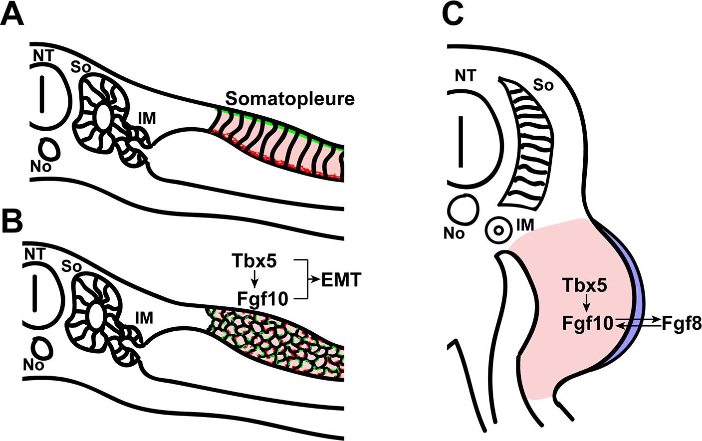
(A) The cells of the somatopleure are initially arranged in a regular, columnar, pattern with apical Actin (green) and basal Laminin (red). (B) Tbx5 induces Fgf10 in the limb forming region which causes an epithelial to mesenchymal transition. Actin and Laminin lose their polarized expression patterns. (C) Fgf10 in the mesenchyme induces Fgf8 in the ectoderm of the limb bud, setting up a feedback loop which leads to limb outgrowth. So – Somite, IM – Intermediate mesoderm, NT – Neural tube, No – Notochord. Pink: Somatopleure, Blue: Ectoderm, Red: Actin, Green: Laminin. See text for references.
While spatially distinct, the underlying tissues of the limb forming region and the flank have similar properties prior to induction. In experiments conducted almost 100 years ago, Balinsky observed that implanting the otic vesicle, nasal placode, or pituitary gland in the flank of a developing newt embryo could induce ectopic limb formation.8 The important interpretation of this experiment is that the whole flank is competent to form a limb and that there must be an inductive factor which drives limb formation. This result raises two questions: what are the factors which induce limb formation, and what are the mechanisms which restrict the limbs to their correct position, despite the limb forming potential in the flank.
Many different fibroblast growth factors (Fgfs) can induce an ectopic limb in the flank of a chick embryo.9–12 However, only two Fgfs are expressed in the right place at the right time to be involved in endogenous limb initiation. These are Fgf8, which is detected in the ectoderm of the limb field prior to limb budding,12,13 and Fgf10, which is restricted to the prospective limb mesoderm.11
Importantly, Fgf10 is expressed prior to the time Fgf8 is detectable. Moreover, while Fgf10 directly triggers downstream events of limb initiation (within 17 hours), Fgf8 only does so (in 28 hours) after it first induces Fgf10 expression.11 Although mice that carry mutations in Fgf8 still initiate formation, mice which are deficient for Fgf10 fail to form limbs or even express Fgf8 in the pre-limb ectoderm.14,15
The ability of the downstream gene Fgf8 to induce Fgf10 when ectopically applied is explained by a feedback loop that is normally establish in the subsequent step of limb patterning. Once expressed, Fgf10 in the mesoderm also induces Fgf8 in the overlying ectoderm, leading to apical ectodermal ridge (AER) formation.11 In turn, this ectopically-derived Fgf8 reciprocally induces Fgf10 in the mesoderm, setting up a setting up a positive feedback loop which leads to continued proliferation and limb outgrowth (Fig. 1C).11,16,17
From the standpoint of limb initiation, the key role of Fgf10 is not its relationship with Fgf8, but rather its effect on the process of EMT. Chick embryos with reduced Fgf10 expression show a more stabilised basement membrane and a higher proportion of epithelial cells in the limb forming region when compared to WT embryos6 and Fgf10 has been previously implicated in EMTs in both development and metastasis.18 Snail and Twist are expressed in a similar pattern to Tbx5/4 and Fgf10.19 Snail is a crucial upstream regulator of EMT in vertebrates.20 Twist is also a regulator of EMT and is involved in mesoderm formation,21 but is thought to be redundant with Snail in normal oxygen levels22 and is not crucial for vertebrate development.20 Manipulations in the level of Snail in the chick did not have an effect on the EMT which formed the limb, possibly due to redundancy.6
Activation of Fgf10 expression in the presumptive limb forming domains is thus a key event in limb initiation. Critical transcription factors acting upstream of Fgf10 are Tbx5 and Tbx4. Tbx5 and Tbx4 are T-box genes which are expressed in the forelimb and hindlimb respectively and are present in these respective fields before the limb buds themselves have formed.23–26 Ectopic limbs induced by Fgf proteins will express Tbx5 or Tbx4/Pitx1 depending on the anterior-posterior position of the limb is induced.24,27
If Tbx5 is deleted prior to, or during, limb initiation then no forelimb forms.28,29 Depletion of Tbx5 in the chick results in an over-stabilised epithelium in the limb forming region,6 mirroring the Fgf10−/− phenotype, as Tbx5 directly induces Fgf10 in the lateral plate mesoderm.28,30 Tbx4 expression can rescue Tbx5−/− phenotypes and induce a forelimb,31 suggesting Tbx4 and Tbx5 play mirror roles in the two limb pairs. Indeed, dominant negative Tbx5 and Tbx4 both led to limbless phenotypes and a downregulation of Fgf10 in chick.32 In mice, despite being upstream of Fgf10,33 and in contrast to the role of Tbx5 in the forelimb, Tbx4 appears to be required for continued hindlimb outgrowth and Fgf10 expression, but not initiation.33 Fgf10 is induced in the hindlimb at low levels without Tbx4 expression, suggesting there is a co-regulator of Fgf10 expressed in the hindlimb but not the forelimb, at least at the earliest stages of expression.34 Wnts are likely suspects as canonical Wnt signalling is required to maintain Fgf10 expression in concert with Tbx5 during the limb outgrowth period.28 Another potential co-factor is retinoic acid, which is also thought to act cooperatively with Tbx5 and Tbx4 in activating Fgf10.35
Another difference between Tbx5 and Tbx4 is that whilst Tbx5 appears to be directly induced by Hox genes at the forelimb level36,37 (see below for further details), Tbx4 appears to be downstream of another hindlimb gene, the OTX-related homeobox gene Pitx1.38–42 Pitx1 binding sites are present in one of the two Tbx4 hindlimb enhancers43 and ectopic Pitx1 can induce Tbx4 in the forelimb of the chick.38 Tbx4 expression is reduced in Pitx1-null mice,44,45 confirming that Tbx4 expression in the hindlimb is dependent on Pitx1 expression. However, Pitx1 alone is not sufficient to rescue Tbx5 deletion,31 perhaps because by the time Tbx4 expression is induced the critical period for T-box expression in limb formation32 is over.
Besides their roles in the initiation of limb development, Tbx5 and Tbx4 play additional key roles in patterning the limb bud and in imparting distinct morphologies to the fore and hindlimb.32,46 Interestingly, the two genes are thought to have arisen from a single gene during genome duplication.47 This ancestral gene is still present in amphioxus, a basal chordate, and the amphioxus Tbx4/5 gene can rescue forelimb formation in a Tbx5−/− mouse.48 For greater detail on the roles of Tbx5 and Tbx4 in limb identity and morphology, see Duboc and Logan.49
Determining Limb Position
The tissue fated to become the limb is specified much earlier than Tbx5 expression becomes apparent, as tissue taken from chick embryos up to 20 hours before Tbx5 expression is apparent will begin to express Tbx5 during explant culture.50 In fact, the LPM which will become the limb is specified during gastrulation.51 Intriguingly, this is the same time as the collinear expression of Hox genes is set up as cells ingress through the primitive streak.52 The Hox genes establish regional identity along the anterior-posterior body axis. Limb buds form at the cervical-thoracic boundary and the lumbar-sacral boundary (Fig. 2).53 The correlation between limb position and axial identity suggests the involvement of Hox genes in limb positioning and, consistent with this, some Hox mutants do show subtle shifts in forelimb and hindlimb location.54,55 The lack of a more dramatic effect, and indeed the lack of any change in limb positioning in most Hox mutants, is likely explained by redundancy. Six Hox genes from paralogous groups 4 and 5 overlap with the earliest forelimb associated Tbx5 expression.36 These same Hox genes have been shown to drive Tbx5 expression by directly binding to an enhancer in intron 2 of the Tbx5 gene.36 In contrast, Hox9 genes have been shown to directly repress Tbx5 by binding at the same locus.37
Figure 2 – The Gene Regulatory Networks Involved in Limb Positioning in Amniotes –

The position of the limb bud is determined by gene regulatory networks which integrate signals from the somites and the lateral plate mesoderm. The above schemes show the major players in determining the position of the limb along the flank. The indications for Wnt genes are specific for experiments carried out in chick embryos. Upregulation and activation are demonstrated by a pointed arrow (→) whereas downregulation and repression are demonstrated by a blunt ended arrow (⊣). See text for references.
While the Hox genes may be responsible for the positional information placing limb bud at the proper axial level, their expression within the LPM is not sufficient to initiate limb formation. A physical barrier inserted between the somatopleure and axial structures can preclude limb formation,56–58 implying there is additionally a necessary inductive signal emanating from the medial tissues. Fgf8 expressed in the intermediate mesoderm was once thought to be one such inductive signal in the forelimb region.13,59 However, the finding that blocking signals from, or ablating Fgf8 in, intermediate mesoderm had no effect on limb formation17,60 finally eliminated that hypothesis. It is now the somites, or paraxial mesoderm, which is considered to be the source of the inductive signal.35
Retinoic acid (RA) is a strong contender for the inductive signal. RA has been proposed to have multiple roles in both the patterning of the LPM and in limb initiation. One of RA’s proposed roles is refining the Hox expression levels to determine the location of Tbx5 expression, as RA has been shown to be upstream of the Hox genes involved in forelimb patterning.61–64 In the post-gastrulation embryo, Hox genes are regulated by a balance between RA expression anteriorly and Fgf8 expression posteriorly.65–67 Changes to RA expression during gastrulation can affect axial levels of Hox expression and forelimb positioning.51 Genes which regulate RA can also have an effect on limb positioning. Cux2 is expressed in the forelimb field and directly regulates Raldh, an RA synthesis gene, and Hoxb genes68 thus refining the position of Tbx5 expression.
Gdf11 is a secreted TGFβ family member which is involved in the trunk to tail transition. As the hindlimbs form at the trunk-tail boundary, Gdf11 is thought to play an important role in hindlimb positioning.72 Indeed, mutations in Gdf11 are associated with axial shifts in the position of the hindlimb.71,72 A direct target of Gdf11 is Islet1, which is transiently expressed in the LPM before hindlimb formation. When Islet1 is inactivated, Tbx4 is downregulated and Fgf10 is not expressed,74 suggesting it may be the primary regulator of hindlimb positioning, not Hox genes. Other evidence suggests Gdf11 activates posterior Hox genes,69,70 possibly directly or possibly by modulating RA expression through Cyp26a1, an enzyme which inactivates RA.73
Another role for RA may be as a direct regulator of limb induction. Some data suggest RA acts as a permissive factor for forelimb development, repressing pre-limb expression of Fgf8,67 and allowing the forelimb bud to initiate.75,76 Others argue that RA acts to directly induce limb formation. Application of a RA inhibitor at pre-limb bud stages prevents formation of a limb bud in chick embryos.77 Similarly, no forelimbs were formed in mice where an RA synthesis gene (Raldh2) was deleted and Tbx5 and Fgf10 expression was also lost.75,78,79 Maternal RA application was sufficient to rescue hypoplastic forelimbs as well as Tbx5 and Fgf10 expression.79,80 A dominant negative Fgf receptor was also sufficient to rescue Raldh2−/− embryos,76 suggesting that RA may be antagonising Fgf8 to allow Tbx5 expression. In all of these studies, the hindlimb appeared normal. This could be because RA is required only to repress Fgf8 to permit forelimb development, but another explanation could be that there is a compensatory source of Raldh3 expression near the hindlimb of the mouse.80
Possibly the most compelling evidence for a direct requirement for RA is through a classical barrier insertion experiment. An impermeable barrier inserted between the paraxial mesoderm and LPM in the chick was sufficient to abrogate limb initiation and Fgf10 expression, despite strong expression of Tbx5 and Tbx435. Application of RA to these embryos was sufficient to rescue both Fgf10 expression and limb formation. Inserting the barrier earlier, only a few hours after somite formation has begun, prevents Tbx5 expression completely.35 Tbx5 expression can be rescued by subsequent application of RA soaked beads. Taken together, these results imply there is an early phase of RA which induces Tbx5 and then a later phase where Tbx5 and RA work in concert to induce Fgf10. The Tbx5 forelimb regulatory element36 also contains RA response elements (RAREs) which directly regulate Tbx5 expression in vivo,35 although this was found to be non-essential in mutation studies.81
The Tbx5 forelimb regulatory element also contains a TCF/LEF site in the mouse.35 TCF and LEF are genes involved in the β-catenin signalling cascade, suggesting a role for Wnt signalling in the limb initiation process. In the chick embryo, Wnt2b is expressed in the LPM at the forelimb level and Wnt8b is expressed at the hindlimb level.82 Wnt2b is upstream of Tbx5 in chick.30 Grafting of cells which overexpress Wnt2b or Wnt8c to the flank of a chick embryo is sufficient to induce Fgf10 in the flank and thus an ectopic limb.82 Both Wnt2b and Wnt8c signal through the canonical Wnt/β-catenin signalling pathway. Further, β-catenin is sufficient to induce Fgf10 and a limb in the flank of the chick embryo and antagonising β-catenin blocks limb formation.35,74,82 LEF and TCF knockouts in the mouse lead to significantly smaller limb buds, due to failure of Fgf8 and AER induction.83
Canonical Wnt signalling maintains Fgf10 expression in concert with Tbx5 during the limb outgrowth period of mice.28 However, the mechanism is not fully clear. Agarwal et al.28 demonstrated that Tbx5 is upstream of Wnt signalling as opposed to downstream, while other experiments using a dominant negative Tbx5 demonstrated that Wnt signalling is downstream of Tbx5.32 Dominant negative LEF did not affect Tbx5 expression initially, but Tbx5 levels did decrease after an extended period. An explanation for the delay could be that loss of Wnt2b reduced levels of Fgf10 which subsequently led to the reduction in Tbx5.32 A feedback loop may exist between Tbx5 and Wnt genes to maintain Tbx5 expression in the forelimb.
Whilst Wnt2b may play a role in chick forelimb development, Wnt2b is not expressed in the limb in mouse until digits have formed.84 Another canonical Wnt gene involved in limb induction in the chick is Wnt3a, which mediates the induction of Fgf8 by Fgf10 in chick.82,85 Wnt3a is not present in the AER of mice,86 however, Wnt3 is present in the limb ectoderm84,87 and removal of Wnt3 in the mouse limb ectoderm prevents AER formation and Fgf8 expression.87 Thus, Wnt3 and Wnt3a appear to have taken on similar roles in the mammalian and avian lineages. It is not clear which, if either, had this activity in the ancestral form.
Variation in Limb Positioning within Amniotes
Amniotes come in a much wider variety of shapes and sizes than represented by the research above. Accordingly, there are some interesting variations where limb initiation differs from the ‘standard’ pattern described above.
In both birds and mammals, the forelimb is placed at the cervical-thoracic transition. In mammals, the vertebral formula is relatively conserved in the cervical region and so the forelimb is almost always formed at the level of the 8th vertebra. In contrast, the bird forelimb varies tremendously in position, from the 10th to the 25th vertebrae. A shift in the border between Hox4 and Hox9 appears to be responsible for changing the posterior extent of Tbx5 expression and thus forelimb position.51 These spatial shifts in Hox expression are caused by temporal changes in Hox regulation, resulting in a longer period of expression of Hoxb4 and a concomitant delay in the onset of Hoxb9 expression.
The hindlimbs are located lateral to the sacral vertebrae, which are specified by Hox11 genes.88 Overexpression of the secreted factor Gdf11 is sufficient to induce posterior Hox genes, including Hox11 paralogues, and the timing of Gdf11 expression appears to be tightly correlated with the position of the hindlimbs in tetrapods.69 This is probably related to the role of Gdf11 in regulating RA expression. In fact, the timing of RA expression was correlated with the position of the forelimb as well, and modulating RA expression levels was sufficient to shift the expression borders of both Hox genes and Tbx5.51
As stated above, almost all mammals have seven cervical vertebrae. Any variation in length or kinematics of the neck is achieved by morphological changes to the vertebrae, but not through a change in vertebral number. There exist a few notable exceptions to this rule, these are the two-toed (Choloepus) and three-toed (Bradypus) sloths and the manatees (Trichechus) (Fig. 3). Choloepus can possess 5–8 cervical vertebrae, Bradypus 8–10, and Trichechus 6. It is thought that the slow metabolism and lifestyle of these animals allows them to avoid the extreme selection against homeotic transformations in most mammals.89–91 However, the developmental explanation for this shift in the position of the forelimb, which lies at the cervical-thoracic boundary, is somewhat unclear. One hypothesis suggests that there has been a homeotic transformation of thoracic vertebrae to cervical, or vice versa,90 while another suggests that there has been a decoupling of the patterning of the vertebral column (primaxial tissues) from the distal ribs and limb girdles (abaxial tissues).92,93 The ossification patterns of the last two cervical vertebrae in the long necked Bradypus were similar to thoracic vertebrae, suggesting the distal ribs and limbs had been shifted, whilst the vertebral column remained the same.92 However, morphometric analysis suggested that a homeotic transformation is more likely, as the same number of morphometric ‘modules’ are present in the sloth neck, just with displaced boundaries.94
Figure 3 – Summary of Amniote Variations in Limb Initiation and Positioning –

The phylogenetic relationships between the species discussed in this review and a summary of the changes which have occurred in the initiation of their limbs. See text for references.
Modifying Limb Pattern by Delaying Limb Formation
There has been convergent flight loss in many paleognaths, an early branching bird lineage, associated with a decrease in limb size and, in many cases, an increase in body size.95 In the emu (Dromaius novaehollandiae), the reduction in limb size is associated with a delay in the formation and development of the limb bud.96,97
A cardiac marker, Nkx2.5, is upregulated in the emu forelimb, possibly repressing forelimb development,98 but this expression does not appear until after the emu limb is already delayed in its formation,99 suggesting it is not a primary driver of the change in timing of limb formation. Nkx2.5 expressing cells also express Pax399 and so this could be a myoblast specific change.
Tbx5 expression has also been described as being delayed in the LPM of the emu compared to the chick, although it appeared at the expected time in the heart field and Pitx1 is expressed as expected in the hindlimb,100 consistent with the possibility that emu forelimb might be initiated later than the chick. However, further analysis has demonstrated that the emu limb is initiated at the same stage as in the chick, and the EMT takes place as expected,99 but subsequent proliferation and outgrowth is delayed. Indeed, Tbx5 expression is comparable between chick and emu and Fgf10 is also expressed at similar stages. However, the Fgf10 domain is smaller in the emu. Supplemental application of Fgf10 can rescue outgrowth of the limb, showing that the LPM is competent to form a limb at early stages.99 This suggests that Fgf10 expression is sufficient to induce the limb but is initially at too low a level to activate the Fgf10-Fgf8 positive feedback loop and form a bud. This causes a the delay in limb formation and thereby contributes to the size reduction in the emu wing.99
When Limbs are Not Initiated at All
Many amniote clades have secondarily lost their fore or hindlimbs. Within mammals, whales and dolphins have vestigial pelvises but lack hindlimbs. Developmentally, the hindlimb buds initiate and the AER is set up but Fgf8 expression is not maintained in the AER and the limb buds falter.101
Manatees also have a vestigial pelvis, which is larger on the left-hand side than the right. This phenotype is very reminiscent of the pelvic reduction seen in mice and stickleback fish deficient for Pitx1 (See below). The asymmetric morphology in these mutants is attributed to partial compensation by the related gene Pitx2 which is asymmetrically expressed on the left side of the embryo. The similarity of the rudiment of the pelvis in the manatee to these mutants suggests that reduction in Pitx1 may be implicated in the manatee.102
The clade which has by far the highest incidence of limb loss is the squamates, encompassing lizards and snakes. Limb reduction and loss has been documented at least 62 times in 53 lineages in squamates alone.103 Limblessness in squamates is correlated with axial extension and increased uniformity in vertebral shape,104 exemplified most notably in the snakes. Within squamates, forelimb loss is more common than hindlimb loss.105 All snakes have lost their pectoral skeleton106 and whilst many limbless lizards and basal snakes initiate a hindlimb bud which then fails, some more derived snakes do not initiate hindlimb buds at all. Pythons have a rudimentary hindlimb. The python hindlimb bud does not form the correct signalling centres and is subsequently severely truncated, similar to the cetaceans.107
Early analysis suggested that expanded, deregionalised Hox domains would preclude the correct, restricted Hox expression patterns which specify limb position.107 In fact, snakes show the same collinear Hox expression patterns as seen in other amniotes.108 In the corn snake (Pantherophis guttatus), a derived snake with no limb elements, the Tbx5 expression domain is expanded, with homogeneous expression throughout the somatopleure.108 As the Hox genes are expressed colinearly, with regionalised expression patterns, the broad Tbx5 domain is interpreted as a decoupling of Hox patterning from the typical limb induction pathways. Whilst Tbx5 deficient mice fail to form a pectoral girdle at all,29,100 Fgf10−/− mice form a sternum and pectoral girdle.15,100 These phenotypic differences between Tbx5 and Fgf10 mutants recapitulate the differences between the forelimb rudiment in limbless lizards and the complete lack of a forelimb in snakes, as well as the differences between the fore and hindlimb in basal snakes.106 But, as Leal and Cohn note in their 2018 review on limb loss in snakes, vertebrates have taken many paths to limb loss and so inference from a phenotype in a model system or comparing convergent phenotypes may lead us down the wrong path.
Evolution at the enhancers of limb genes provides an attractive mechanism for modulating limb initiation. Accordingly, there has been accelerated evolution at the ZRS, a limb specific sonic hedgehog enhancer, in advanced snakes.109,110 This accelerated evolution is only seen in derived snakes with a complete loss of hindlimb elements. Given that complete loss of Shh expression in mice or chicks results in well formed, albeit reduced, proximal limb elements,111 it is unlikely that the loss of the ZRS in advanced snakes is responsible for their complete absence of limbs. Rather the accelerated evolution of the ZRS is likely a result of relaxed selection leading to degradation. The boa ZRS appears fully functional, further supporting the idea that there is a separate mechanism which led to limb reduction and loss and the degradation of the ZRS may be incidental.109 Considering the ZRS does not play a role in limb initiation and advanced snakes do not initiate a limb bud, it is likely that the cause of limblessness in snakes has not fully been determined.
Enhancers located close to other limb initiation genes may provide insights. Tbx4 appears to be involved in development of the genital tubercle in amniotes. A common mesenchymal cell population gives rise to the hemipenes and hindlimbs in squamates.112 This developmental constraint may explain why hindlimb reduction is less common than forelimb reduction. Tbx4 has a limb specific enhancer, HLEB, which is conserved throughout vertebrates,43 and this enhancer drives Tbx4 expression in both the hindlimb and genital tubercle in mice.110 The python HLEB enhancer sequence is able to drive reporter expression in the genital tubercle and weak expression in the hindlimb of the mouse. The corn snake HLEB sequence can only drive reporter expression in the genital tubercle of the mouse.110 These enhancers appear to have progressively lost the ability to drive Tbx4 in the hindlimb, and could be responsible for, or at least contribute to, the loss of hindlimbs throughout snake evolution. This pattern has been seen in other genes expressed in both the hindlimb and genital tubercle.113
Fish and Fins
Zebrafish are teleost fish, and teleost fish have undergone an extra round of genome duplication beyond that of other vertebrates.114 They consequently have two Tbx5 paralogues, tbx5a and tbx5b, which have evolved distinct roles in teleost development.115 Both paralogues are involved in heart and pectoral fin development, similar to Tbx5 expression in amniotes, but they have separate roles in the development of these organs and cannot substitute for each other.116
Similar to the absence of Tbx5 in amniotes, tbx5a loss leads to a complete loss of pectoral fins in zebrafish.117,118 In a wild type embryo, tbx5a expression appears as a stripe along the flank which condenses into a fin forming region (Fig. 4). In a tbx5a morpholino treated embryo, where the mRNA is present but not translated, tbx5a expression does not condense but remains dispersed and the fin bud fails to form.117 Further research also showed that tbx5a expressing cells migrated to form the pectoral fin bud and that knock down of tbx5a prevented cells converging.119 tbx5b expression is not seen in the pectoral fin itself,115 but tbx5b loss does result in delayed pectoral fins which are reduced in size.116 This suggests that tbx5b may have a later role in fin growth as opposed to initiation.
Figure 4 – There Exist Fundamental Differences in Limb Initiation Across Vertebrates –

Despite similarities in the gene regulatory networks which lead to limb formation in zebrafish and amniote models, the mechanism of initiation is quite different. Whilst the zebrafish limb forms through collective cell migration, the amniote limb forms by a prolonged EMT. Zebrafish limb initiation also requires Fgf24, whereas amniote limb initiation does not. The mechanism of limb initiation in amphibians remains unclear. The evolutionary time since the divergence of amphibians and amniotes is so vast that one cannot assume that amphibians will follow the amniote model. The lack of a clear outgroup means inference of an ancestral state of limb initiation is unwise. A: Anterior, P: Posterior, Red: Actin, Green: Laminin. See text for references.
Fgf24 is another fibroblast growth factor gene, in the same subfamily as Fgf8. Deletion of fgf24 leads to complete pectoral fin loss in zebrafish embryos.120 It appears that fgf24 lies between Tbx5 and Fgf10 in the zebrafish limb induction cascade and is also required for Tbx5 cells to converge in the pectoral fin bud.120 Interestingly, fgf24 does not appear to be involved in pelvic fin development.
Although there is some evidence for cell movement into the limb bud in amniotes,121 this occurs only after the limb bud has been initiated and Tbx5 expression is not associated with cell movement in amniotes. This suggests Tbx5 may have acquired a new role between zebrafish and amniotes.29 It is unclear whether EMT or cell migration is the ancestral mode of limb initiation as no outgroup state is known. Tbx5 expression is seen in the pectoral fin of the two sharks studied, the dogfish (Scyliorhinus canicula)122 and the skate (Leucoraja erinacea),123 but its mode of action is unknown. Further research in sharks is required before an ancestral mode of appendage initiation can be determined.
Tbx4 and the Loss of Pelvic Fins
The threespine stickleback (Gasterosteus aculeatus) is a fish with a broad geographic and ecological distribution. It is deemed a species complex, rather than a species, because many thousands of isolated or semi-isolated populations exist which show extreme phenotypic diversity.124 These phenotypic differences are heritable and include the number of spines, the presence of body armour, body size and behavioural traits. Some populations have pelvic spines, modified from the pelvic fin, and others lack pelvic spines and the pelvic girdle entirely.
Whilst spined morphs form a pelvic fin bud as normal, the spine-deficient morphs never initiate a pelvic fin bud.125 The spine-deficient morphs also do not express Tbx4 in the pelvic region, whilst the spined morphs express Tbx4 as normal. Pitx1 also shares this expression pattern.125 A genetic mapping experiment, which crossed spined and spine-deficient fish to examine the phenotype of the progeny, found that most of the variation in spine length in the progeny mapped to the Pitx1 gene.126 No consequential coding mutations were found in the Pitx1 gene, implying that the spine-deficient phenotype was probably a result of regulatory changes which removed the pelvic region-specific enhancer for Pitx1. Indeed, Pitx1 is expressed identically in the thymus and cranial and trunk neuromasts of both the spined and spine-deficient fish, but is absent in the pelvic region of the spine-deficient morphs.126 Later work identified the pelvic spine specific enhancer which lies upstream of Pitx1 and is inactivated in spine-deficient lineages.127 Sequence features of the Pitx1 locus in the stickleback genome, specifically its flexibility and susceptibility to double-stranded DNA breaks, may predispose this region to mutations.127 This could explain why similar genetic changes underlie the loss of pelvic spines in multiple, independent spine-deficient lineages.125–127
In the pufferfish (Takifugu rubripes), Tbx4 and Pitx1 expression are also absent in the pelvic region.128 However, the mode of Pitx1 loss appears to be different from the sticklebacks. In fish, pelvic fins form during metamorphosis rather than during embryogenesis. In the sticklebacks, Hoxd9 expression reappears in the pelvic region during metamorphosis, whereas in the pufferfish it does not.128 It’s hypothesised that the pufferfish have lost their hindlimbs due to a loss of peri-metamorphosis expression of posterior Hoxd9 genes,128 whereas the sticklebacks lost the ability of Pitx1 to respond to the patterning by Hox genes.
Amphibians
Frogs, or anurans, are probably the most easily recognised group within the amphibians, and are known for their highly specialised morphology and locomotory style, as well as their drastic metamorphosis. The embryo first develops into a primarily aquatic tadpole, which uses tail-driven locomotion and requires gills for respiration and filter feeding. After this transient larval stage, the frog metamorphoses into its typically terrestrial adult form with powerful, hindlimb-driven locomotion. This life history is ancestral for extant amphibians, but is not thought to represent the ancestral state for tetrapods.129 A major consequence of this biphasic life history is that limb buds develop just before metamorphosis.130 Formation of the limb long after embryogenesis presents a striking contrast to early, embryonic formation of the amniote limb.
Most work on frog limb buds has been focussed on Xenopus laevis. The X. laevis limb bud is first visible during the tadpole stage, before metamorphosis, as ‘a slight concentration of mesenchyme cells dorsal and lateral to the anal tube’.130 At this stage, embryogenesis is complete, the tadpole has begun feeding, and the somatopleure, the embryonic tissue which gives rise to the limb bud in amniotes, has already differentiated. The mechanism of limb initiation in frogs therefore differs from the ‘standard’ pattern described in amniotes. This is similar to the pelvic fins of fish, which arise during metamorphosis rather than during embryogenesis.128
Reinterpretation of early work on the frog Rana pipiens suggests an EMT occurs in the epithelium surrounding the proctodeum followed by migration of these mesenchymal cells to the limb bud,131,132 but this has not been re-examined since 1971, when conclusive cellular markers were unavailable.133 There is also evidence that the lateral plate mesoderm is the ultimate origin of the limb mesenchyme in X. laevis.69,133,134 Neither the EMT, as seen in limb initiation in amniotes, nor the migration of presumptive limb cells, as seen in zebrafish, is ruled out by existing data (Fig 4).6,117,131 Further research into the mechanism of limb formation in X. laevis may help to determine which limb formation mechanism is ancestral and which is derived. The mechanisms that allow the limb initiation module to be delayed are yet to be resolved.
Little is known about the genetics of limb initiation in frogs, but many of the limb patterning genes are expressed in conserved patterns later in development.132 Tbx5 is expressed in the prospective forelimb region just prior to limb bud initiation in Xenopus tropicalis,135 hinting that similar regulatory networks may form the limb in frogs as in other vertebrates. It is also worth noting that limb initiation in the metamorphosing tadpole is independent of thyroid hormone, the hormone which triggers metamorphosis in frogs.136
Despite the similarities to other vertebrates, X. tropicalis appears to have a regulatory pathway which has not yet been observed in amniotes or fish. The charmingly named xenopus de milo (xdm) mutation results in frogs which completely lack all forelimb elements.135 This mutation lies within the nephronectin gene, an integrin gene involved in epithelial-mesenchymal interactions in the mouse kidney.137 The forelimb never initiates in these mutants, and Tbx5 is absent from the LPM, suggesting nephronectin lies upstream of Tbx5 in frogs.135 The role of nephronectin in Xenopus limb initiation is particularly interesting as the limb initiation signal in amniotes was originally thought to be Fgf activity originating from the intermediate mesoderm, the embryonic tissue which forms the kidney.12,13,138 However, the intermediate mesoderm was later shown to express Fgf8 after Tbx5 is already expressed in the presumptive forelimb,28 and blocking posterior migration of the mesonephros via barrier insertion had no effect on limb formation.60 The kidney is unaffected in xdm mutants,135 suggesting nephronectin may have a novel role in anuran metamorphosis, perhaps related to its role in cell migration.139,140 Abnormalities in the kidney in direct developing frogs have also been reported in conjunction with forelimb loss, but this is likely due to RA deficiency.141 The role of nephronectin, or integrins more widely, in amniote or fish limb initiation has not been studied.
Direct Developers
Not all frogs have a tadpole stage. Some frog species have lost the tadpole stage and develop an adult-like head and limbs during embryogenesis, mirroring amniote development 142. It is unclear whether these direct developing frogs have reverted to a more ancestral patterning process or evolved a new mechanism of limb initiation and development. One species which has served as a quasi-model system for direct developing frogs is Eleutherodactylus coqui, a species native to Puerto Rico that has spread to California and Hawaii 143. E. coqui has allowed researchers to learn more about developmental conservation and novelties and has also led to hypotheses as to how the amniotic egg evolved 144.
The limb buds of E. coqui form just after the neural folds close and neurulation ends, similar to the timing of limb bud formation in amniotes.143 Once the limb bud has formed, the patterning of the limb bud is largely conserved when compared to other model systems, with a few variations.145 But, similar to the metamorphosing frogs, very little is known about the initiation of the limb bud. Pitx1 expression is seen in the E. coqui hindlimb once it has formed,146 suggesting conservation of the hindlimb initiation pathway, but there is no definitive evidence. More compelling perhaps is the observation that a lack of retinoic acid (RA) during E. coqui development leads to a loss of forelimb buds and abnormalities in the pronephros.141 Treatment with a compound that inhibits Raldh at early neurulation stages leads to forelimb loss completely, whereas at later stages only digits are lost. This suggests that a Raldh gene is required in E. coqui during early neurulation to initiate forelimbs, as is the case in amniotes.35 Indeed, Raldh2 is expressed in the E. coqui gastrula in a pattern which supports this hypothesis.147 Examining the E. coqui limb initiation pathway may tell us about whether the early initiation of the limbs is a novel or rederived ancestral trait in these frogs and other direct developers.
Conclusions
The process of limb initiation seems relatively conserved and well understood in amniotes, with only minor variation seen in the emu or the advanced snakes. This process seems to be less constrained in earlier branching groups, with different cellular mechanisms for limb bud formation and variation in the timing of limb initiation. With limited reference points on the phylogeny, it is hard to make statements about which mechanism of limb formation is ancestral or derived.
Shared mechanisms or regulatory networks may tell us about the fundamental pathways needed to form a paired appendage. Tbx5 and Tbx4 appear to be a necessity for limb initiation in all classes of animals studied. Another required gene for pectoral appendage initiation is RA, but its lack of effect on the pelvic fin or limb reminds us that the two sets of appendages cannot be considered identical in their formation. In fact, the significant delay between fish pectoral and pelvic fin development is an interesting area which has not received much attention. How the fish delays its pelvic fin formation relative to its pectoral fins may help answer the question of how frogs delay all limb formation until metamorphosis. Whilst limb morphogenesis remains one of the best understood aspects of vertebrate embryogenesis, the lack of knowledge of limb initiation outside of amniote model systems shows us that nature still has a lot to reveal to us. Therefore, before we close the chapter on limb development and evolution, we need to learn to walk before we run.
References
- 1.Young JJ, Tabin CJ. Saunders’s framework for understanding limb development as a platform for investigating limb evolution. Dev Biol. 2017;429(2):401–408. doi: 10.1016/j.ydbio.2016.11.005 [DOI] [PMC free article] [PubMed] [Google Scholar]
- 2.Brazeau MD, Friedman M. The origin and early phylogenetic history of jawed vertebrates. Nature. 2015;520(7548):490–497. doi: 10.1038/nature14438 [DOI] [PMC free article] [PubMed] [Google Scholar]
- 3.Benton MJ, Donoghue PCJ, Asher RJ, Friedman M, Near TJ, Vinther J. Constraints on the timescale of animal evolutionary history. Palaeontol Electron. 2015;18(1):1–107. Accessed October 1, 2020. https://research-information.bris.ac.uk/en/publications/constraints-on-the-timescale-of-animal-evolutionary-history [Google Scholar]
- 4.Zuniga A, Zeller R. Dynamic and self-regulatory interactions among gene regulatory networks control vertebrate limb bud morphogenesis. In: Current Topics in Developmental Biology. Vol 139. Academic Press Inc.; 2020:61–88. doi: 10.1016/bs.ctdb.2020.02.005 [DOI] [PubMed] [Google Scholar]
- 5.Searls RL, Janners MY. The initiation of limb bud outgrowth in the embryonic chick. Dev Biol. 1971;24(2):198–213. doi: 10.1016/0012-1606(71)90095-9 [DOI] [PubMed] [Google Scholar]
- 6.Gros J, Tabin CJ. Vertebrate limb bud formation is initiated by localized epithelial-to-mesenchymal transition. Science (80- ). 2014;343(6176):1253–1256. doi: 10.1126/science.1248228 [DOI] [PMC free article] [PubMed] [Google Scholar]
- 7.Nakaya Y, Sukowati EW, Wu Y, Sheng G. RhoA and microtubule dynamics control cell–basement membrane interaction in EMT during gastrulation. Nat Cell Biol. 2008;10(7):765–775. doi: 10.1038/ncb1739 [DOI] [PubMed] [Google Scholar]
- 8.Balinsky BI. Transplantation des Ohrblaschens bei Triton. Arch Mikrosk Anat Enwicklmech. 1925;(105):718–731. [DOI] [PubMed] [Google Scholar]
- 9.Ohuchi H, Nakagawa T, Yamauchi M, et al. An additional limb can be induced from the flank of the chick embryo by FGF4. Biochem Biophys Res Commun. 1995;209(3):809–816. doi: 10.1006/bbrc.1995.1572 [DOI] [PubMed] [Google Scholar]
- 10.Cohn MJ, Izpisúa-Belmonte JC, Abud H, Heath JK, Tickle C. Fibroblast growth factors induce additional limb development from the flank of chick embryos. Cell. 1995;80(5):739–746. doi: 10.1016/0092-8674(95)90352-6 [DOI] [PubMed] [Google Scholar]
- 11.Ohuchi H, Nakagawa T, Yamamoto A, et al. The mesenchymal factor, FGF10, initiates and maintains the outgrowth of the chick limb bud through interaction with FGF8, an apical ectodermal factor. Development. 1997;124(11):2235–2244. Accessed December 16, 2018. http://www.ncbi.nlm.nih.gov/pubmed/8674413 [DOI] [PubMed] [Google Scholar]
- 12.Vogel A, Rodriguez C, Izpisúa-Belmonte JC. Involvement of FGF-8 in initiation, outgrowth and patterning of the vertebrate limb. Development. 1996;122(6):1737–1750. http://www.ncbi.nlm.nih.gov/pubmed/8674413 [DOI] [PubMed] [Google Scholar]
- 13.Crossley PH, Minowada G, MacArthur CA, Martin GR. Roles for FGF8 in the induction, initiation, and maintenance of chick limb development. Cell. 1996;84(1):127–136. doi: 10.1016/S0092-8674(00)80999-X [DOI] [PubMed] [Google Scholar]
- 14.Min H, Danilenko DM, Scully SA, et al. Fgf-10 is required for both limb and lung development and exhibits striking functional similarity to Drosophila branchless. Genes Dev. 1998;12(20):3156–3161. doi: 10.1101/gad.12.20.3156 [DOI] [PMC free article] [PubMed] [Google Scholar]
- 15.Sekine K, Ohuchi H, Fujiwara M, et al. Fgf10 is essential for limb and lung formation. Nat Genet. 1999;21(1):138–141. doi: 10.1038/5096 [DOI] [PubMed] [Google Scholar]
- 16.Amaya E, Musci TJ, Kirschner MW. Expression of a dominant negative mutant of the FGF receptor disrupts mesoderm formation in xenopus embryos. Cell. 1991;66(2):257–270. doi: 10.1016/0092-8674(91)90616-7 [DOI] [PubMed] [Google Scholar]
- 17.Boulet AM, Moon AM, Arenkiel BR, Capecchi MR. The roles of Fgf4 and Fgf8 in limb bud initiation and outgrowth. Dev Biol. 2004;273(2):361–372. doi: 10.1016/j.ydbio.2004.06.012 [DOI] [PubMed] [Google Scholar]
- 18.Abolhassani A, Riazi GH, Azizi E, et al. FGF10: Type III epithelial mesenchymal transition and invasion in breast cancer cell lines. J Cancer. 2014;5(7):537–547. doi: 10.7150/jca.7797 [DOI] [PMC free article] [PubMed] [Google Scholar]
- 19.Isaac A, Cohn MJ, Ashby P, et al. FGF and genes encoding transcription factors in early limb specification. Mech Dev. 2000;93(1–2):41–48. doi: 10.1016/S0925-4773(00)00261-6 [DOI] [PubMed] [Google Scholar]
- 20.Thiery JP, Acloque H, Huang RYJ, Nieto MA. Epithelial-Mesenchymal Transitions in Development and Disease. Cell. 2009;139(5):871–890. doi: 10.1016/j.cell.2009.11.007 [DOI] [PubMed] [Google Scholar]
- 21.Yang J, Mani SA, Donaher JL, et al. Twist, a master regulator of morphogenesis, plays an essential role in tumor metastasis. Cell. 2004;117(7):927–939. doi: 10.1016/j.cell.2004.06.006 [DOI] [PubMed] [Google Scholar]
- 22.Yang M-H, Wu K-J. Cell Cycle TWIST activation by hypoxia inducible factor-1 (HIF-1): Implications in metastasis and development. Cell Cycle. 2008;7(14):14. doi: 10.4161/cc.7.14.6324 [DOI] [PubMed] [Google Scholar]
- 23.Gibson-Brown JJ, Agulnik SI, Chapman DL, et al. Evidence of a role for T-☐ genes in the evolution of limb morphogenesis and the specification of forelimb/hindlimb identity. Mech Dev. 1996;56(1–2):93–101. doi: 10.1016/0925-4773(96)00514-X [DOI] [PubMed] [Google Scholar]
- 24.Logan M, Simon HG, Tabin C. Differential regulation of T-box and homeobox transcription factors suggests roles in controlling chick limb-type identity. Development. 1998;125(15). [DOI] [PubMed] [Google Scholar]
- 25.Tamura K, Yonei-Tamura S, Belmonte JCI. Differential expression of Tbx4 and Tbx5 in Zebrafish Fin buds. Mech Dev. 1999;87(1–2):181–184. doi: 10.1016/S0925-4773(99)00126-4 [DOI] [PubMed] [Google Scholar]
- 26.Chapman DL, Garvey N, Hancock S, et al. Expression of the T-box family genes, Tbx1-Tbx5, during early mouse development. Dev Dyn. 1996;206(4):379–390. doi: 10.1002/(SICI)1097-0177(199608)206:4<379::AID-AJA4>3.0.CO;2-F [DOI] [PubMed] [Google Scholar]
- 27.Ohuchi H, Takeuchi J, Yoshioka H, et al. Correlation of wing-leg identity in ectopic FGF-induced chimeric limbs with the differential expression of chick Tbx5 and Tbx4. Development. 1998;125(1). [DOI] [PubMed] [Google Scholar]
- 28.Agarwal P, Wylie JN, Galceran J, et al. Tbx5 is essential for forelimb bud initiation following patterning of the limb field in the mouse embryo. Development. 2003;130(3):623–633. doi: 10.1242/dev.00191 [DOI] [PubMed] [Google Scholar]
- 29.Rallis C, Bruneau BG, Del Buono J, et al. Tbx5 is required for forelimb bud formation and continued outgrowth. Development. 2003;130(12):2741–2751. Accessed October 18, 2018. http://www.ncbi.nlm.nih.gov/pubmed/12736217 [DOI] [PubMed] [Google Scholar]
- 30.Ng JK, Kawakami Y, Büscher D, et al. The limb identity gene Tbx5 promotes limb initiation by interacting with Wnt2b and Fgf10. Development. 2002;129(22). [DOI] [PubMed] [Google Scholar]
- 31.Minguillon C, Del Buono J, Logan MP. Tbx5 and Tbx4 Are Not Sufficient to Determine Limb-Specific Morphologies but Have Common Roles in Initiating Limb Outgrowth. Dev Cell. 2005;8(1):75–84. doi: 10.1016/J.DEVCEL.2004.11.013 [DOI] [PubMed] [Google Scholar]
- 32.Takeuchi JK, Koshiba-Takeuchi K, Suzuki T, Kamimura M, Ogura K, Ogura T. Tbx5 and Tbx4 trigger limb initiation through activation of the Wnt/Fgf signaling cascade. Development. 2003;130(12):2729 LP – 2739. doi: 10.1242/dev.00474 [DOI] [PubMed] [Google Scholar]
- 33.Naiche LA, Papaioannou VE. Loss of Tbx4 blocks hindlimb development and affects vascularization and fusion of the allantois. Development. 2003;130(12):2681–2693. doi: 10.1242/dev.00504 [DOI] [PubMed] [Google Scholar]
- 34.Naiche LA, Papaioannou VE. Tbx4 is not required for hindlimb identity or post-bud hindlimb outgrowth. Development. 2007;134(1):93–103. doi: 10.1242/dev.02712 [DOI] [PubMed] [Google Scholar]
- 35.Nishimoto S, Wilde SM, Wood S, Logan MPO. RA Acts in a Coherent Feed-Forward Mechanism with Tbx5 to Control Limb Bud Induction and Initiation. Cell Rep. 2015;12(5):879–891. doi: 10.1016/j.celrep.2015.06.068 [DOI] [PMC free article] [PubMed] [Google Scholar]
- 36.Minguillon C, Nishimoto S, Wood S, Vendrell E, Gibson-Brown JJ, Logan MPO. Hox genes regulate the onset of Tbx5 expression in the forelimb. Development. 2012;139(17):3180–3188. doi: 10.1242/dev.084814 [DOI] [PMC free article] [PubMed] [Google Scholar]
- 37.Nishimoto S, Minguillon C, Wood S, Logan MPO. A Combination of Activation and Repression by a Colinear Hox Code Controls Forelimb-Restricted Expression of Tbx5 and Reveals Hox Protein Specificity. Duboule D, ed. PLoS Genet. 2014;10(3):e1004245. doi: 10.1371/journal.pgen.1004245 [DOI] [PMC free article] [PubMed] [Google Scholar]
- 38.Logan M, Tabin CJ. Role of Pitx1 upstream of Tbx4 in specification of hindlimb identity. Science (80- ). 1999;283(5408):1736–1739. doi: 10.1126/science.283.5408.1736 [DOI] [PubMed] [Google Scholar]
- 39.Duboc V, Logan MPO. Pitx1 is necessary for normal initiation of hindlimb outgrowth through regulation of Tbx4 expression and shapes hindlimb morphologies via targeted growth control. Development. 2011;138(24):5301–5309. doi: 10.1242/dev.074153 [DOI] [PMC free article] [PubMed] [Google Scholar]
- 40.DeLaurier A, Schweitzer R, Logan M. Pitx1 determines the morphology of muscle, tendon, and bones of the hindlimb. Dev Biol. 2006;299(1):22–34. doi: 10.1016/j.ydbio.2006.06.055 [DOI] [PubMed] [Google Scholar]
- 41.Shang J, Li X, Ring HZ, Clayton DA, Francke U. Backfoot, a novel homeobox gene, maps to human chromosome 5 (BFT) and mouse chromosome 13 (Bft). Genomics. 1997;40(1):108–113. doi: 10.1006/geno.1996.4558 [DOI] [PubMed] [Google Scholar]
- 42.Szeto DP, Rvan AK, O’Connell SM, Rosenfeld MG. P-OTX: A PIT-1-interacting homeodomain factor expressed during anterior pituitary gland development. Proc Natl Acad Sci U S A. 1996;93(15):7706–7710. doi: 10.1073/pnas.93.15.7706 [DOI] [PMC free article] [PubMed] [Google Scholar]
- 43.Menke DB, Guenther C, Kingsley DM. Dual hindlimb control elements in the Tbx4 gene and region-specific control of bone size in vertebrate limbs. Development. 2008;135(15):2543–2553. doi: 10.1242/dev.017384 [DOI] [PubMed] [Google Scholar]
- 44.Szeto DP, Rodriguez-Esteban C, Ryan AK, et al. Role of the Bicoid-related homeodomain factor Pitx1 in specifying hindlimb morphogenesis and pituitary development. Genes Dev. 1999;13(4):484–494. doi: 10.1101/gad.13.4.484 [DOI] [PMC free article] [PubMed] [Google Scholar]
- 45.Lanctot C, Moreau A, Chamberland M, Tremblay ML, Drouin J. Hindlimb patterning and mandible development require the Ptx1 gene. Development. 1999;126(9):1805 LP – 1810. http://dev.biologists.org/content/126/9/1805.abstract [DOI] [PubMed] [Google Scholar]
- 46.Hasson P, Del Buono J, Logan MPO. Tbx5 is dispensable for forelimb outgrowth. Development. 2007;134(1):85–92. doi: 10.1242/dev.02622 [DOI] [PubMed] [Google Scholar]
- 47.Ruvinsky I, Silver LM, Gibson-Brown JJ. Phylogenetic Analysis of T-Box Genes Demonstrates the Importance of Amphioxus for Understanding Evolution of the Vertebrate Genome. Genetics. 2000;156(3). [DOI] [PMC free article] [PubMed] [Google Scholar]
- 48.Minguillon C, Gibson-Brown JJ, Logan MP. Tbx4/5 gene duplication and the origin of vertebrate paired appendages. Proc Natl Acad Sci U S A. 2009;106(51):21726–21730. doi: 10.1073/pnas.0910153106 [DOI] [PMC free article] [PubMed] [Google Scholar]
- 49.Duboc V, Logan MPO. Regulation of limb bud initiation and limb-type morphology. Dev Dyn. 2011;240(5):1017–1027. doi: 10.1002/dvdy.22582 [DOI] [PubMed] [Google Scholar]
- 50.Saito D, Yonei-Tamura S, Kano K, Ide H, Tamura K. Specification and determination of limb identity: evidence for inhibitory regulation of Tbx gene expression. Development. 2002;129(1):211 LP – 220. http://dev.biologists.org/content/129/1/211.abstract [DOI] [PubMed] [Google Scholar]
- 51.Moreau C, Caldarelli P, Rocancourt D, et al. Timed Collinear Activation of Hox Genes during Gastrulation Controls the Avian Forelimb Position. Curr Biol. 2019;29(1):35–50.e4. doi: 10.1016/j.cub.2018.11.009 [DOI] [PMC free article] [PubMed] [Google Scholar]
- 52.Iimura T, Pourquié O. Collinear activation of Hoxb genes during gastrulation is linked to mesoderm cell ingression. Nature. 2006;442(7102):568–571. doi: 10.1038/nature04838 [DOI] [PubMed] [Google Scholar]
- 53.Burke A, Nelson C, Morgan B, Tabin C. Hox genes and the evolution of vertebrate axial morphology. Development. 1995;121(2):333–346. Accessed January 30, 2016. http://dev.biologists.org/content/121/2/333.long [DOI] [PubMed] [Google Scholar]
- 54.Rancourt DE, Tsuzuki T, Capecchi MR. Genetic interaction between hoxb-5 and hoxb-6 is revealed by nonallelic noncomplementation. Genes Dev. 1995;9(1):108–122. doi: 10.1101/gad.9.1.108 [DOI] [PubMed] [Google Scholar]
- 55.van Den Akker E, Fromental-Ramain C, de Graaff W, et al. Axial skeletal patterning in mice lacking all paralogous group 8 Hox genes. Development. 2001;128(10):1911 LP – 1921. http://dev.biologists.org/content/128/10/1911.abstract [DOI] [PubMed] [Google Scholar]
- 56.Murillo-Ferrol NL. Étude causale de la diffÉrenciation la plus prÉcoce de l’Ébauche morphologique des membres. analyse expÉrimentale chez les embryons d’oiseaux. Cells Tissues Organs. 1965;62(1):80–103. doi: 10.1159/000142745 [DOI] [PubMed] [Google Scholar]
- 57.Sweeney RM, Watterson RL. Rib development in chick embryos analyzed by means of tantalum foil blocks. Am J Anat. 1969;126(2):127–149. doi: 10.1002/aja.1001260202 [DOI] [PubMed] [Google Scholar]
- 58.Stephens TD, McNulty TR. Evidence for a metameric pattern in the development of the chick humerus. Development. 1981;61(1). [PubMed] [Google Scholar]
- 59.Capdevila J, Belmonte JCI. Patterning Mechanisms Controlling Vertebrate Limb Development. Annu Rev Cell Dev Biol. 2001;17(1):87–132. doi: 10.1146/annurev.cellbio.17.1.87 [DOI] [PubMed] [Google Scholar]
- 60.Fernandez-Teran M, Piedra ME, Simandl BK, Fallon JF, Ros MA. Limb initiation and development is normal in the absence of the mesonephros. Dev Biol. 1997;189(2):246–255. doi: 10.1006/dbio.1997.8680 [DOI] [PubMed] [Google Scholar]
- 61.Lo PCH, Frasch M. Establishing A-P polarity in the embryonic heart tube: A conserved function of Hox genes in Drosophila and vertebrates? Trends Cardiovasc Med. 2003;13(5):182–187. doi: 10.1016/S1050-1738(03)00074-4 [DOI] [PubMed] [Google Scholar]
- 62.Waxman JS, Keegan BR, Roberts RW, Poss KD, Yelon D. Hoxb5b Acts Downstream of Retinoic Acid Signaling in the Forelimb Field to Restrict Heart Field Potential in Zebrafish. Dev Cell. 2008;15(6):923–934. doi: 10.1016/j.devcel.2008.09.009 [DOI] [PMC free article] [PubMed] [Google Scholar]
- 63.Conlon RA, Rossant J. Exogenous retinoic acid rapidly induces anterior ectopic expression of murine Hox-2 genes in vivo. Development. 1992;116(2):357 LP - 368. http://dev.biologists.org/content/116/2/357.abstract [DOI] [PubMed] [Google Scholar]
- 64.Kessel M, Gruss P. Homeotic Transformations of Murine Vertebrae and Concomitant Alteration of Hox Codes Induced by Retinoic Acid. Vol 67.; 1991. [DOI] [PubMed] [Google Scholar]
- 65.Abu-Abed S, Dollé P, Metzger D, et al. Developing with lethal RA levels: genetic ablation of Rarg can restore the viability of mice lacking Cyp26a1. Development. 2003;130(7):1449 LP – 1459. doi: 10.1242/dev.00357 [DOI] [PubMed] [Google Scholar]
- 66.Bel-Vialar S, Itasaki N, Krumlauf R. Initiating Hox gene expression: in the early chick neural tube differential sensitivity to FGF and RA signaling subdivides the HoxB genes in two distinct groups. Development. 2002;129(22):5103 LP – 5115. http://dev.biologists.org/content/129/22/5103.abstract [DOI] [PubMed] [Google Scholar]
- 67.Shimozono S, Iimura T, Kitaguchi T, Higashijima SI, Miyawaki A. Visualization of an endogenous retinoic acid gradient across embryonic development. Nature. 2013;496(7445):363–366. doi: 10.1038/nature12037 [DOI] [PubMed] [Google Scholar]
- 68.Ueda S, Cordeiro IR, Moriyama Y, et al. Cux2 refines the forelimb field by controlling expression of Raldh2 and Hox genes. Biol Open. 2019;8(2):bio040584. doi: 10.1242/bio.040584 [DOI] [PMC free article] [PubMed] [Google Scholar]
- 69.Matsubara Y, Hirasawa T, Egawa S, et al. Anatomical integration of the sacral-hindlimb unit coordinated by GDF11 underlies variation in hindlimb positioning in tetrapods. Nat Ecol Evol. 2017;1(9):1392–1399. doi: 10.1038/s41559-017-0247-y [DOI] [PubMed] [Google Scholar]
- 70.Liu JP, Laufer E, Jessell TM. Assigning the positional identity of spinal motor neurons: Rostrocaudal patterning of Hox-c expression by FGFs, Gdf11, and retinoids. Neuron. 2001;32(6):997–1012. doi: 10.1016/S0896-6273(01)00544-X [DOI] [PubMed] [Google Scholar]
- 71.McPherron AC, Lawler AM, Lee SJ. Regulation of anterior/posterior patterning of the axial skeleton by growth/differentiation factor 11. Nat Genet. 1999;22(3):260–264. doi: 10.1038/10320 [DOI] [PubMed] [Google Scholar]
- 72.Jurberg AD, Aires R, Varela-Lasheras I, Nóvoa A, Mallo M. Switching axial progenitors from producing trunk to tail tissues in vertebrate embryos. Dev Cell. 2013;25(5):451–462. doi: 10.1016/j.devcel.2013.05.009 [DOI] [PubMed] [Google Scholar]
- 73.Lee YJ, McPherron A, Choe S, et al. Growth differentiation factor 11 signaling controls retinoic acid activity for axial vertebral development. Dev Biol. 2010;347(1):195–203. doi: 10.1016/j.ydbio.2010.08.022 [DOI] [PMC free article] [PubMed] [Google Scholar]
- 74.Kawakami Y, Marti M, Kawakami H, et al. Islet1-mediated activation of the β-catenin pathway is necessary for hindlimb initiation in mice. Development. 2011;138(20):4465–4473. doi: 10.1242/dev.065359 [DOI] [PMC free article] [PubMed] [Google Scholar]
- 75.Zhao X, Sirbu IO, Mic FA, et al. Retinoic Acid Promotes Limb Induction through Effects on Body Axis Extension but Is Unnecessary for Limb Patterning. Curr Biol. 2009;19(12):1050–1057. doi: 10.1016/j.cub.2009.04.059 [DOI] [PMC free article] [PubMed] [Google Scholar]
- 76.Cunningham TJ, Zhao X, Sandell LL, Evans SM, Trainor PA, Duester G. Antagonism between Retinoic Acid and Fibroblast Growth Factor Signaling during Limb Development. Cell Rep. 2013;3(5):1503–1511. doi: 10.1016/j.celrep.2013.03.036 [DOI] [PMC free article] [PubMed] [Google Scholar]
- 77.Stratford T, Horton C, Maden M. Retinoic acid is required for the initiation of outgrowth in the chick limb bud. Curr Biol. 1996;6(9):1124–1133. doi: 10.1016/S0960-9822(02)70679-9 [DOI] [PubMed] [Google Scholar]
- 78.Niederreither K, Subbarayan V, Dollé P, Chambon P. Embryonic retinoic acid synthesis is essential for early mouse post- implantation development. Nat Genet. 1999;21(4):444–448. doi: 10.1038/7788 [DOI] [PubMed] [Google Scholar]
- 79.Mic FA, Sirbu IO, Duester G. Retinoic acid synthesis controlled by Raldh2 is required early for limb bud initiation and then later as a proximodistal signal during apical ectodermal ridge formation. J Biol Chem. 2004;279(25):26698–26706. doi: 10.1074/jbc.M401920200 [DOI] [PubMed] [Google Scholar]
- 80.Mic FA, Haselbeck RJ, Cuenca AE, Duester G. Novel retinoic acid generating activities in the neural tube and heart identified by conditional rescue of Raldh2 null mutant mice. Development. 2002;129(9). [DOI] [PMC free article] [PubMed] [Google Scholar]
- 81.Cunningham TJ, Lancman JJ, Berenguer M, Dong PDS, Duester G. Genomic Knockout of Two Presumed Forelimb Tbx5 Enhancers Reveals They Are Nonessential for Limb Development. Cell Rep. 2018;23(11):3146–3151. doi: 10.1016/j.celrep.2018.05.052 [DOI] [PMC free article] [PubMed] [Google Scholar]
- 82.Kawakami Y, Capdevila J, Büscher D, Itoh T, Esteban CR, Belmonte JCI. WNT signals control FGF-dependent limb initiation and AER induction in the chick embryo. Cell. 2001;104(6):891–900. doi: 10.1016/S0092-8674(01)00285-9 [DOI] [PubMed] [Google Scholar]
- 83.Galceran J, Fariñas I, Depew MJ, Clevers H, Grosschedl R. Wnt3a(−/−) -like phenotype and limb deficiency in Lef1(−/−)Tcf1(−/−) mice. Genes Dev. 1999;13(6):709–717. doi: 10.1101/gad.13.6.709 [DOI] [PMC free article] [PubMed] [Google Scholar]
- 84.Witte F, Dokas J, Neuendorf F, Mundlos S, Stricker S. Comprehensive expression analysis of all Wnt genes and their major secreted antagonists during mouse limb development and cartilage differentiation. Gene Expr Patterns. 2009;9(4):215–223. doi: 10.1016/j.gep.2008.12.009 [DOI] [PubMed] [Google Scholar]
- 85.Kengaku M, Capdevila J, Rodriguez-Esteban C, et al. Distinct WNT pathways regulating AER formation and dorsoventral polarity in the chick limb bud. Science (80- ). 1998;280(5367):1274–1277. doi: 10.1126/science.280.5367.1274 [DOI] [PubMed] [Google Scholar]
- 86.Parr BA, Shea MJ, Vassileva G, McMahon AP. Mouse Wnt genes exhibit discrete domains of expression in the early embryonic CNS and limb buds. Development. 1993;119(1):247–261. [DOI] [PubMed] [Google Scholar]
- 87.Barrow JR, Thomas KR, Boussadia-Zahui O, et al. Ectodermal Wnt3β-catenin signaling is required for the establishment and maintenance of the apical ectodermal ridge. Genes Dev. 2003;17(3):394–409. doi: 10.1101/gad.1044903 [DOI] [PMC free article] [PubMed] [Google Scholar]
- 88.Wellik DM, Capecchi MR. Hox10 and Hox11 genes are required to globally pattern the mammalian skeleton. Science. 2003;301(5631):363–367. doi: 10.1126/science.1085672 [DOI] [PubMed] [Google Scholar]
- 89.Galis F, Dooren TJM Van, Feuth JD, et al. Extreme Selection in Humans Against Homeotic Transformations of Cervical Vertebrae. Evolution (N Y). 2006;60(12):2643–2654. doi: 10.1111/j.0014-3820.2006.tb01896.x [DOI] [PubMed] [Google Scholar]
- 90.Varela-Lasheras I, Bakker AJ, van der Mije SD, Metz JAJ, van Alphen J, Galis F. Breaking evolutionary and pleiotropic constraints in mammals: On sloths, manatees and homeotic mutations. Evodevo. 2011;2(1):1–27. doi: 10.1186/2041-9139-2-11 [DOI] [PMC free article] [PubMed] [Google Scholar]
- 91.Buchholtz EA. Crossing the frontier: A hypothesis for the origins of meristic constraint in mammalian axial patterning. Zoology. 2014;117(1):64–69. doi: 10.1016/j.zool.2013.09.001 [DOI] [PubMed] [Google Scholar]
- 92.Hautier L, Weisbecker V, Sánchez-Villagra MR, Goswami A, Asher RJ. Skeletal development in sloths and the evolution of mammalian vertebral patterning. Proc Natl Acad Sci U S A. 2010;107(44):18903–18908. doi: 10.1073/pnas.1010335107 [DOI] [PMC free article] [PubMed] [Google Scholar]
- 93.Buchholtz EA, Stepien CC. Anatomical transformation in mammals: developmental origin of aberrant cervical anatomy in tree sloths. Evol Dev. 2009;11(1):69–79. doi: 10.1111/j.1525-142X.2008.00303.x [DOI] [PubMed] [Google Scholar]
- 94.Böhmer C, Amson E, Arnold P, Van Heteren AH, Nyakatura JA. Homeotic transformations reflect departure from the mammalian “rule of seven” cervical vertebrae in sloths: Inferences on the Hox code and morphological modularity of the mammalian neck. BMC Evol Biol. 2018;18(1):1–11. doi: 10.1186/s12862-018-1202-5 [DOI] [PMC free article] [PubMed] [Google Scholar]
- 95.Sackton TB, Grayson P, Cloutier A, et al. Convergent regulatory evolution and loss of flight in paleognathous birds. Science (80- ). 2019;364(6435):74–78. doi: 10.1126/science.aat7244 [DOI] [PubMed] [Google Scholar]
- 96.Faux C, Field DJ. Distinct developmental pathways underlie independent losses of flight in ratites. Biol Lett. 2017;13(7):20170234. doi: 10.1098/rsbl.2017.0234 [DOI] [PMC free article] [PubMed] [Google Scholar]
- 97.Nagai H, Mak S-S, Weng W, Nakaya Y, Ladher R, Sheng G. Embryonic development of the emu, Dromaius novaehollandiae. Dev Dyn. 2011;240(6):spcone-spcone. doi: 10.1002/dvdy.22664 [DOI] [PubMed] [Google Scholar]
- 98.Farlie PG, Davidson NM, Baker NL, et al. Co-option of the cardiac transcription factor Nkx2.5 during development of the emu wing. Nat Commun. 2017;8(1):1–12. doi: 10.1038/s41467-017-00112-7 [DOI] [PMC free article] [PubMed] [Google Scholar]
- 99.Young JJ, Grayson P, Edwards SV, Tabin CJ. Attenuated Fgf Signaling Underlies the Forelimb Heterochrony in the Emu Dromaius novaehollandiae. Curr Biol. 2019;29(21):3681–3691.e5. [DOI] [PMC free article] [PubMed] [Google Scholar]
- 100.Bickley SRB, Logan MPO. Regulatory modulation of the T-box gene Tbx5 links development, evolution, and adaptation of the sternum. Proc Natl Acad Sci U S A. 2014;111(50):17917–17922. doi: 10.1073/pnas.1409913111 [DOI] [PMC free article] [PubMed] [Google Scholar]
- 101.Thewissen JGM, Cohn MJ, Stevens LS, Bajpai S, Heyning J, Horton WE. Developmental basis for hind-limb loss in dolphins and origin of the cetacean bodyplan. Proc Natl Acad Sci U S A. 2006;103(22):8414–8418. doi: 10.1073/pnas.0602920103 [DOI] [PMC free article] [PubMed] [Google Scholar]
- 102.Shapiro MD, Bell MA, Kingsley DM. Parallel genetic origins of pelvic reduction in vertebrates. Proc Natl Acad Sci U S A. 2006;103(37):13753–13758. doi: 10.1073/pnas.0604706103 [DOI] [PMC free article] [PubMed] [Google Scholar]
- 103.Greer AE. Limb Reduction in Squamates: Identification of the Lineages and Discussion of the Trends. J Herpetol. 1991;25(2):166. doi: 10.2307/1564644 [DOI] [Google Scholar]
- 104.Caldwell MW. “Without a leg to stand on”: on the evolution and development of axial elongation and limblessness in tetrapods. Can J Earth Sci. 2003;40:573–588. doi: 10.1139/e02-081 [DOI] [Google Scholar]
- 105.Brandley MC, Huelsenbeck JP, Wiens JJ. Rates and patterns in the evolution of snake-like body form in squamate reptiles: Evidence for repeated re-evolution of lost digits and long-term persistence of intermediate body forms. Evolution (N Y). 2008;62(8):2042–2064. doi: 10.1111/j.1558-5646.2008.00430.x [DOI] [PubMed] [Google Scholar]
- 106.Leal F, Cohn MJ. Developmental, genetic, and genomic insights into the evolutionary loss of limbs in snakes. genesis. 2018;56(1):e23077. doi: 10.1002/dvg.23077 [DOI] [PubMed] [Google Scholar]
- 107.Cohn MJ, Tickle C. Developmental basis of limblessness and axial patterning in snakes. Nature. 1999;399(6735):474–479. doi: 10.1038/20944 [DOI] [PubMed] [Google Scholar]
- 108.Woltering JM, Vonk FJ, Müller H, et al. Axial patterning in snakes and caecilians: evidence for an alternative interpretation of the Hox code. Dev Biol. 2009;332(1):82–89. doi: 10.1016/j.ydbio.2009.04.031 [DOI] [PubMed] [Google Scholar]
- 109.Kvon EZ, Kamneva OK, Melo US, et al. Progressive Loss of Function in a Limb Enhancer during Snake Evolution. Cell. 2016;167(3):633–642.e11. doi: 10.1016/j.cell.2016.09.028 [DOI] [PMC free article] [PubMed] [Google Scholar]
- 110.Infante CR, Mihala AG, Park S, et al. Shared Enhancer Activity in the Limbs and Phallus and Functional Divergence of a Limb-Genital cis-Regulatory Element in Snakes. Dev Cell. 2015;35(1):107–119. doi: 10.1016/j.devcel.2015.09.003 [DOI] [PMC free article] [PubMed] [Google Scholar]
- 111.Sagai T, Hosoya M, Mizushina Y, Tamura M, Shiroishi T. Elimination of a long-range cis-regulatory module causes complete loss of limb-specific Shh expression and truncation of the mouse limb. Development. 2005;132(4):797–803. doi: 10.1242/dev.01613 [DOI] [PubMed] [Google Scholar]
- 112.Tschopp P, Sherratt E, Sanger TJ, et al. A relative shift in cloacal location repositions external genitalia in amniote evolution. Nature. 2014;516(7531):391–394. doi: 10.1038/nature13819 [DOI] [PMC free article] [PubMed] [Google Scholar]
- 113.Guerreiro I, Gitto S, Novoa A, et al. Reorganisation of Hoxd regulatory landscapes during the evolution of a snake-like body plan. Elife. 2016;5(AUGUST). doi: 10.7554/eLife.16087 [DOI] [PMC free article] [PubMed] [Google Scholar]
- 114.Amores A, Force A, Yan YL, et al. Zebrafish hox clusters and vertebrate genome evolution. Science(80- ). 1998;282(5394):1711–1714. doi: 10.1126/science.282.5394.1711 [DOI] [PubMed] [Google Scholar]
- 115.Albalat R, Baquero M, Minguillón C. Identification and characterisation of the developmental expression pattern of tbx5b, a novel tbx5 gene in zebrafish. Gene Expr Patterns. 2010;10(1):24–30. doi: 10.1016/j.gep.2009.11.003 [DOI] [PubMed] [Google Scholar]
- 116.Parrie LE, Renfrew EM, Wal A Vander, Mueller RL, Garrity DM. Zebrafish tbx5 paralogs demonstrate independent essential requirements in cardiac and pectoral fin development. Dev Dyn. 2013;242(5):485–502. doi: 10.1002/dvdy.23953 [DOI] [PubMed] [Google Scholar]
- 117.Ahn DG, Kourakis MJ, Rohde LA, Slivert LM, Ho RK. T-box gene Tbx5 is essential for formation of the pectoral limb bud. Nature. 2002;417(6890):754–758. doi: 10.1038/nature00814 [DOI] [PubMed] [Google Scholar]
- 118.Garrity DM, Childs S, Fishman MC. The heartstrings mutation in zebrafish causes heart/fin Tbx5 deficiency syndrome. Development. 2002;129(19):4635 LP – 4645. http://dev.biologists.org/content/129/19/4635.abstract [DOI] [PubMed] [Google Scholar]
- 119.Mao Q, Stinnett HK, Ho RK. Asymmetric cell convergence-driven zebrafish fin bud initiation and pre-pattern requires Tbx5a control of a mesenchymal Fgf signal. Dev. 2015;142(24):4329–4339. doi: 10.1242/dev.124750 [DOI] [PMC free article] [PubMed] [Google Scholar]
- 120.Fischer S, Draper BW, Neumann CJ. The zebrafish fgf24 mutant identifies and additional level of Fgf signaling involved in vertebrate forelimb initiation. Development. 2003;130(15):3515–3524. doi: 10.1242/dev.00537 [DOI] [PubMed] [Google Scholar]
- 121.Wyngaarden LA, Vogeli KM, Ciruna BG, Wells M, Hadjantonakis AK, Hopyan S. Oriented cell motility and division underlie early limb bud morphogenesis. Development. 2010;137(15):2551–2558. doi: 10.1242/dev.046987 [DOI] [PMC free article] [PubMed] [Google Scholar]
- 122.Tanaka M, Münsterberg A, Anderson WG, Prescott AR, Hazon N, Tickle C. Fin development in a cartilaginous fish and the origin of vertebrate limbs. Nature. 2002;416(6880):527–531. doi: 10.1038/416527a [DOI] [PubMed] [Google Scholar]
- 123.Adachi N, Robinson M, Goolsbee A, Shubin NH. Regulatory evolution of Tbx5 and the origin of paired appendages. Proc Natl Acad Sci U S A. 2016;113(36):10115–10120. doi: 10.1073/pnas.1609997113 [DOI] [PMC free article] [PubMed] [Google Scholar]
- 124.Bell MA, Foster SA. The Evolutionary Biology of the Threespine Stickleback. Oxford University Press; 1994. [Google Scholar]
- 125.Cole NJ, Tanaka M, Prescott A, Tickle C. Expression of limb initiation genes and clues to the morphological diversification of threespine stickleback. Curr Biol. 2003;13(24):R951–R952. doi: 10.1016/j.cub.2003.11.039 [DOI] [PubMed] [Google Scholar]
- 126.Shapiro MD, Marks ME, Peichel CL, et al. Genetic and developmental basis of evolutionary pelvic reduction in threespine sticklebacks. Nature. 2004;428(6984):717–723. doi: 10.1038/nature02415 [DOI] [PubMed] [Google Scholar]
- 127.Chan YF, Marks ME, Jones FC, et al. Adaptive evolution of pelvic reduction in sticklebacks by recurrent deletion of a pitxl enhancer. Science (80- ). 2010;327(5963):302–305. doi: 10.1126/science.1182213 [DOI] [PMC free article] [PubMed] [Google Scholar]
- 128.Tanaka M, Hale LA, Amores A, et al. Developmental genetic basis for the evolution of pelvic fin loss in the pufferfish Takifugu rubripes. Dev Biol. 2005;281(2):227–239. doi: 10.1016/j.ydbio.2005.02.016 [DOI] [PubMed] [Google Scholar]
- 129.Schoch RR. The evolution of metamorphosis in temnospondyls. 2002;12:309–327. [Google Scholar]
- 130.Nieuwkoop PDand Faber J Normal Table of Xenopus Laevis (Daudin). North-Holland Publishing Company; 1967. [Google Scholar]
- 131.Taylor AC. Development of the innervation pattern in the limb bud of the frog. Anat Rec. 1943;87(4):379–413. doi: 10.1002/ar.1090870409 [DOI] [Google Scholar]
- 132.Keenan SR, Beck CW. Xenopus Limb bud morphogenesis. Dev Dyn. 2016;245(3):233–243. doi: 10.1002/dvdy.24351 [DOI] [PubMed] [Google Scholar]
- 133.Tarin D, Sturdee AP. Early limb development of Xenopus laevis. J Embryol Exp Morphol. 1971;26(2):169–179. http://dev.biologists.org/content/26/2/169.abstract [PubMed] [Google Scholar]
- 134.Tschumi PA. The growth of the hindlimb bud of Xenopus laevis and its dependence upon the epidermis. J Anat. 1957;91(2):149–173. Accessed January 14, 2020. http://www.ncbi.nlm.nih.gov/pubmed/13416124 [PMC free article] [PubMed] [Google Scholar]
- 135.Abu-Daya A, Nishimoto S, Fairclough L, Mohun TJ, Logan MPO, Zimmerman LB. The secreted integrin ligand nephronectin is necessary for forelimb formation in Xenopus tropicalis. Dev Biol. 2011;349(2):204–212. doi: 10.1016/j.ydbio.2010.10.015 [DOI] [PMC free article] [PubMed] [Google Scholar]
- 136.Rot-Nikcevic I, Wassersug RJ. Arrested development in Xenopus laevis tadpoles: how size constrains metamorphosis. J Exp Biol. 2004;207(12):2133 LP – 2145. doi: 10.1242/jeb.01002 [DOI] [PubMed] [Google Scholar]
- 137.Linton JM, Martin GR, Reichardt LF. The ECM protein nephronectin promotes kidney development via integrin α8β1-mediated stimulation of Gdnf expression. Development. 2007;134(13):2501–2509. doi: 10.1242/dev.005033 [DOI] [PMC free article] [PubMed] [Google Scholar]
- 138.Geduspan JS, Solursh M. A growth-promoting influence from the mesonephros during limb outgrowth. Dev Biol. 1992;151(1):242–250. doi: 10.1016/0012-1606(92)90230-E [DOI] [PubMed] [Google Scholar]
- 139.Kuphal S, Wallner S, Bosserhoff AK. Loss of nephronectin promotes tumor progression in malignant melanoma. Cancer Sci. 2008;99(2):229–233. doi: 10.1111/j.1349-7006.2007.00678.x [DOI] [PMC free article] [PubMed] [Google Scholar]
- 140.Eckhardt BL, Parker BS, Van Laar RK, et al. Genomic Analysis of a Spontaneous Model of Breast Cancer Metastasis to Bone Reveals a Role for the Extracellular Matrix; 2005. Accessed September 15, 2020. http://mcr.aacrjournals.org/ [PubMed] [Google Scholar]
- 141.Lee SY, Elinson RP. Abnormalities of forelimb and pronephros in a direct developing frog suggest a retinoic acid deficiency. Appl Herpetol. 2008;5(1):33–46. doi: 10.1163/157075408783489202 [DOI] [Google Scholar]
- 142.Elinson RP. Leg development in a frog without a tadpole (Eleutherodactylus coqui). J Exp Zool. 1994;270(2):202–210. doi: 10.1002/jez.1402700209 [DOI] [PubMed] [Google Scholar]
- 143.Townsend DS, Stewart MM. Direct Development in Eleutherodactylus coqui (Anura: Leptodactylidae): A Staging Table. Copeia. 1985;1985(2):423. doi: 10.2307/1444854 [DOI] [Google Scholar]
- 144.Elinson RP. Development of a non-amphibious amphibian: views of a coquí. Int J Dev Biol. Published online August 20, 2020. doi: 10.1387/ijdb.190386re [DOI] [PubMed] [Google Scholar]
- 145.Gross JB, Kerney R, Hanken J, Tabin CJ. Molecular anatomy of the developing limb in the coquí frog, Eleutherodactylus coqui. Evol Dev. 2011;13(5):415–426. doi: 10.1111/j.1525-142X.2011.00500.x [DOI] [PMC free article] [PubMed] [Google Scholar]
- 146.Chang WY, KhosrowShahian F, Wolanski M, et al. Conservation of Pitx1 expression during amphibian limb morphogenesis. Biochem Cell Biol. 2006;84(2):257–262. doi: 10.1139/o06-036 [DOI] [PubMed] [Google Scholar]
- 147.Elinson RP, Walton Z, Nath K. Raldh expression in embryos of the direct developing frog Eleutherodactylus coqui and the conserved retinoic acid requirement for forelimb initiation. J Exp Zool Part B Mol Dev Evol. 2008;310B(7):588–595. doi: 10.1002/jez.b.21229 [DOI] [PubMed] [Google Scholar]