Abstract
The two 2-hydroxy-6-oxohepta-2,4-dienoate (HOHD) hydrolase genes, etbD1 and etbD2, were cloned from a strong polychlorinated biphenyl (PCB) degrader, Rhodococcus sp. strain RHA1, and their nucleotide sequences were determined. The etbD2 gene was located in the vicinity of bphA gene homologs and encoded an enzyme whose amino-terminal sequence was very similar to the amino-terminal sequence of the HOHD hydrolase which was purified from RHA1. Using the etbD2 gene fragment as a probe, we cloned the etbD1 gene encoding the purified HOHD hydrolase by colony hybridization. Both genes encode a product having 274 amino acid residues and containing the nucleophile motif conserved in α/β hydrolase fold enzymes. The deduced amino acid sequences were quite similar to the amino acid sequences of the products of the single-ring aromatic hydrolase genes, such as dmpD, cumD, todF, and xylF, and not very similar to the amino acid sequences of the products of bphD genes from PCB degraders, including RHA1. The two HOHD hydrolase genes and the RHA1 2-hydroxy-6-oxo-6-phenylhexa-2,4-dienoate (HPDA) hydrolase gene, bphD, were expressed in Escherichia coli, and their relative enzymatic activities were examined. The product of bphD was very specific to HPDA, and the products of etbD1 and etbD2 were specific to HOHD. All of the gene products exhibited poor activities against the meta-cleavage product of catechol. These results agreed with the results obtained for BphD and EtbD1 hydrolases purified from RHA1. The three hydrolase genes exhibited similar induction patterns both in an RNA slot blot hybridization analysis and in a reporter gene assay when a promoter probe vector was used. They were induced by biphenyl, ethylbenzene, benzene, toluene, and ortho-xylene. Strain RCD1, an RHA1 mutant strain lacking both the bphD gene and the etbD2 gene, grew well on ethylbenzene. This result suggested that the etbD1 gene product is involved in the meta-cleavage metabolic pathway of ethylbenzene.
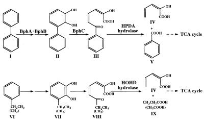
Polychlorinated biphenyls (PCBs) are some of the most serious environmental pollutants because of their exceptional stability. Removal of PCBs from the environment is very desirable. Recently, elimination of environmental contaminants with degradative microorganisms has been studied. The bacteria that have been isolated and examined in aerobic PCB degradation studies are mainly gram-negative bacteria, and these bacteria degrade PCBs through cometabolism with biphenyl. We isolated a gram-positive biphenyl/PCB degrader, Rhodococcus sp. strain RHA1, from a γ-hexachlorocyclohexane-contaminated upland soil (25). RHA1 has a great capacity to degrade highly chlorinated PCBs. In the biphenyl metabolic pathway (Fig. 1), biphenyl is transformed to 2,3-dihydroxy-1-phenylcyclohexa-4,6-diene (dihydrodiol) by a multicomponent biphenyl dioxygenase (BphA). Dihydrodiol is converted to 2,3-dihydroxybiphenyl (23DHBP) by dihydrodiol dehydrogenase (BphB). 23DHBP is cleaved at the 1,2 position (meta-ring cleavage) by 23DHBP dioxygenase (BphC). The ring cleavage product (2-hydroxy-6-oxo-6-phenylhexa-2,4-dienoate [HPDA]) is hydrolyzed to benzoate and 2-hydroxypenta-2,4-dienoate by HPDA hydrolase (BphD), and the resulting 2-hydroxypenta-2,4-dienoate is further converted to tricarboxylic acid cycle intermediates by 2-hydroxypenta-2,4-dienoate hydratase, 4-hydroxy-2-oxovalerate aldolase, and acetaldehyde dehydrogenase (BphE, BphF, and BphG, respectively). Thus, the products of a set of catabolic genes, bphA1A2A3A4BCDEFG, are responsible for the aerobic metabolism of biphenyl. Furukawa et al. indicated that the meta-ring cleavage hydrolase encoded by bphD is critical for successful metabolism because of its discrete substrate specificity (7). We have isolated biphenyl/PCB-degradative genes (bphA1A2A3A4CB) from RHA1 (19). It has been shown that these genes are essential for both growth on biphenyl as a sole source of carbon and energy and degradation of PCBs. In addition to biphenyl, RHA1 can degrade and grow well on ethylbenzene. The metabolic pathway for ethylbenzene seems to be separate from the metabolic pathway for biphenyl in RHA1, because RHA1 mutants lacking either bphA or bphD grew on ethylbenzene. RHA1 transiently accumulates a yellow metabolite, suggesting that a meta-ring cleavage product is produced and subsequently transformed during ethylbenzene metabolism (Fig. 1).
FIG. 1.
Proposed metabolic pathway for aerobic degradation of biphenyl and ethylbenzene in Rhodococcus sp. strain RHA1. Compound I, biphenyl; compound II, 23DHBP; compound III, HPDA; compound IV, 2-hydroxypenta-2,4-dienoate; compound V, benzoic acid; compound VI, ethylbenzene (toluene); compound VII, 3-ethylcatechol (3-methylcatechol); compound VIII, HOHD; compound IX, propanoic acid (acetic acid). TCA, tricarboxylic acid.
Recently, we purified and characterized two different kinds of aromatic compound hydrolases from RHA1, the hydrolase specific to HPDA, the meta-ring cleavage product of 23DHBP, and the hydrolase specific to 2-hydroxy-6-oxohepta-2,4-dienoate (HOHD), the meta-ring cleavage product of 3-methylcatechol (10). These HPDA and HOHD hydrolases were induced during growth on biphenyl and ethylbenzene and were thought to be involved in the metabolism of biphenyl and ethylbenzene, respectively. The bphD gene encoding HPDA hydrolase was cloned and sequenced (20). The amino acid sequence of 29 amino-terminal residues deduced from the bphD gene nucleotide sequence agreed completely with the amino acid sequence of the amino-terminal residues of the purified HPDA hydrolase.
In this study, the structures, activities, and expression of the genes encoding HOHD and HPDA hydrolases were examined to determine the functional significance of these enzymes in the catabolism of aromatic compounds, including ethylbenzene, biphenyl, and PCBs, by strain RHA1.
MATERIALS AND METHODS
Bacterial strains, plasmids, and culture conditions.
A PCB degrader, Rhodococcus sp. strain RHA1, was grown in Luria broth (LB) (10 g of Bacto Tryptone [Difco] per liter, 5 g of yeast extract per liter, 5 g of NaCl per liter) and W minimal medium (19) containing one of the following carbon sources; 0.2% biphenyl, 0.2% sodium benzoate, 0.2% sodium succinate, ethylbenzene, toluene, benzene, or ortho-xylene. Ethylbenzene, toluene, benzene, and ortho-xylene were supplied in vapor form. The two spontaneous mutant strains of RHA1 used, RCD1 and RCAD1, are deficient in growth on biphenyl and were grown in LB. Strain RCD1 cannot grow on biphenyl but can grow on ethylbenzene (Bph− Etb+). Isolation of RCD1 has been described previously (26). Strain RCAD1 grows on neither biphenyl nor ethylbenzene (Bph− Etb−) (unpublished data). Escherichia coli JM109 {recA1 endA1 gyrA96 thi hsdR17 supE44 relA1, Δ(lac-proAB)/F′[traD36 proAB+ lacIq lacZΔM15]} was used as a host strain and was grown in LB. The plasmids used in this study are listed in Table 1.
TABLE 1.
Plasmids used in this study
| Plasmid | Relevant characteristic(s) | Reference or source |
|---|---|---|
| pUC18 | Cloning vector, Apr | 29 |
| pUC118 | Cloning vector, Apr | 27 |
| pK4 | Rhodococcus-E. coli shuttle vector, Kmr | 9 |
| pK4HKcos | pK4 containing cos region | This study |
| pVK100 | Cosmid vector, Kmr Tcr | 17 |
| pKHD1 | pUC118 with 3.0-kb EcoRI fragment of RHA1 carrying etbD1; the direction of etbD1 is identical to that of the lac promoter of pUC118 | This study |
| pKHD2 | pUC118 with 3.0-kb EcoRI fragment of RHA1 carrying etbD1; the direction of etbD1 is opposite that of the lac promoter of pUC118 | This study |
| pUAD1 | pUC18 with 1.2-kb StuI-HindIII fragment of RHA1 carrying bphD | This study |
| pUAD2 | pUC18 with 1.0-kb ScaI-SphI fragment of RHA1 carrying etbD1 | This study |
| pUAD3 | pUC18 with 1.3-kb DNA fragment of RHA1 carrying etbD2 | This study |
| pAC1 | pUC19 with 2.1-kb PstI-SacI fragment of RHA1 carrying bphC | 19 |
| pKLA1 | pK4 with 2.4-kb luciferase structural gene, luxAB from Vibrio harveyi | 28 |
| pKLABD1 | pKLA1 with 1.7-kb BamHI-SacI fragment of RHA1 carrying promoter region of bphD | This study |
| pKLAED1 | pKLA1 with 1.2-kb EcoRI-XhoI fragment of RHA1 carrying promoter region of etbD1 | This study |
| pKLAED2 | pKLA1 with 1.2-kb EcoRI fragment of RHA1 carrying promoter region of etbD2 | This study |
Southern hybridization with DIG-labeled probe.
RHA1, RCD1, and RCAD1 total DNAs were prepared as described previously (19). Each total DNA was completely digested with appropriate restriction enzymes. DNA fragments were separated by agarose gel electrophoresis and transferred to a nylon membrane (Hybond N; Amersham International plc, Buckinghamshire, United Kingdom). Southern hybridization was carried out by using a probe labeled with the digoxigenin (DIG) system (Boehringer Mannheim Biochemicals, Indianapolis, Ind.).
Cloning of the etbD1 gene.
RHA1 total DNA was partially digested with EcoRI, and the fragments obtained were inserted into pK4HKcos. The resultant library DNA was introduced into E. coli JM109 by in vitro packaging by using Gigapack II gold packaging extract (Stratagene, La Jolla, Calif.). The E. coli-Rhodococcus shuttle cosmid vector pK4HKcos was constructed as follows. The unique BglII recognition sequence (AGATCT) of E. coli-Rhodococcus shuttle vector pK4 (9) was altered to the sequence AGATCA by using site-directed mutagenesis to remove the BglII site without disrupting the essential function encoded in this region. Then the unique XbaI site was converted to a BglII site by blunting the XbaI ends and adding the BglII linker, and this new BglII site was used to insert the 1.9-kb BglII fragment containing the cos region of the cosmid vector pVK100 (17).
The cosmid library was screened by colony hybridization by using a DIG-labeled etbD2 gene probe according to the instructions of the manufacturer (Boehringer Mannheim Biochemicals). The plasmids of the positive clones were isolated and examined for the presence of a 3.0-kb EcoRI insert containing the etbD1 gene.
Nucleotide sequence.
A series of deletion clones were constructed by using a Kilo-sequence deletion kit (Takara shuzo, Kyoto, Japan), and the nucleotide sequences of these clones were determined by using the dideoxy termination method (24) and an ALFred DNA sequencer (Pharmacia, Milwaukee, Wis.). The nucleotide sequence analysis was carried out with GeneWorks software (IntelliGenetics, Inc., Mountain View, Calif.) and the FASTA program provided by the National Institute of Genetics, Japan.
Activity assays and analysis of gene products.
E. coli cells were grown in LB containing ampicillin (250 μg/ml) at 37°C for 2 h and then for 4 h in the presence of isopropyl-β-d-thiogalactopyranoside (final concentration, 1 mM). The cells were washed with 50 mM potassium phosphate buffer (pH 7.5) and resuspended in the same buffer containing 10% glycerol. They were disrupted by sonication, and the cell debris was removed by centrifugation at 18,400 × g for 15 min at 4°C. The supernatant (cell extract) was used immediately.
Hydrolase activities were determined at 25°C in 50 mM potassium phosphate buffer (pH 7.5) containing substrates at the concentrations indicated in Table 2. The decrease in absorbance specific to each meta-cleavage product was measured with a Beckman model DU-640 spectrophotometer. The molar extinction coefficients used for the meta-cleavage products of catechol (2-hydroxymuconic semialdehyde [HMSA]), HOHD, and HPDA were 36,000 cm−1 M−1 at 375 nm, 32,000 cm−1 M−1 at 388 nm, and 13,200 cm−1 M−1 at 434 nm, respectively (3). The meta-cleavage products were prepared from catechol, 3-methylcatechol, and 23DHBP in 50 mM potassium phosphate buffer (pH 7.5) by using a crude extract of E. coli carrying the RHA1 bphC gene (pAC1) (19). One unit of enzyme activity was defined as the amount of enzyme that catalyzed the disappearance of 1 μmol of substrate per min. The protein concentration was determined with a protein assay kit (Bio-Rad Laboratories, Richmond, Calif.) by using the method of Bradford (5). Sodium dodecyl sulfate (SDS)-polyacrylamide gel electrophoresis was performed as described previously (20).
TABLE 2.
Substrate preferences of the three RHA1 hydrolases expressed in E. coli
| Substrate | Concn (μM)a | % Activity (U/mg of protein)b
|
||
|---|---|---|---|---|
| EtbD1 | EtbD2 | BphD | ||
| HOHD | 15.6 | 100 (65) | 100 (9.8) | 2.9 |
| HPDA | 37.8 | 3.3 | 11 | 100 (210) |
| HMSA | 13.8 | 12 | 18 | <1 |
Substrate concentrations were determined by using extinction coefficients as described in Materials and Methods.
The relative activities are expressed as percentages of the specific activity either on HOHD (EtbD1 and EtbD2) or on HPDA (BphD). The values in parentheses are the absolute specific activities on the most preferred substrates. The enzyme activities were measured by using cell extracts from E. coli strains carrying plasmids pUAD1 (BphD), pUAD2 (EtbD1), and pUAD3 (EtbD2), as described in Materials and Methods.
RNA slot blot hybridization.
RHA1 total RNA was prepared as described by Ausubel et al. (4). RNAs (2 μg each) were blotted onto a nylon membrane by using slot blot apparatus (Bio-Rad), and hybridization was carried out with a DIG-labeled specific probe.
Luciferase assay.
Recombinant plasmids were introduced into RHA1 cells by electroporation (19). Cells grown in LB containing 50 μg of kanamycin per ml were washed with 50 mM sodium phosphate buffer (pH 7.0) and suspended in 10 ml of 0.2× LB containing 50 μg of kanamycin per ml at an A600 of 1.0. Each cell suspension was incubated at 30°C for 5 h in the absence or in the presence of an inducer compound. The following compounds were used as inducers: sodium succinate (0.2%), sodium benzoate (0.2%), biphenyl (0.2%), benzene, toluene, ethylbenzene, and ortho-xylene. After 10 μl of 1-decanal diluted 1/1,000 in lux buffer (23) was added to a mixture containing 480 μl of lux buffer and 10 μl of each culture, the luciferase activity was measured with a luminometer (lumitester K-100; Kikkoman, Noda, Japan). The total light generated during the initial 15 s was recorded, and the activity was expressed as light units per milliliter of culture per unit of A600.
Nucleotide sequence accession numbers.
The nucleotide sequences determined in this study have been deposited in the DDBJ, EMBL, and GenBank databases under accession no. AB004320 (etbD1) and AB004321 (etbD2).
RESULTS
Nucleotide sequence of etbD2.
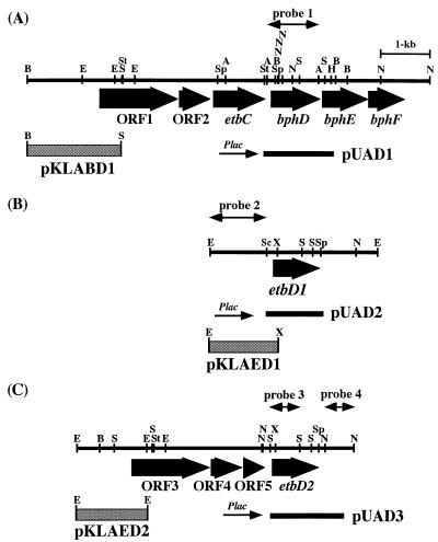
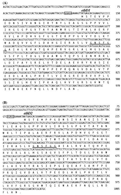
In RHA1, the HPDA hydrolase gene, bphD, was preceded by the etbC gene, which encodes an alternative meta-cleavage dioxygenase, and was preferentially induced by ethylbenzene (11, 20). Two open reading frames (ORFs), ORF1 and ORF2, were located adjacent to and upstream from etbC and were homologous to bphA1 and bphA2, respectively (unpublished data). These ORFs seem to constitute an operon with etbC and bphD (Fig. 2A). Using part of ORF1 and ORF2 as a hybridization probe, we discovered and cloned another bphA homolog (unpublished data). The nucleotide sequence of the region containing this newly identified bphA homolog indicated that there was an ORF product (Fig. 2C) whose amino-terminal amino acid sequence was significantly similar to the amino-terminal amino acid sequence of purified RHA1 HOHD hydrolase (Fig. 3B). The ORF was designated etbD2. In the amino-terminal sequence consisting of 50 amino acid residues, 47 residues were the same in the etbD2 product and the HOHD hydrolase which was purified from RHA1. The putative ribosome binding site, GGAGG, was separated by seven bases from the ATG initiation codon. The entire etbD2 gene encodes 274 amino acid residues, which include the nucleophile motif Gly-Xaa-Ser-Xaa-Gly-Gly conserved in α/β hydrolase fold enzymes (2). The great similarity between the amino-terminal amino acid sequences of the deduced etbD2 product and the purified HOHD hydrolase prompted us to clone the gene encoding the purified enzyme.
FIG. 2.
Gene organizations of the DNA fragments containing the bphD (A), etbD1 (B), and etbD2 (C) hydrolase genes. Coding regions of the genes or ORFs possibly involved in aromatic compound metabolism are indicated by broad arrows. The solid bars below the gene organizations represent the fragments used to construct subclones of each hydrolase gene, whose designations are indicated on the right. The transcriptional direction of each subclone from the lac promoter of the vector plasmid is indicated by a thin arrow. Double-headed arrows indicate the fragments used for hybridization experiments. The shaded boxes represent the fragments used to construct reporter plasmids, whose designations are given below the boxes. Abbreviations: A, ApaI; B, BamHI; E, EcoRI; H, HindIII; N, NarI; S, SacI; Sc, ScaI; Sp, SphI; St, StuI; X, XhoI.
FIG. 3.
Nucleotide and deduced amino acid sequences of the etbD1 (A) and etbD2 (B) genes of Rhodococcus sp. strain RHA1 and their products. Putative ribosome binding sites and nucleophile motifs of α/β hydrolase fold enzymes are enclosed in boxes and underlined, respectively. Stop codons are indicated by dots.
Cloning of etbD1.
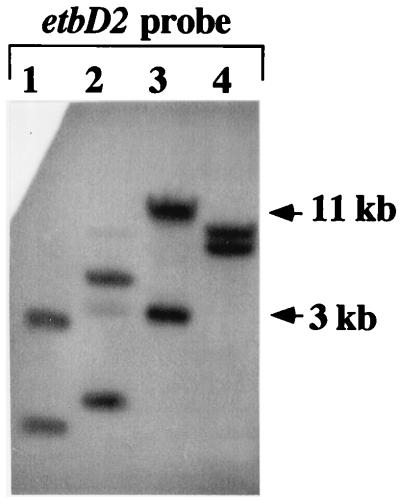
The hybridization experiment was conducted by using a 0.5-kb SacI DNA probe of the etbD2 internal DNA region (Fig. 2, probe 3). In addition to the 11-kb band derived from the etbD2 locus, a 3-kb band hybridizing to the etbD2 probe was observed among the EcoRI fragments of RHA1 total DNA (Fig. 4, lane 3). It was presumed that this 3-kb band originated from the gene encoding the purified HOHD hydrolase. A cosmid library of RHA1 was constructed by using the partial EcoRI digest of total RHA1 DNA and the Rhodococcus-E. coli shuttle cosmid vector pK4HKcos, as described in Materials and Methods. A colony hybridization experiment in which a 0.5-kb SacI fragment of the etbD2 gene was used as a probe yielded 14 positive clones. Only one of these clones contained the 3-kb EcoRI fragment hybridizing with the etbD2 probe that was subcloned into pUC118. The resulting plasmids were designated pKHD1 and pKHD2 and differed from each other in the orientation of the 3-kb insert with respect to the lac promoter of pUC118. The nucleotide sequence of the 3-kb EcoRI fragment was determined by using deletion derivatives of these subclones. The complete nucleotide sequence of the 970-bp ScaI-SphI fragment containing the entire HOHD hydrolase gene is presented in Fig. 3A. The deduced amino acid sequence from the ATG codon at nucleotide position 131 completely agreed with the 50-amino-acid amino-terminal sequence of the purified HOHD hydrolase. Thus, we concluded that the ORF starting at position 131 certainly encodes the RHA1 HOHD hydrolase that was purified, and we designated this ORF etbD1.
FIG. 4.
Southern hybridization analysis of genomic DNA from Rhodococcus sp. strain RHA1 performed with the etbD2 gene probe. The etbD2 gene probe used is shown in Fig. 2 (probe 3). Genomic DNA was digested with SacI, SalI, EcoRI, and BamHI (lanes 1 to 4, respectively).
The etbD1 gene contained an 822-bp sequence encoding a 274-amino-acid polypeptide. The nucleotide sequences of the etbD1 and etbD2 genes are significantly similar (97% identical), and the deduced amino acid sequences also exhibit 97% identity. In addition to the coding region, the 29 bases preceding the initiation codon and the 72 bases following the stop codon were almost identical. The putative ribosome binding site GGAGG separated by seven bases from the ATG initiation codon and the α/β hydrolase fold enzyme nucleophile motif Gly-Xaa-Ser-Xaa-Gly-Gly are also present in the etbD1 gene product. The deduced amino acid sequences of the etbD1 and etbD2 gene products were similar to the amino acid sequences of the products of the single-ring aromatic hydrolase genes (see below).
Substrate preference of RHA1 hydrolases produced in E. coli.
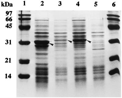
Three hydrolase genes were expressed in E. coli to determine the substrate preferences of their products. The entire coding regions of three hydrolase genes were subcloned into pUC18, and the resulting plasmids (pUAD1, pUAD2, and pUAD3 containing bphD, etbD1, and etbD2, respectively) (Fig. 2) were transformed into E. coli JM109. Crude extracts prepared from each transformant were analyzed by SDS-polyacrylamide gel electrophoresis, and each extract was reacted with HOHD, HPDA, and HMSA, which is the meta-cleavage product of catechol. The proteins with molecular masses of 31.5, 35.0, and 34.0 kDa were identified as bphD, etbD1, and etbD2 products, respectively (BphD, EtbD1, and EtbD2, respectively) (Fig. 5). The etbD1 gene product was poorly expressed. The molecular masses of these products were slightly different from the molecular masses calculated from the deduced amino acid sequences of BphD (31.6 kDa), EtbD1 (30.0 kDa), and EtbD2 (30.0 kDa). The relative enzymatic activities of the crude extracts are shown in Table 2. BphD was highly specific to HPDA, the meta-cleavage product of 23DHBP, and EtbD1 and EtbD2 were specific to HOHD, the meta-cleavage product of 3-methylcatechol. These results agreed with the results obtained for BphD and EtbD1 hydrolases purified from the RHA1 cells (10). The enzyme activities were very weak on HMSA.
FIG. 5.
Expression of the bphD, etbD1, and etbD2 genes in E. coli. Putative gene products are indicated by arrowheads. Cell extracts of E. coli transformants grown in the presence of isopropyl-β-d-thiogalactopyranoside were subjected to SDS–15% polyacrylamide gel electrophoresis. Lanes 1 and 6, molecular mass markers, including phosphorylase (97 kDa), bovine serum albumin (66 kDa), ovalbumin (45 kDa), carbonic anhydrase (31 kDa), soybean trypsin inhibitor (21 kDa), and lysozyme (14 kDa); lane 2, E. coli JM109(pUAD1 carrying bphD); lane 3, E. coli JM109(pUAD2 carrying etbD1); lane 4, E. coli JM109(pUAD3 carrying etbD2); lane 5, E. coli JM109(pUC18).
Expression of the hydrolase genes in RHA1.
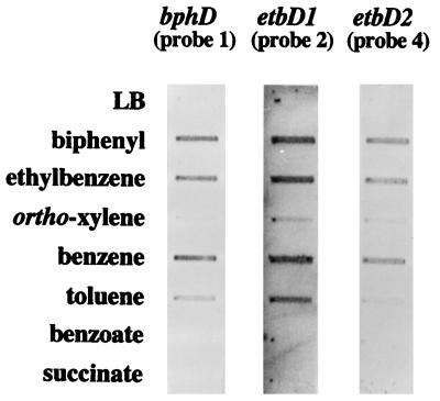
To study the transcription of the three hydrolase genes in RHA1, an RNA slot blot hybridization analysis was performed. Total RNAs were extracted from RHA1 cells grown on various carbon sources and were blotted onto a nylon membrane. DNA probes specific to each of the three hydrolase genes were designed. The outside regions of the etbD1 and etbD2 genes were employed because of the homology between etbD1 and etbD2 (Fig. 2, probes 1, 2, and 4). The results are presented in Fig. 6. Interestingly, all of the hydrolase genes showed almost the same induction pattern. They were induced strongly by biphenyl, ethylbenzene, and benzene and weakly by toluene and ortho-xylene. No induction was observed in the cells grown in LB and on succinate and benzoate.
FIG. 6.
RNA slot blot hybridization analysis of bphD-, etbD1-, and etbD2-specific transcripts in Rhodococcus sp. strain RHA1. Two micrograms of each total RNA from RHA1 cells grown on the substrates indicated was blotted onto a nylon membrane and hybridized with DIG-labeled probes 1, 2, and 4 (Fig. 2) specific to bphD, etbD1, and etbD2, respectively.
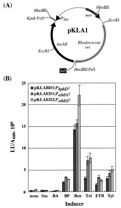
To confirm these results, the promoter activity in the upstream region of each hydrolase gene was examined. Each of the upstream regions indicated in Fig. 2 was inserted into the site preceding the luxAB luciferase reporter gene in the promoter probe vector pKLA1 (Fig. 7A) (28), which was derived from an E. coli-Rhodococcus shuttle vector, pK4. The resultant plasmids were designated pKLABD1, pKLAED1, and pKLAED2 and contained the putative promoter regions of bphD, etbD1, and etbD2, respectively. The RHA1 cells harboring these plasmids were subjected to luciferase assays. The promoter activities are shown in Fig. 7B, and these activities were basically consistent with the results obtained in the RNA slot blot hybridization analyses. The activities were induced by biphenyl, ethylbenzene, toluene, and ortho-xylene. There was strong induction by benzene. No induction was observed in cells grown in LB and on succinate and benzoate.
FIG. 7.
(A) Physical map of the promoter probe vector, pKLA1. (B) Luciferase activities of Rhodococcus sp. strain RHA1 harboring reporter plasmid derivatives. pKLABD1, pKLAED1, and pKLAED2 contain the promoter regions of bphD, etbD1, and etbD2, respectively, which were inserted at the SalI site preceding the luxAB reporter genes of the promoter probe vector, pKLA1. Data are averages from triplicate determinations in at least three independent experiments; error bars are shown. The luciferase activities of RHA1 cells harboring pKLA1 in 0.2× LB, 0.2× LB containing biphenyl, and 0.2× LB supplemented with ethylbenzene were less than 0.1 × 10⁶ light units per A600 unit. Inducer compound abbreviations: Suc, succinate; BA, benzoate; BP, biphenyl; Ben, benzene; Tol, toluene; ETB, ethylbenzene; Xyl, ortho-xylene. LU, light units.
Hydrolase genes in the mutant strains.
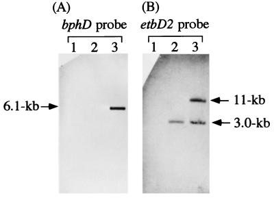
Southern hybridization experiments with the spontaneous growth-defective mutant strains on biphenyl were conducted to obtain insight into the function of the two etbD genes. The deletion of the bphD gene in RCD1 was shown to be responsible for the growth defect on biphenyl in a complementation experiment in which a bphD recombinant plasmid was used (26). RCD1 can grow on ethylbenzene, but RCAD1 cannot. As illustrated in Fig. 8, the etbD2 gene, as well as bphD, was deleted in RCD1. In RCAD1, not only the bphD and etbD2 genes but also etbD1 was deleted. Both strain RHA1 and strain RCD1 grew well on ethylbenzene and transiently accumulated yellow substances, indicating that the meta-ring cleavage pathway responsible for ethylbenzene catabolism was present. These results imply that the etbD1 gene product is involved in the assimilation of ethylbenzene via the meta-ring cleavage catabolic pathway.
FIG. 8.
Southern hybridization analysis of mutant strains defective for growth on biphenyl. Genomic DNAs from RHA1, RCD1, and RCAD1 were digested with EcoRI and hybridized with the bphD gene probe (A) (probe 1 in Fig. 2) and the etbD2 gene probe (B) (probe 3 in Fig. 2). Lanes 1, RCAD1 (Bph− Etb−); lanes 2, RCD1 (Bph− Etb+); lanes 3, RHA1 (Bph+ Etb+).
DISCUSSION
Here we present evidence which indicates that expression of three meta-cleavage compound hydrolase genes is induced by aromatic compounds, including biphenyl, ethylbenzene, and benzene. The bphD gene product is highly specific to HPDA. The etbD1 and etbD2 gene products are specific to HOHD.
In the products of these three genes, the nucleophile motif Gly-Xaa-Ser-Xaa-Gly-Gly of α/β hydrolase fold enzymes is conserved. Inhibition of the BphD and EtbD1 enzymes of RHA1 by a serine-specific inhibitor, phenylmethanesulfonyl fluoride, has been reported previously (10). It has been reported that replacement of a serine residue by alanine in this motif in Pseudomonas putida mt-2 XylF and in Comamonas testosteroni B-356 BphD results in a complete loss of activity (1, 6).
The etbD1 and etbD2 transcripts were induced similarly in RHA1 cells by aromatic compounds, such as biphenyl, ethylbenzene, and benzene. However, only the etbD1-encoded hydrolase was purified from RHA1 by Hatta et al. (10). This may suggest that posttranscriptional hindrance of production occurs or that the etbD2-encoded hydrolase is unstable. In contrast, production of the etbD1-encoded hydrolase was poor in E. coli. This may be because an AGG codon for arginine is used in etbD1 which is known to be recognized by a very rare tRNA in E. coli (15).
The similar induction of bphD, etbD1, and etbD2 genes by aromatic compounds suggested that the expression of these genes is coregulated in RHA1. In RHA1, the biphenyl dioxygenase gene encoding the first step in the biphenyl/PCB degradation pathway is induced by both biphenyl and ethylbenzene (unpublished data). The two meta-ring cleavage enzymes encoded by bphC and etbC are also induced by both biphenyl and ethylbenzene (11). Thus, multiple degradative enzymes that exhibit activities against a variety of aromatic compounds seem to be simultaneously induced by a single substrate in RHA1, although the genes are located in separate operons. Such loosely regulated multiple degradative enzyme systems may provide an advantage in that broader degradation substrate specificity can be acquired by RHA1. The bphD-encoded hydrolase was responsible for biphenyl degradation (20, 26). Based on the substrate specificities of the hydrolases encoded by etbD1 and etbD2, it appears that these two enzymes are involved in toluene and ethylbenzene degradation. Transient accumulation of yellow substances during the growth of RHA1 and RCD1 on ethylbenzene indicated that ethylbenzene catabolism occurred through the meta-cleavage pathway. The hybridization experiment results (Fig. 8) indicated that etbD1 is the only meta-cleavage compound hydrolase gene expressed in RCD1, which grew on ethylbenzene but not on biphenyl. These results suggested that at least the etbD1 gene is involved in the catabolism of ethylbenzene.
In the putative amino acid sequences of the etbD1 and etbD2 gene products, 267 of 274 amino acid residues are conserved. The substrate preferences presented in Table 2 imply that the two enzymes are alike. We suggest that one of the two etbD genes may have been generated recently by gene duplication. The bphD gene is accompanied by ORF1 and ORF2 (homologous to bphA1A2), as well as the etbC, bphE, and bphF genes, while the etbD2 gene is linked to ORF3, ORF4, and ORF5, which are homologs of bphA1, bphA2, and bphA3, respectively. However, no sequence homology to any aromatic-compound degradation gene was found in the vicinity of the etbD1 gene. Duplication of etbD2 in the operon containing bphA homologs and insertion into a separate locus might have been responsible for the formation of the etbD1 gene.
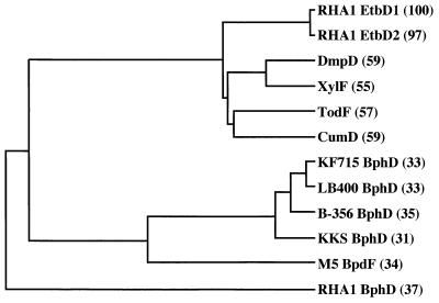
The EtbD1 and EtbD2 hydrolases encoded by the etbD1 and etbD2 genes, respectively, exhibited considerable identity (55 to 59%) with DmpD, CumD, TodF, and XylF hydrolases. These six hydrolases constitute a subfamily on the phylogenetic tree presented in Fig. 9. The BphD hydrolases encoded by bphD genes in gram-negative bacterial strains are closely related and form another subfamily. The BpdF hydrolase from gram-positive Rhodococcus sp. strain M5 seems to be a member of this subfamily, although its similarity with the gram-negative BphD hydrolases is not very significant. In contrast, the RHA1 BphD hydrolase is unique. The biphenyl/PCB degradation pathway genes of RHA1, including bphA1A2A3A4, bphB, bphC, and bphD, would have evolved separately from the genes of gram-negative PCB degraders.
FIG. 9.
Phylogenetic tree for EtbD1, EtbD2, BphD, and related hydrolases. The tree was deduced from pairwise alignments of amino acid sequences by using an unweighted pair group method. Enzyme abbreviations: DmpD, dmpD product of Pseudomonas putida CF600 (22); XylF, xylF product of P. putida mt-2 (14); TodF, todF product of P. putida F1 (21); CumD, cumD product of Pseudomonas fluorescens IP01 (8); KF715 BphD, bphD product of P. putida KF715 (12); LB400 BphD, bphD product of Pseudomonas sp. strain LB400 (13); B-356 BphD, bphD product of Comamonas testosteroni B-356 (1); KKS BphD, bphD product of Pseudomonas sp. strain KKS102 (16); M5 BpdF, bpdF product of Rhodococcus sp. strain M5 (18). The percentages of amino acid sequence identity with EtbD1 are shown in parentheses.
It has been suggested that the bphD, etbD1, and etbD2 genes of RHA1 are coregulated, as mentioned above. It would be interesting to know the regulatory mechanism of these three genes. A detailed study to elucidate the regulatory elements and genes is now in progress, and this study should provide insight into the evolution of degradation pathway genes in RHA1.
ACKNOWLEDGMENTS
We are grateful to W. Mizunashi, Nitto Chemical Industry, and S. Horinouchi, The University of Tokyo, for providing pK4.
This study was supported in part by the Promotion of Basic Research Activities for Innovative Bioscience (PROBRAIN) in Japan.
REFERENCES
- 1.Ahmad D, Fraser J, Sylvestre M, Larose A, Khan A, Bergeron J, Juteau J M, Sondossi M. Sequence of the bphD gene encoding 2-hydroxy-6-oxo-(phenyl/chlorophenyl)hexa-2,4-dienoic acid (HOP/cPDA) hydrolase involved in the biphenyl/polychlorinated biphenyl degradation pathway in Comamonas testosteroni: evidence suggesting involvement of Ser112 in catalytic activity. Gene. 1995;156:69–74. doi: 10.1016/0378-1119(95)00073-f. [DOI] [PubMed] [Google Scholar]
- 2.Arand M, Grant D F, Beetham J K, Friedberg T, Oesch F, Hammock B D. Sequence similarity of mammalian epoxide hydrolases to the bacterial haloalkane dehalogenase and other related proteins. FEBS Lett. 1994;338:251–256. doi: 10.1016/0014-5793(94)80278-5. [DOI] [PubMed] [Google Scholar]
- 3.Asturias J A, Timmis K N. Three different 2,3-dihydroxybiphenyl-1,2-dioxygenase genes in the gram-positive polychlorobiphenyl-degrading bacterium Rhodococcus globerulus P6. J Bacteriol. 1993;175:4631–4640. doi: 10.1128/jb.175.15.4631-4640.1993. [DOI] [PMC free article] [PubMed] [Google Scholar]
- 4.Ausubel F M, Brent R, Kingston R E, Moore D D, Seidman J G, Smith J A, Struhl K. Current protocols in molecular biology. New York, N.Y: John Wiley & Sons, Inc.; 1990. [Google Scholar]
- 5.Bradford M M. A rapid and sensitive method for the quantitation of microgram quantities of protein utilizing the principle of protein-dye binding. Anal Biochem. 1976;72:248–254. doi: 10.1016/0003-2697(76)90527-3. [DOI] [PubMed] [Google Scholar]
- 6.Díaz E, Timmis K N. Identification of functional residues in a 2-hydroxymuconic semialdehyde hydrolase. J Biol Chem. 1995;270:6403–6411. doi: 10.1074/jbc.270.11.6403. [DOI] [PubMed] [Google Scholar]
- 7.Furukawa K, Hirose J, Suyama A, Zaiki T, Hayashida S. Gene components responsible for discrete substrate specificity in the metabolism of biphenyl (bph operon) and toluene (tod operon) J Bacteriol. 1993;175:5224–5232. doi: 10.1128/jb.175.16.5224-5232.1993. [DOI] [PMC free article] [PubMed] [Google Scholar]
- 8.Habe H, Kasuga K, Nojiri H, Yamane H, Omori T. Analysis of cumene (isopropylbenzene) degradation genes from Pseudomonas fluorescens IP01. Appl Environ Microbiol. 1996;62:4471–4477. doi: 10.1128/aem.62.12.4471-4477.1996. [DOI] [PMC free article] [PubMed] [Google Scholar]
- 9.Hashimoto Y, Nishiyama M, Yu F, Watanabe I, Horinouchi S, Beppu T. Development of a host-vector system in a Rhodococcus strain and its use for expression of the cloned nitrile hydratase gene cluster. J Gen Microbiol. 1992;138:1003–1010. doi: 10.1099/00221287-138-5-1003. [DOI] [PubMed] [Google Scholar]
- 10.Hatta T, Shimada T, Yoshihara T, Yamada A, Masai E, Fukuda M, Kiyohara H. Meta-Fission product hydrolases from a strong PCB degrader Rhodococcus sp. RHA1. J Ferment Bioeng. 1998;85:174–179. [Google Scholar]
- 11.Hauschild J E, Masai E, Sugiyama K, Hatta T, Kimbara K, Fukuda M, Yano K. Identification of an alternative 2,3-dihydroxybiphenyl 1,2-dioxygenase in Rhodococcus sp. strain RHA1 and cloning of the gene. Appl Environ Microbiol. 1996;62:2940–2946. doi: 10.1128/aem.62.8.2940-2946.1996. [DOI] [PMC free article] [PubMed] [Google Scholar]
- 12.Hayase N, Taira K, Furukawa K. Pseudomonas putida KF715 bphABCD operon encoding biphenyl and polychlorinated biphenyl degradation: cloning, analysis, and expression in soil bacteria. J Bacteriol. 1990;172:1160–1164. doi: 10.1128/jb.172.2.1160-1164.1990. [DOI] [PMC free article] [PubMed] [Google Scholar]
- 13.Hofer B, Eltis L D, Dowling D N, Timmis K N. Genetic analysis of a Pseudomonas locus encoding a pathway for biphenyl/polychlorinated biphenyl degradation. Gene. 1993;130:47–55. doi: 10.1016/0378-1119(93)90345-4. [DOI] [PubMed] [Google Scholar]
- 14.Horn J M, Harayama S, Timmis K N. DNA sequence determination of the TOL plasmid (pWW0) xylGFJ genes of Pseudomonas putida: implications for the evolution of aromatic catabolism. Mol Microbiol. 1991;5:2459–2474. doi: 10.1111/j.1365-2958.1991.tb02091.x. [DOI] [PubMed] [Google Scholar]
- 15.Ikemura T. Correlation between the abundance of Escherichia coli transfer RNAs and the occurrence of the respective codons in its protein genes: a proposal for a synonymous codon choice that is optimal for the E. coli translational system. J Mol Biol. 1981;151:389–409. doi: 10.1016/0022-2836(81)90003-6. [DOI] [PubMed] [Google Scholar]
- 16.Kimbara K, Hashimoto T, Fukuda M, Koana T, Takagi M, Oishi M, Yano K. Cloning and sequencing of two tandem genes involved in degradation of 2,3-dihydroxybiphenyl to benzoic acid in the polychlorinated biphenyl-degrading soil bacterium Pseudomonas sp. strain KKS102. J Bacteriol. 1989;171:2740–2747. doi: 10.1128/jb.171.5.2740-2747.1989. [DOI] [PMC free article] [PubMed] [Google Scholar]
- 17.Knauf V C, Nester E W. Wide host range cloning vectors: a cosmid clone bank of an Agrobacterium Ti plasmid. Plasmid. 1982;8:45–54. doi: 10.1016/0147-619x(82)90040-3. [DOI] [PubMed] [Google Scholar]
- 18.Lau P C K, Garnon J, Labbé D, Wang Y. Location and sequence analysis of a 2-hydroxy-6-oxo-6-phenylhexa-2,4-dienoate hydrolase-encoding gene (bpdF) of the biphenyl/polychlorinated biphenyl degradation pathway in Rhodococcus sp. M5. Gene. 1996;171:53–57. doi: 10.1016/0378-1119(96)00025-x. [DOI] [PubMed] [Google Scholar]
- 19.Masai E, Yamada A, Healy J M, Hatta T, Kimbara K, Fukuda M, Yano K. Characterization of biphenyl catabolic genes of gram-positive polychlorinated biphenyl degrader Rhodococcus sp. strain RHA1. Appl Environ Microbiol. 1995;61:2079–2085. doi: 10.1128/aem.61.6.2079-2085.1995. [DOI] [PMC free article] [PubMed] [Google Scholar]
- 20.Masai E, Sugiyama K, Iwashita N, Shimizu S, Hauschild J E, Hatta T, Kimbara K, Yano K, Fukuda M. The bphDEF meta-cleavage pathway genes involved in biphenyl/polychlorinated biphenyl degradation are located on a linear plasmid and separated from the initial bphACB genes in Rhodococcus sp. strain RHA1. Gene. 1997;187:141–149. doi: 10.1016/s0378-1119(96)00748-2. [DOI] [PubMed] [Google Scholar]
- 21.Menn F-M, Zylstra G J, Gibson D T. Location and sequence of the todF gene encoding 2-hydroxy-6-oxohepta-2,4-dienoate hydrolase in Pseudomonas putida F1. Gene. 1991;104:91–94. doi: 10.1016/0378-1119(91)90470-v. [DOI] [PubMed] [Google Scholar]
- 22.Nordlund I, Shingler V. Nucleotide sequences of the meta-cleavage pathway enzymes 2-hydroxymuconic semialdehyde dehydrogenase and 2-hydroxymuconic semialdehyde hydrolase from Pseudomonas CF600. Biochim Biophys Acta. 1990;1049:227–230. doi: 10.1016/0167-4781(90)90046-5. [DOI] [PubMed] [Google Scholar]
- 23.Olsson O, Koncz C, Szalay A A. The use of the luxA gene of the bacterial luciferase operon as a reporter gene. Mol Gen Genet. 1988;215:1–9. doi: 10.1007/BF00331295. [DOI] [PubMed] [Google Scholar]
- 24.Sanger F, Nicklen S, Coulson A R. DNA sequencing with chain-terminating inhibitors. Proc Natl Acad Sci USA. 1977;74:5463–5467. doi: 10.1073/pnas.74.12.5463. [DOI] [PMC free article] [PubMed] [Google Scholar]
- 25.Seto M, Kimbara K, Shimura M, Hatta T, Fukuda M, Yano K. A novel transformation of polychlorinated biphenyls by Rhodococcus sp. strain RHA1. Appl Environ Microbiol. 1995;61:3353–3358. doi: 10.1128/aem.61.9.3353-3358.1995. [DOI] [PMC free article] [PubMed] [Google Scholar]
- 26.Seto M, Okita N, Sugiyama K, Masai E, Fukuda M. Growth inhibition of Rhodococcus sp. strain RHA1 in the course of PCB transformation. Biotechnol Lett. 1996;18:1193–1198. [Google Scholar]
- 27.Vieira J, Messing J. Production of single-stranded plasmid DNA. Methods Enzymol. 1987;153:3–11. doi: 10.1016/0076-6879(87)53044-0. [DOI] [PubMed] [Google Scholar]
- 28.Yamada, A., H. Takeda, T. Hatta, K. Nakamura, E. Masai, and M. Fukuda. Unpublished data.
- 29.Yanisch-Perron C, Vieira J, Messing J. Improved M13 phage cloning vectors and host strains: nucleotide sequences of the M13mp18 and pUC19 vectors. Gene. 1985;33:103–119. doi: 10.1016/0378-1119(85)90120-9. [DOI] [PubMed] [Google Scholar]


