



Abstract
Patients with extensive and complex wounds due to Necrotizing Soft-Tissue Infections (NSTI) may be referred to a burn center. This study describes the characteristics, outcomes, as well as diagnostic challenges of these patients. Patients admitted to three hospitals with a burn center for the treatment of NSTI in a 5-year period were included. Eighty patients (median age 54 years, 60% male) were identified, of whom 30 (38%) were referred by other centers, usually after survival of the initial septic phase. Those referred from other centers, compared to those primarily admitted to the study hospitals, were more likely to have group A streptococcal involvement (62% vs 35%, p = .02), larger wounds (median 7% vs 2% total body surface area, p < .001), and a longer length of stay (median 49 vs 22 days, p < .001). Despite a high incidence of septic shock (50%), the mortality rate was low (12%) for those primarily admitted. Approximately half (53%) of the patients were initially misdiagnosed upon presentation, which was associated with delay to first surgery (16 hours vs 4 hours, p < .001). Those initially misdiagnosed had more (severe) comorbidities, and less frequently reported pain or blue livid discoloration of the skin. This study underlines the burn centers’ function as referral centers for extensively affected patients with NSTI. Besides the unique wound and reconstructive expertise, the low mortality rate indicates these centers provide adequate acute care as well. A major remaining challenge remains recognition of the disease upon presentation. Future studies in which factors associated with misdiagnosis are explored are needed.
INTRODUCTION
Necrotizing Soft-Tissue Infections (NSTI) are severe infections characterized by progressive soft-tissue destruction, and often systemic toxicity that may progress to septic shock. It may be monomicrobial (type 2 NSTI), most frequently due to a Group A Streptococcus (GAS), or polymicrobial (type 1 NSTI) in origin.1 While it may affect all body areas, it most often affects the legs, followed by the anogenital and abdominal area.2 When NSTI is suspected, emergency surgical inspection is indicated, followed by surgical debridement when the diagnosis is conformed.3 Adjacently, broad-spectrum intravenous antibiotics must be started, and most will be admitted to the Intensive Care Unit (ICU) as part of multimodal treatment. Mortality has historically been around 30%, though advances in treatment seem to indicate decreasing mortality,4 ranging from 10 to 18% in recent studies.2,5–8
NSTI and its management bear many similarities with those of patients with severe burns, including extensive wounds, reconstructive challenges, sometimes amputation, generalized weakness, and post-traumatic stress symptoms. This is why patients with NSTI in The Netherlands may be referred to a burn center for multidisciplinary treatment by a team that includes general surgeons, burn surgeons, plastic surgeons, intensivists, medical microbiologists, general and burn specialized nurses, occupational therapists, psychologists, rehabilitation physicians and aftercare nurses. The national Dutch guideline on NSTI recommends considering referral to a specialized center (burn center) in case of wounds in a complex area (head/neck, genital, hands, feet) or in case of >10% TBSA wound size.9 These patients can be classified as secondary referred, in contrast to those admitted after presentation to the Emergency Department of the community hospitals these burn centers are located in (primary referred). Hence, there is a focus of the multidisciplinary teams involved these centers on both acute, lifesaving care, as well as reconstructive and rehabilitative care to improve long-term quality-of-life.
Due to the experience with NSTI in these centers, as well as burn expertise, the question arises whether this results in differences in regards to the patient population, treatment characteristics, and outcomes, when compared to existing literature. Furthermore, it is interesting to explore whether the focus on NSTI care may decrease the rate of misdiagnosis, which has been reported to range from 41 to 96% in other studies.10 These questions formed the basis of this study.
METHODS
A retrospective cohort study was conducted in all three Dutch Burn Centers and the community hospitals they are part of (Red Cross Hospital, Maasstad Hospital and Martini Hospital). In Table 1, the unique characteristics of care in these centers are displayed. The Medical Ethical Committee of the Amsterdam University Medical Centers agreed this study is not subject to the Medical Research Involving Human Subjects Act (WMO) and that the nature of the study does not require written informed consent. Institutional Review Board (IRB) approval was obtained in all three hospitals.
Table 1.
Overview of characteristics of care in burn centers contributing to its unique properties to care for patients affected by NSTI.
| • One department combining wound-, reconstructive-, and ICU care |
| • Complete infrastructure designed to treat patients with complex and extensive wounds, including its own operating theatre and planning |
| • Nurses specialized in the daily care of complex wounds with increased attention fort he patient (1 nurse per 2 patients) |
| • Specialized pain management team able to deal with pain due to extensive wounds |
| • Specialized early reconstructive care for complex and/or extensive wounds |
| • Multidisciplinary teams to meet all physical, psychological, rehabilitative, and social needs of patiënts with complex and/or large wounds with multi-disciplinary evaluations three times a week |
| • Specialized aftercare nurse and the availability of peer support groups |
| • Multi-disciplinary outpatient follow-up for long-term reconstructive, rehabilitative and psychosocial care |
Patient Identification
All patients diagnosed with, and treated for acute NSTI between January 1, 2013 until December 31, 2017, were eligible for inclusion. Patients were identified according to local possibilities, with up to four strategies; (1) ICD-10 diagnosis codes necrotizing fasciitis (M72.6), inflammatory diseases of the scrotum (N49.2) and gas gangrene (A48.0) from the Dutch version of the ICD-10 codebook,11 (2) Dutch Hospital Data (DHD) registration,12 (3) The Dutch Burn Centres Registry13 (R3), and (4) free text terms. Only those who had NSTI according to the involved surgeon(s) based on perioperative findings or unequivocal clinical signs, or based on the judgment of the pathologist in case of frozen section biopsy, were included.
Data Collection
Data were collected from electronic patient records by three researchers (JS, LFW, and AE) and registered in an online Clinical Report Form (e-CRF), using Castor EDC (www.castoredc.com). Relevant data concerning patient-, disease-, presentation-, and treatment characteristics, various local outcomes (skin defect size, amputations) and general outcomes (mortality, length of stay (LOS)) were collected. To facilitate comparison regarding overall comorbidity severity, the Charlson Comorbidity Index (CCI) was calculated for each patient, including age variables.14 Different comorbidities can contribute one up to six points, depending on the severity, up to a maximum of 29 points. For age, one point is attributed for each decade over 50 years old, with a maximum of four points for patients of 80 years and older. The Laboratory Risk Indicator for Necrotizing Fasciitis (LRINEC) score, developed to differentiate diagnostically between low risk (≤5) and high risk (≥8) of NSTI, was calculated based on the relevant laboratory values at presentation (C-reactive protein, leukocyte count, hemoglobin, sodium, glucose and creatinine).15
Definitions
Regarding recognition, patients were classified as correctly diagnosed when the diagnosis of NSTI was considered upon presentation, and diagnostic surgical inspection and systemic treatment for NSTI was initiated. When NSTI was not considered, they were labelled as “misdiagnosed.” NSTI was classified as in-hospital developed when patients were admitted electively (e.g., planned surgery), when admitted acutely for another diagnosis (e.g., cellulitis) that initially improved without surgery, or when surgery was performed for a soft tissue infection (e.g., drainage of an abscess) with no signs of NSTI during that surgery. Time of presentation was defined as the moment the patient presented to the ER before being admitted, based on the first note made or measurement (vital parameters or lab) performed. Alternatively, in case of in-hospital developed NSTI, as the moment the patient needed unplanned assessment due to the onset of symptoms consistent with NSTI (increasing pain, expanding redness, signs of sepsis). The Sepsis-3 definition was used for septic shock.16 Mortality was defined as death due to any cause within 30 days from admission.
Analysis
For analysis, IBM Statistical Package for the Social Sciences (SPSS) statistics version 27.0 was used. Descriptive statistics were used to describe the cohort. Since the vast majority of data was not normally distributed, continuous data were described by the median and interquartile range (IQR), and discrete data were expressed as number and percentage. For each variable the number and proportion of missing data was reported in the study tables. Differences between the subgroups (primary vs referred and correctly diagnosed vs misdiagnosed) were tested by means of complete case analysis according to data type by the Mann–Whitney U test and the Chi-squared test. Statistical significance was set at alpha <0.05. In addition, independent predictors of misdiagnosis were investigated by a complete case multivariable logistic regression analysis with backward elimination, with P criteria = 0.10.
Results
Eighty patients were identified, of which 50 (62%) were primary referred (i.e., admitted upon first presentation, prediagnosis), and 30 (38%) secondary referred (i.e., referred after initial presentation in another center). As follows from Figure 1, the vast majority (94%) of patients referred to a burn center had undergone at least one surgery in the referring hospital, and most (71%) were past the acute disease phase.
Figure 1.
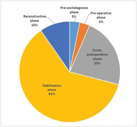
The distribution of different phases in which (n = 30) patients were referred to the burn centers. Five different phases were discerned. Pre-work diagnosis (patient referred before NSTI was suspected), Pre-operative (NSTI suspected, but not yet confirmed surgically), Acute post-operative (Phase from first surgery until detubation, stop of vasopression and CRP <100), Stabilization phase (detubated, no vasopression needed, CRP <100 mg/L until fist reconstructive surgery) and Reconstructive phase (from first reconstructive surgery until discharge).
Patient and Disease Characteristics
As displayed in Table 2, the cohort comprised patients with median age of 54 (IQR 44–69) of whom 51 (64%) were male and about half was overweight (BMI >25). Most were classified as either ASA II (46%) or ASA III (38%). The majority (n = 51, 64%) had one or more comorbidities, most often cardiac (40%), followed by diabetes mellitus (19%), pulmonary (13%), immunosuppression (13%), and active malignancy (6%). The median CCI was 3.0 (IQR 1.0–5.0), and the maximum CCI was 12.0. Of patients of whom the smoking status was known, 36% were active smokers. The location where NSTI most often started were legs (40%) and the anogenital and abdominal area (39%). Culture results, of which a detailed overview of different pathogens in relation to type of NSTI can be found in Supplementary Table 1, revealed that Monomicrobial NSTI was more common (55%) compared to Polymicrobial NSTI (41%). The percent of cases in which GAS was cultured was the only statistically significant difference between primary referred (35%) and secondary referred (62%) patients (p = .02). Other characteristics that differed between primary referred and secondary referred patients were respectively age (median 57 vs 50 years), ASA classification ≥3 (median 48% vs 26%), diabetes mellitus (26% vs 7%) and pulmonary comorbidity (18% vs 3%). However, none of these differences was statistically significant. As more in detail displayed in Table 2, the proportion of missing data was small and varied somewhat for patient characteristics (range 0–16%) and disease characteristics (range 0–5%).
Table 2.
Baseline and disease characteristics of the complete cohort (n = 80), as well as for primary referred (n = 50), and secondary referred (n = 30) patients.
| Total (80) | Primary referred (50) | Secondary referred (30) | P-value | |
|---|---|---|---|---|
| Age, (years) median (IQR) | 54 (44 - 69) | 57 (46 - 73) | 50 (39 - 68) | .08 |
| Male, n (%) | 51 (64%) | 31 (62%) | 20 (67%) | .67 |
| BMI, (kg/m2) median (IQR) | 26 (23–30) (m=5) | 27 (24–31) (m=2) | 25 (23–29) (m=3) | .11 |
| ASA status, n (%) | ||||
| I | 11 (14%) | 7 (14%) | 4 (13%) | .20 |
| II | 37 (46%) | 19 (38%) | 18 (60%) | |
| III | 30 (38%) | 23 (46%) | 7 (23%) | |
| IV | 2 (3%) | 1 (2%) | 1 (3%) | |
| Comorbidities, n (%) | ||||
| Any comorbidity | 51 (64%) | 33 (66%) | 18 (60%) | .59 |
| Cardiac | 32 (40%) | 22 (44%) | 10 (33%) | .35 |
| Diabetes mellitus | 15 (19%) | 13 (26%) | 2 (7%) | .03 |
| Pulmonary | 10 (13%) | 9 (18%) | 1 (3%) | .06 |
| Active malignancy | 5 (6%) | 3 (6%) | 2 (7%) | .91 |
| Immunodeficiency | 10 (13%) | 6 (12%) | 4 (13%) | .86 |
| CCI score, median (IQR) | 3.0 (1.0 – 5.0) | 3.0 (1.0 – 6.0) | 3.0 (1.0 – 4.0) | .16 |
| NSTI in history, n (%) | 0 (0%) | 0 (0%) | 0 (0%) | 1.00 |
| Actively smoking, n (%) | 29 (36%) (m=13) | 19 (38%) (m=9) | 10 (33%) (m=4) | .70 |
| Location start NSTI, n (%) | ||||
| Head/neck | 1 (1%) | 0 (0%) | 1 (3%) | .61 |
| Arm/chest | 16 (20%) | 10 (20%) | 6 (20%) | |
| Anogenital/abdominal | 31 (39%) | 19 (38%) | 12 (40%) | |
| Leg | 32 (40%) | 21 (42%) | 11 (37%) | |
| Type of NSTI, n (%) | m=4 | m=1 | m=3 | |
| Monomicrobial | 42 (55%) | 25 (51%) | 17 (63%) | .60 |
| Polymicrobial | 31 (41%) | 22 (45%) | 9 (33%) | |
| No growth | 3 (4%) | 2 (4%) | 1 (4%) | |
| GAS cultured, n (%) | 35 (45%) (m=2) | 17 (35%) (m=1) | 18 (62%) (m=1) | .02 |
Note: m, missing; ASA, American Society of Anesthesiologists; BMI, Body Mass Index; CCI, Charlson Comorbidity Index; NSTI, Necrotizing Soft-Tissue Infection; GAS, Group A Streptococcus.
Presentation Characteristics
As displayed in Table 3, the majority of the patients (n = 72, 90%) developed NSTI outside of the hospital setting. Pain was the symptom most often described as being present (82%), followed by swelling (76%) and erythema (72%). Pain was less frequently reported in those with anogenital or abdominal involvement (72%) compared to those with leg involvement (87%), though not significantly (p = .17). No association with type of NSTI (Monomicrobial 81% vs Polymicrobial 83%) and or presence of GAS (present 79% vs absent 82%) was found either. Late signs of NSTI (bullae, blue livid discoloration, ecchymosis, skin necrosis), as well as fever, were absent in the majority. The medical records of secondary referred patients, compared to primary referred patients, more often reported a blunt trauma (26% vs 4%) preceding the start of symptoms, and less often (58% vs 80%) erythema. Since the rates of missing data for fever, laboratory values and combined LRINEC score were extensive (range 57–77%) for secondary referred patients, this data is only reported for primary referred patients. Of those, most had a portal of entry (81%). Approximately half (53%) had a LRINEC score indicating a high risk of NSTI (≥8), while 30% had a score corresponding with a low risk (≤5). Imaging studies were performed (ultrasound, CT-scan, MRI-scan) in 18, 28, and 1% of patients, respectively, with rate of findings consistent with NSTI being 57% for ultrasound (false negative rate 43%), and 86% for CT-scans (false negative rate 14%).
Table 3.
Presentation characteristics of the complete cohort (n = 80) of patients included, as well as for primary referred (n = 50) and secondary referred (n = 30) patients.
| Total (80) | Primary referred (50) | Secondary referred (30) | P-value | |
|---|---|---|---|---|
| Hospital acquired NSTI, n (%) | 8 (10%) (m=1) | 4 (8%) (m=1) | 3 (10%) | .72 |
| Portal of entry present, n (%) | 56 (81%) (m=11) | 38 (79%) (m=2) | 18 (86%) (m=9) | .52 |
| Blunt trauma, n (%) | 9 (11%) (m=3) | 2 (4%) | 7 (26%) (m=3) | .004 |
| Localized symptom present at presentation, n (%) | (m=4) | (m=4) | ||
| Pain | 62 (82%) | 41 (82%) | 21 (81%) | .90 |
| Erythema | 55 (72%) | 40 (80%) | 15 (58%) | .04 |
| Swelling | 58 (76%) | 41 (82%) | 17 (65%) | .11 |
| Bullae | 11 (15%) | 6 (12%) | 5 (19%) | .40 |
| Blue livid discoloration | 10 (13%) | 6 (12%) | 4 (15%) | .68 |
| Skin necrosis | 11 (15%) | 7 (14%) | 4 (15%) | .87 |
| Ecchymosis | 4 (5%) | 3 (6%) | 1 (4%) | .69 |
| Crepitations | 4 (5%) | 2 (4%) | 2 (8%) | .49 |
| Any early localized symptom,* n (%) | 73 (96%)(m=4) | 48 (96%) | 25 (96%) (m=4) | .97 |
| Any late localized symptom,** n (%) | 26 (34%)(m=4) | 16 (32%) | 10 (39%) (m=4) | .57 |
| Only from primary referred (n=50) *** | ||||
| Fever (>38.5 degrees Celsius), n (%) | 10 (21%) (m=2) | |||
| Leukocyte count (109/litre) median (IQR) | 17 (13–24) (m=2) | |||
| CRP (milligram/litre) median (IQR), | 305 (179–392) (m=2) | |||
| Creatinine (µmoll/litre) median (IQR), | 119 (88–211) (m=3) | |||
| LRINEC score at presentation | (m=10) | |||
| Total score, median (IQR) | 8 (4.3–9.0) | |||
| Low risk (score ≤5), n (%) | 12 (30%) | |||
| Intermediate risk (score 6-7), n (%) | 7 (18%) | |||
| High risk (score ≥8), n (%) | 21 (53%) |
Note: NSTI, Necrotizing soft-tissue infection; m, missing; HR, Heart rate; IQR, Interquartile range; MAP, Mean arterial pressure; CRP, C-reactive protein; GAS, Group A streptococcus; NA, = Not applicable.
*Early symptoms: pain, swelling, erythema.
**Late symptoms: blue livid discoloration, ecchymosis, blisters, necrosis.
***Since this data lacked for most secondary referred patients, only those directly admitted are described here.
Treatment and Outcomes
All patients received antibiotics according to local protocols, first empiric, and subsequently tailored based on gram staining and later definite culture results, as outlined in Supplementary Table 2. All patients underwent diagnostic surgical inspection to confirm the diagnosis of NSTI, except for two (3%) patients with unequivocal clinical signs of NSTI in which direct amputation was performed without diagnostic surgical inspection of affected tissues. In most patients the diagnosis was made based on perioperative findings, in three (4%) based on frozen section biopsy. As displayed in Table 4, most patients (n = 68, 85%) were admitted to the ICU. More than half of the patients (55%) had septic shock, with secondary referred patients having a higher rate (63%). Thirty-day in-hospital mortality was 10% (n = 8) for the whole cohort, and 12% (n = 6) within the subgroup of primary referred patients. The two (7%) patients who died in the secondary referred group, died due to the consequences of acute NSTI (irreversible septic shock), and were transferred before the diagnosis NSTI was made in one, and in the acute postoperative phase in the other (Figure 1). Amputation was performed in 13 (17%) patients. The median size of the skin defect remaining after the last debridement surgery was performed, was 6.0% Total Body Surface Area (TBSA) (IQR 1.6–10.0%), ranging from 0.0 to 44.0% TBSA. In 13 (17%) patients, complete delayed primary closure was achieved, meaning no grafts were needed. In others, either partial primary closure, skin transpositions, or skin grafting was performed. The median TBSA of transplanted skin was 5.5% (IQR 2.0–8.0%). Patients referred to a burn center (secondary referred), compared to primary referred patients, had more extensive disease progression in the fascial layer (median 9.0% vs 4.0% TBSA, p < .001) and more extensive skin defects (median 7.0% vs 2.0% TBSA, p < .001). They also had a longer ICU LOS (median 12 vs 4 days, p < .001), total LOS (median 49 vs 22 days, p < .001), underwent more surgeries (p < .001) and required more skin transplants (median 7.0 vs 2.0 TBSA, p < .001). Missing data varied for both treatment characteristics (range 0–18%) and outcomes (0–27%) (Table 4).
Table 4.
Treatment characteristics and outcomes of the complete cohort (n = 80), as well as for primary referred (n = 50) and secondary referred (n = 30) patients.
| Total (80) | Primary referred (50) | Secondary referred (30) | P-value | |
|---|---|---|---|---|
| ICU admission, n (%) | 68 (85%) | 40 (80%) | 28 (93%) | .11 |
| ICU LOS* (days), median (IQR) | 6.0 (3.0–15.0) (m=1) | 4.0 (1.0–8.0) | 12.0 (6.0–21.0) (m=1) | <.001 |
| Septic Shock, n (%) | 44 (55%) | 25 (50%) | 19 (63%) | .08 |
| Total LOS (days), median (IQR) | 33 (16 - 52) | 22 (11-42) | 49 (30-57) | <.001 |
| 30-day mortality, n (%) | 8 (10%) | 6 (12%) | 2 (7%) | .44 |
| Debridement performed, n (%) | 78** (98%) | 48** (96%) | 30 (100%) | .27 |
| Time until first debridement (hours), median (IQR) | 6.1 (3.6–16.9) (m=18) | 5.5 (3.7–17.0) (m=2) | 8.5 (3.3–17.4) (m=16) | .87 |
| Skin-sparing approach to debridement, n (%) | 23 (30%) | 19 (40%) | 4 (13%) | .01 |
| Number of surgeries | ||||
| Total (n = 78), median (IQR) | 4.0 (2.0–7.0) (m=3) | 3.0 (2.0–4.0) (m=2) | 7.0 (4.5–10.5) (m=1) | <.001 |
| Debridement surgeries (n = 78), median (IQR) (m=3) | 2.5 (2.0–5.0) (m=3) | 2.0 (1.0–3.0) (m=2) | 5.0 (3.0–6.0) (m=2) | <.001 |
| Reconstructive surgeries (n=60), median (IQR) | 1.0 (1.0–3.0) | 1.00 (1.00–1.75) | 2.00 (1.00–5.50) | <.001 |
| Amputation, n (%) | ||||
| Total | 13 (17%) | 8 (16.7%) | 5 (16.7%) | 1.00 |
| On lower extremity | 7 (9%) | 4 (8.3%) | 3 (10.0%) | .80 |
| On upper extremity | 1 (1%) | 1 (2.1%) | 0 (0.0%) | .43 |
| Genital (penis/testis) | 3 (4%) | 2 (4.2%) | 1 (3.3%) | .85 |
| Breast | 2 (3%) | 1 (2.1%) | 1 (3.3%) | .73 |
| Size of fascial wound (TBSA), median (IQR) | 8.0 (2.5–11.0) (m=21) | 4.0 (1.6–8.0) (m=20) | 9.0 (7.0–13.5) (m=1) | <.001 |
| Size of skin defect (TBSA), median (IQR) | 6.0 (1.6–10.0) (m=18) | 2.0 (0.1–7.0) (m=17) | 7.0 (6.0–12.0) (m=1) | <.001 |
| Total delayed primary closure, n (%) | 13 (16%) | 12 (24%) | 1 (3%) | .02 |
| Area grafted***(TBSA), median (IQR) | 5.5 (2.0–8.0) (m=12) | 2.0 (0.0–5.0) (m=9) | 7.0 (6.0–11.5) (m=3) | <.001 |
Note: ICU, Intensive Care Unit; LOS, Length of Stay; IQR, Interquartile range; m, missing; TBSA, Total Body Surface Area.
*Among n = 68 patients that were admitted to the ICU.
**In two patients debridement was not performed due to advanced, inoperable NSTI.
***Among n = 60 patients that did not die or did not undergo amputation.
Exploration of Misdiagnosis and Related Factors
Of all patients included in this study, half (n = 40, 53%) were initially misdiagnosed upon first admission, and this rate was similar for primary referred (n = 26, 53%) and secondary referred patients (n = 14, 52%). Therefore, no distinction between primary referred and secondary referred patient was made for the analysis of misdiagnosis. In those initially misdiagnosed, the alternative diagnosis was most often another skin or soft tissue infection (n = 29, 73%), usually erysipelas, cellulitis, or an abscess. An infection with different localization (i.e., gastro-enteritis, pneumonia, urinary tract infection, or viral infection) was the alternative diagnosis in eight (20%). Five (12%) patients were initially diagnosed with a noninfectious disease, which were limb ischemia, soft tissue tumor, acromioclavicular arthrosis, deep venous thrombosis, and kidney failure.
Comparison of all variables described in Tables 2–4, between those correctly diagnosed upon admission (n = 36) and those initially misdiagnosed (n = 40), revealed some statistically significant differences. Patients classified as initially misdiagnosed had a longer interval to first debridement (16.0 vs 4.1 hours, p < .001), and more frequently developed NSTI in-hospital. These patients had more frequently comorbidities (73% vs 50%, p = .04), of increased severity (CCI score 3.0 vs 2.0, p < .01), and an especially large difference for immunosuppression (23% vs 3%, p = .01). Pain (73% vs 92%, p = .04) and blue livid skin discoloration (5% vs 22%, p = .04) were less frequently reported in those initially misdiagnosed compared to those correctly diagnosed (Figure 2). Although not statistically significant, there were considerable differences in the areas primarily affected between both groups. Those initially misdiagnosed, compared to those correctly diagnosed, had significantly more often NSTI of the leg (50% vs 28%), and less often of the anogenital and abdominal area (33% vs 68%). In addition, no significant difference for mortality was observed, although the total number of deaths was higher in those initially misdiagnosed (n = 5, 13%) compared to those correctly diagnosed (n = 2, 6%). A complete overview of the comparison of all these characteristics and their p-values can be found in Supplementary Table 3a-c.
Figure 2.
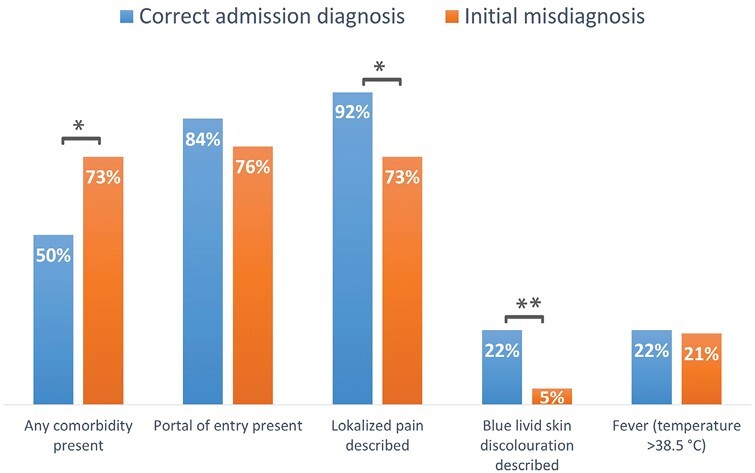
Prevalence of various relevant characteristics of patients presenting with NSTI, for those that were directly diagnosed correctly (n = 36) vs those initially misdiagnosed (n = 40). Differences for those marked with one asterisk (*) are statistically significant with p < .05 when comparing groups, while those marked with two asterisks (**) were also independent, significant predictors within the multivariable regression analysis.
Using multivariable logistic regression analysis the robustness of identified factors associated with misdiagnosis was assessed. Complete case (n = 71, 89%) multivariable logistic regression analysis including data on whether NSTI developed in-hospital or not, CCI comorbidity score, presence of pain, presence of late signs of NSTI, anatomical location of onset of the disease, and GAS involvement, revealed that the absence of reported pain (1.7, p = .04) and a higher CCI comorbidity score (0.19, p = .05) remained independently associated with initial misdiagnosis.
DISCUSSION
This retrospective multicenter cohort study of patients treated in the three Dutch hospitals with a dedicated Burn Centre in which patients with NSTI are regularly treated, confirms that NSTI is a severe disease that affects a heterogeneous population and causes extensive morbidity due to often prolonged ICU admissions, amputations and considerable wounds. Based on the recently estimated yearly number of patients diagnosed with NSTI in the Netherlands (193–238 patients per year), approximately 6.7–8.3% are treated in a burn centre.17 Those referred to a burn center for treatment, are a subgroup of patients who in most cases survived the initial septic shock, and have above average sizes of skin defects for which specialized treatment in a burn center is requested. This selection of survivors may explain the even lower incidence of mortality (7%) in those secondary referred, compared to those primary referred (12%). However, 12% mortality is still low compared to the estimated national Dutch average of 23–29% in the study period,17 as well compared to international literature.2,4–8
Interestingly, although the study centers do have increased exposure to NSTI due to their referral function, misdiagnosis was considerable, although in line (and on the lower end) of reported rates in previous studies of 41–96%.10 Although patients are usually diagnosed before transfer to a burn center, one could have expected misdiagnosis to be lower in the hospitals where these centers are located since the surgical teams working on the ER and ward the same as those involved at the burn centers. Since there was no difference in the rate of misdiagnosis between these centers and referring centers, the findings of this study indicate the diagnosis of NSTI remains a challenging diagnosis despite increased experience. Recognition of NSTI is especially difficult since pathognomonic signs (blue livid skin discoloration, hemorrhagic bullae, skin necrosis) become evident later in the disease process.18 According to a survey among clinicians, misdiagnosis was named as a main modifiable prognostic factor, since it may result in a delay to the first surgery.19 Although misdiagnosis was indeed associated with delay to first surgery in this study, misdiagnosis was not found to be statistically significantly associated with outcomes (mortality or morbidity), although the absolute number of deaths was higher in those initially misdiagnosed. This lack of a statistically different difference is unlikely an indication that delaying the interval to first debridement does not affect outcomes.3,17 It is more likely these lack of differences in outcomes are due to insufficient power of this study do detect these differences, or due to differences in characteristics of those correctly diagnosed vs those misdiagnosed. One such difference in characteristics was patients comorbidity status, with those being misdiagnosed having more often, more severe comorbidity, with the biggest difference observed for immunosuppression. Although one would expect increased comorbidity to be associated with higher mortality, something else may be at play here. Those who are healthy and are infected, may be infected with very virulent pathogens, often Monomicrobial infections such as due to group A streptococcus. Similarly, those with decreased immunity may be infected with less virulent bacteria or bacterial stems, and more often Polymicrobial infections. The finding that legs, associated with Monomicrobial infection, were more often, and anogenital and abdominal area, associated with Polymicrobial infection, less often the anatomical region of onset in those initially misdiagnosed, further supports this.
Besides comorbidity status being independently associated with misdiagnosis, pain was less frequently reported as presenting symptom in charts of patients who were initially misdiagnosed. Although pain is often present, and extreme pain requiring analgesics is accepted as indicative for the presence of NSTI, pain is not always severe and may even be absent. This is supported by findings of a recent study, in which severe pain requiring analgesics upon presentation was noted in less than half of the patients (42%) with NSTI.2 In our study, pain (of any intensity) was reported in 82% of the patients, indicating not all patients with NSTI experience (extreme) pain.
Though not found to be significantly different between those correctly diagnosed and misdiagnosed, some findings are relevant to discuss. First, fever (>38.5 °C) is often absent in NSTI. Although NSTI are severe bacterial infections, which may lead clinicians to expect fever, only 21% of the patients in this study had fever upon presentation. This is in line with previous reports in which only 40% of patients with NSTI had a temperature of >37.5 °C.10 Therefore, clinicians should realize the absence of fever cannot rule out the diagnosis NSTI. This is also true for the presence of a portal of entry; in approximately one in five patients in this study, no portal of entry was observed. This is in concordance with the up to half of the patients with GAS NSTI in which no portal of entry is present. In these patients, NSTI may be associated with blunt trauma, which was present in 11% in this study, or follow sport-related muscle injuries, or without clear local trauma or injury.1 The LRINEC score, which is intended as a tool to help differentiating between patients with a high risk and low risk of NSTI,15 was found to have a considerable false negative rate in this current study of 30%. Therefore, when using this score in practice, clinicians should be aware that it cannot be used to rule out NSTI.20–22 The same is true for ultrasound,23 which had a false negative rate of >40% in our study. CT scans and MRI scans do perform better, which was also true in this study. These imaging modalities may therefore be valuable in case of low suspicion in stable patients, as well as for operative planning in anatomically challenging areas (pelvic area, neck). However, these imaging studies are time-consuming, and should not lead to delay of emergency surgical treatment, which also remains the golden standard for the diagnosis of NSTI.9,24,25
Strengths of this study are its multicenter character, the inclusion of primary and secondary referred patients, and the amount of relevant data collected, especially regarding presentation and diagnostic characteristics. Furthermore, the extensive identification strategy performed, which makes it likely that a representative sample was identified, adds to the strength of this study.
A limitation of this study is it’s retrospective nature, leading to missingness of varying degrees, which may influence this studies’ findings. For example, it could be true that skin defect size and size of the wound in the fascial plane are more often described in those with bigger defects, leading to an overestimation of the severity of this outcome parameter in NSTI patients. Similarly, the estimations made of the fascial/subcutaneous wounds and skin defects when the exact TBSA was not described, could lead to either under- or overestimation. Furthermore, data based on written reports by clinicians remains partly subjective, since not describing a symptom or operative finding, does not mean it was not present. Therefore, the findings of this study, especially the observed differences between those correctly diagnosed and misdiagnosed, should not be regarded as definite proof, and confirmation in future studies is needed. In addition, it remains difficult to be completely sure that those classified as “misdiagnosed” did have NSTI upon presentation, and did not develop shortly after.
Despite the limitations, we believe this study presents a valuable contribution in improving knowledge on NSTI. Several studies regarding NSTI in the burn center setting have been published, but were often smaller,26–31 or had a different focus as this current study.32–34 Therefore, despite not yet providing definite answers, our study is of added value and underlines the relevance of burn centers in the treatment of NSTI. We believe burn centers could have a central role in the care for, and improvement of care and knowledge on NSTI internationally. We believe the findings from this current study indicates the need for the description of larger samples, in which more routinely and systematically collected data regarding presentation and early treatment characteristics are included. In those studies, the relation between various outcomes and patient-, disease-, presentation- and treatment characteristics should be studied. Since RCTs are impractical, or even unethical in case of rare diseases like NSTI, this may initially be done by larger cohort studies. In this light, nationwide prospective registries would be preferable, in which a core set of data are collected, and outcomes (including patient related outcomes) are systematically collected.
CONCLUSIONS
In conclusion, NSTI remains a severe potentially lethal disease that causes extensive morbidity. Burn centers have an important role in the management of patients with extensive wounds due to NSTI, and also provide adequate acute care. Improved outcomes may be achieved by improving recognition, which is currently poor, with half of the patients with NSTI being initially misdiagnosed. We believe the findings from this study indicate the need for future, bigger, preferably prospective studies in which burn centers play a central role. In those studies, the relation between presentation characteristics, recognition and outcomes should further explored.
Supplementary Material
Acknowledgments
The authors would like to thank those who facilitated patient identification and/or access to data in the different centers, including Margriet van Baar, Matthea Stoop, Sonja Scholten, Nicole Trommel, Annette Vink, Robert Jan van Olphen, and Peter Klingeler.
Contributor Information
Jaco Suijker, Burn Centre, Red Cross Hospital, Beverwijk, The Netherlands; Association of Dutch Burn Centres, Beverwijk, The Netherlands; Amsterdam UMC location Vrije Universiteit Amsterdam, Department of Plastic, Reconstructive and Hand Surgery, De Boelelaan 1117, Amsterdam, The Netherlands; Amsterdam Movement Sciences, Tissue Function and Regeneration, Amsterdam, The Netherlands.
Lisca Wurfbain, Burn Centre, Red Cross Hospital, Beverwijk, The Netherlands.
Anouk M L H Emmen, Department of Surgery, Rode Kruis Ziekenhuis, Beverwijk, The Netherlands.
Anouk Pijpe, Burn Centre, Red Cross Hospital, Beverwijk, The Netherlands; Association of Dutch Burn Centres, Beverwijk, The Netherlands; Amsterdam UMC location Vrije Universiteit Amsterdam, Department of Plastic, Reconstructive and Hand Surgery, De Boelelaan 1117, Amsterdam, The Netherlands.
Kelly A A Kwa, Burn Centre, Red Cross Hospital, Beverwijk, The Netherlands.
Cornelis H van der Vlies, Burn Centre, Maasstad Ziekenhuis, Rotterdam, The Netherlands; Trauma Research Unit, Department of Surgery, Erasmus MC, University Medical Centre Rotterdam, Rotterdam, The Netherlands.
Marianne K Nieuwenhuis, Association of Dutch Burn Centers, Burn Centre, Martini Hospital, Groningen, The Netherlands; Centre for Human Movement Sciences, University Medical Centre Groningen, Groningen, The Netherlands; Research group Healthy Ageing, Allied Health Care and Nursing, Hanze University of Applied Sciences Groningen, Groningen, Nederland.
Paul P M van Zuijlen, Burn Centre, Red Cross Hospital, Beverwijk, The Netherlands; Amsterdam UMC location Vrije Universiteit Amsterdam, Department of Plastic, Reconstructive and Hand Surgery, De Boelelaan 1117, Amsterdam, The Netherlands; Amsterdam Movement Sciences, Tissue Function and Regeneration, Amsterdam, The Netherlands; Amsterdam UMC location University of Amsterdam, Pediatric Surgical Centre, Emma Children's Hospital, Meibergdreef 9, Amsterdam, Netherlands.
Annebeth Meij-de Vries, Burn Centre, Red Cross Hospital, Beverwijk, The Netherlands; Department of Surgery, Rode Kruis Ziekenhuis, Beverwijk, The Netherlands; Amsterdam UMC location University of Amsterdam, Pediatric Surgical Centre, Emma Children's Hospital, Meibergdreef 9, Amsterdam, Netherlands.
Source of Funding:
Salary of J. Suijker depended on grant 17.109 by the Dutch Burns Foundation. No other conflicts of interest.
Conflicts of Interest: J. Suijker: Except for the above mentioned funding, none declared. L. Fockema-Wurfbain: None declared
A.M.L.H. Emmen: None declared
A. Pijpe: None declared
K.A.A. Kwa: None declared
C.H. van der Vlies: None declared
M.K. Nieuwenhuis: None declared
P.P.M. van Zuijlen: None declared
A. Meij-de Vries: None declared
References
- 1. Stevens DL, Bryant AE.. Necrotizing soft-tissue infections. N Engl J Med 2017;377:2253–65. doi: 10.1056/NEJMra1600673. [DOI] [PubMed] [Google Scholar]
- 2. Madsen MB, Skrede S, Perner Aet al. ; INFECT study group. Patient’s characteristics and outcomes in necrotising soft-tissue infections: results from a Scandinavian, multicentre, prospective cohort study. Intensive Care Med 2019;45:1241–51. doi: 10.1007/s00134-019-05730-x. [DOI] [PubMed] [Google Scholar]
- 3. Nawijn F, Smeeing DPJ, Houwert RM, Leenen LPH, Hietbrink F.. Time is of the essence when treating necrotizing soft tissue infections: a systematic review and meta-analysis. World J Emerg Surg. 2020;15:4. doi: 10.1186/s13017-019-0286-6. [DOI] [PMC free article] [PubMed] [Google Scholar]
- 4. Ustin JS, Malangoni MA.. Necrotizing soft-tissue infections. Crit Care Med 2011;39:2156–62. doi: 10.1097/CCM.0b013e31821cb246. [DOI] [PubMed] [Google Scholar]
- 5. Tom LK, Maine RG, Wang CS, Parent BA, Bulger EM, Keys KA.. Comparison of traditional and skin-sparing approaches for surgical treatment of necrotizing soft-tissue infections. Surg Infect (Larchmt). 2020;21:363–9. doi: 10.1089/sur.2019.263. [DOI] [PubMed] [Google Scholar]
- 6. Al-Qurayshi Z, Nichols RL, Killackey MT, Kandil E.. Mortality risk in necrotizing fasciitis: national prevalence, trend, and burden. Surg Infect (Larchmt). 2020;21:840–52. doi: 10.1089/sur.2019.277. [DOI] [PMC free article] [PubMed] [Google Scholar]
- 7. Horn DL, Shen J, Roberts Eet al. Predictors of mortality, limb loss, and discharge disposition at admission among patients with necrotizing skin and soft tissue infections. J Trauma Acute Care Surg. 2020;89:186–91. doi: 10.1097/TA.0000000000002636. [DOI] [PMC free article] [PubMed] [Google Scholar]
- 8. Suijker J, de Vries A, de Jong VM, Schepers T, Ponsen KJ, Halm JA.. Health-related quality of life is decreased after necrotizing soft-tissue infections. J Surg Res 2020;245:516–22. doi: 10.1016/j.jss.2019.07.097. [DOI] [PubMed] [Google Scholar]
- 9. Federatie Medisch Specialisten. Richtlijn Necrotiserende wekedeleninfecties. 2018accessed 28 Feb. 2019; Available from: https://richtlijnendatabase.nl/richtlijn/necrotiserende_wekedeleninfecties/startpagina_-_nwdi.html.
- 10. Goh T, Goh LG, Ang CH, Wong CH.. Early diagnosis of necrotizing fasciitis. Br J Surg 2014;101:e119–25. doi: 10.1002/bjs.9371. [DOI] [PubMed] [Google Scholar]
- 11. Nictiz. ProbleemNaamCodelijst. 2017. accessed 19 Dec. 2021; Available from: http://terminologie.nictiz.nl/art-decor/claml?collection=icd10-nl-data.
- 12. Landelijke Basisregistratie Ziekenhuiszorg (LBZ). 2021. accessed 23 Aug. 2021; Available from: https://www.dhd.nl/producten-diensten/LBZ/Paginas/Dataverzameling-LBZ.aspx.
- 13. Dokter J, Vloemans AF, Beerthuizen GIet al. ; Dutch Burn Repository Group. Epidemiology and trends in severe burns in the Netherlands. Burns 2014;40:1406–14. doi: 10.1016/j.burns.2014.03.003. [DOI] [PubMed] [Google Scholar]
- 14. Charlson ME, Pompei P, Ales KL, MacKenzie CR.. A new method of classifying prognostic comorbidity in longitudinal studies: development and validation. J Chronic Dis. 1987;40:373–83. doi: 10.1016/0021-9681(87)90171-8. [DOI] [PubMed] [Google Scholar]
- 15. Wong CH, Khin LW, Heng KS, Tan K-C, Low C-O.. The LRINEC (Laboratory Risk Indicator for Necrotizing Fasciitis) score: a tool for distinguishing necrotizing fasciitis from other soft tissue infections. Crit Care Med 2004;32:1535–41. [DOI] [PubMed] [Google Scholar]
- 16. Singer M, Deutschman CS, Seymour CWet al. The third international consensus definitions for sepsis and septic shock (Sepsis-3). JAMA 2016;315:801–10. doi: 10.1001/jama.2016.0287. [DOI] [PMC free article] [PubMed] [Google Scholar]
- 17. Nawijn F, de Gier B, Brandwagt DAH, Groenwold RHH, Keizer J, Hietbrink F.. Incidence and mortality of necrotizing fasciitis in The Netherlands: the impact of group A Streptococcus. BMC Infect Dis 2021;21:1217. doi: 10.1186/s12879-021-06928-5. [DOI] [PMC free article] [PubMed] [Google Scholar]
- 18. Wong CH, Wang YS.. The diagnosis of necrotizing fasciitis. Curr Opin Infect Dis 2005;18:101–6. [DOI] [PubMed] [Google Scholar]
- 19. de Prost N, Sbidian E, Chosidow O, Brun-Buisson C, Amathieu R; Henri Mondor Hospital Necrotizing Fasciitis Group. Management of necrotizing soft tissue infections in the intensive care unit: results of an international survey. Intensive Care Med 2015;41:1506–8. doi: 10.1007/s00134-015-3916-9. [DOI] [PubMed] [Google Scholar]
- 20. Hsiao CT, Chang CP, Huang TY, Chen Y-C, Fann W-C.. Prospective validation of the Laboratory Risk Indicator for Necrotizing Fasciitis (LRINEC) score for necrotizing fasciitis of the extremities. PLoS One 2020;15:e0227748. doi: 10.1371/journal.pone.0227748. [DOI] [PMC free article] [PubMed] [Google Scholar]
- 21. Neeki MM, Dong F, Au Cet al. Evaluating the laboratory risk indicator to differentiate cellulitis from necrotizing fasciitis in the emergency department. West J Emerg Med. 2017;18:684–9. doi: 10.5811/westjem.2017.3.33607. [DOI] [PMC free article] [PubMed] [Google Scholar]
- 22. Fernando SM, Tran A, Cheng Wet al. Necrotizing soft tissue infection: diagnostic accuracy of physical examination, imaging, and LRINEC score: a systematic review and meta-analysis. Ann Surg 2019;269:58–65. doi: 10.1097/SLA.0000000000002774. [DOI] [PubMed] [Google Scholar]
- 23. Castleberg E, Jenson N, Dinh VA.. Diagnosis of necrotizing faciitis with bedside ultrasound: the STAFF Exam. West J Emerg Med. 2014;15:111–3. doi: 10.5811/westjem.2013.8.18303. [DOI] [PMC free article] [PubMed] [Google Scholar]
- 24. Malghem J, Lecouvet FE, Omoumi P, Maldague BE, Vande Berg BC.. Necrotizing fasciitis: contribution and limitations of diagnostic imaging. Joint Bone Spine. 2013;80:146–54. doi: 10.1016/j.jbspin.2012.08.009. [DOI] [PubMed] [Google Scholar]
- 25. Nawijn F, Hietbrink F, Peitzman AB, Leenen LPH.. Necrotizing soft tissue infections, the challenge remains. Front Surg. 2021;8:721214. doi: 10.3389/fsurg.2021.721214. [DOI] [PMC free article] [PubMed] [Google Scholar]
- 26. Barillo DJ, McManus AT, Cancio LC, Sofer A, Goodwin CW.. Burn center management of necrotizing fasciitis. The Journal of Burn Care & Rehabilitation. 2003;24:127–32. doi: 10.1097/01.bcr.0000066790.57127.61. [DOI] [PubMed] [Google Scholar]
- 27. Redman DP, Friedman B, Law E, Still JM.. Experience with necrotizing fasciitis at a burn care center. South Med J 2003;96:868–70. doi: 10.1097/00007611-200309000-00007. [DOI] [PubMed] [Google Scholar]
- 28. Louro JM, Albano M, Baltazar Jet al. Fournier’s gangrene: 10-year experience of a plastic surgery and burns department at a tertiary hospital. Acta Med Port 2019;32:368–74. doi: 10.20344/amp.11003. [DOI] [PubMed] [Google Scholar]
- 29. Ward JA, Gibson JAG, Nguyen DQ.. Management of necrotising fasciitis within a burns centre: do outcomes differ? Scars Burn Heal. 2020;6:2059513120924749. doi: 10.1177/2059513120924749. [DOI] [PMC free article] [PubMed] [Google Scholar]
- 30. Cui Z, Lu S, Bai Yet al. Necrotizing soft tissue infection: clinical characteristics, diagnosis, and management of 32 cases in Beijing. J Int Med Res 2021;49:3000605211018442. doi: 10.1177/03000605211018442. [DOI] [PMC free article] [PubMed] [Google Scholar]
- 31. Faucher LD, Morris SE, Edelman LS, Saffle JR.. Burn center management of necrotizing soft-tissue surgical infections in unburned patients. Am J Surg 2001;182:563–9. doi: 10.1016/s0002-9610(01)00785-1. [DOI] [PubMed] [Google Scholar]
- 32. Light TD, Choi KC, Thomsen TAet al. Long-term outcomes of patients with necrotizing fasciitis. J Burn Care Res 2010;31:93–9. doi: 10.1097/BCR.0b013e3181cb8cea. [DOI] [PubMed] [Google Scholar]
- 33. Endorf FW, Klein MB, Mack CD, Jurkovich GJ, Rivara FP.. Necrotizing soft-tissue infections: differences in patients treated at burn centers and non-burn centers. J Burn Care Res 2008;29:933–8. doi: 10.1097/BCR.0b013e31818ba112. [DOI] [PMC free article] [PubMed] [Google Scholar]
- 34. Burnett E, Gawaziuk JP, Shek K, Logsetty S.. Healthcare resource utilization associated with burns and necrotizing fasciitis: a single-center comparative analysis. J Burn Care Res 2017;38:e886–91. doi: 10.1097/bcr.0000000000000513. [DOI] [PubMed] [Google Scholar]
Associated Data
This section collects any data citations, data availability statements, or supplementary materials included in this article.