Abstract
Salmonella enterica serovar Enteritidis that grows to a higher cell density (SE-HCD) than wild type while retaining O-chain lipopolysaccharide was isolated by transforming wild type serovar Enteritidis with the cell density sensor plasmid pSB402 and selecting for bioluminescence. A luminescent strain, SE-HCD, that emitted light in proportion with cell density and opacity through stationary phase was isolated. After a peak cell density of 1.5 × 10¹¹ CFU/ml was observed, luminescence decreased, although opacity continued to increase. Scanning electron microscopy revealed that changes in luminescence and opacity past peak cell density were associated with lysis of a swarming hyperflagellated coccobacillary cell type and emergence of a 10-to-30-fold-elongated rod cell type that lacked cell surface structures. Vigorous aeration was required to induce this dramatic cellular differentiation. The virulence of two isogenic variants with different patterns of light emission at an opacity of 0.2 after the culture was diluted 10-fold (1/10 OD) was assessed in animal models. Whereas SE-HCD1 killed 70% of 6-day-old chicks challenged subcutaneously, the same dose of SE-HCD2 did not kill any chicks. Conversely, subcutaneous challenge of hens with SE-HCD2 contaminated eggs five and seven times more often, respectively, than did SE-HCD1 or wild type serovar Enteritidis. Intravenous challenge with SE-HCD2 contaminated 22% of eggs versus 0.5% with wild type, depressed egg production for 4 weeks, and caused clinical signs of Gallinarum Disease (Fowl Typhoid) in hens. SE-HCD2 produced no contaminated eggs following oral infection, whereas wild type contaminated 1.3% of eggs. Thus, SE-HCD2 is better at contaminating eggs than wild type, but only by parenteral challenge. These results suggest that it may be possible to separate luminescent serovar Enteritidis into groups that infect different age groups and organs and contaminate eggs.
Salmonella enterica serovar Enteritidis is the cause of a worldwide increase in human salmonellosis associated with the consumption of contaminated eggs (17, 25). Research indicates that serovar Enteritidis that is organ invasive and efficiently contaminates eggs produces glucosylated high-molecular-weight lipopolysaccharide (HMW LPS) and grows to a cell density greater than 3.0 × 10⁹ with aeration in brain heart infusion (BHI) broth (11, 13, 21). Virulent strains grow to high numbers in the spleens of chicks and sometimes undergo swarming migration on inhibitory agar, but they are unstable and can lose their ability to grow to higher cell density (HCD) and produce HMW LPS, sometimes following a single passage (11, 12). The most common pathotype (wild type) of serovar Enteritidis is avirulent and differs from virulent strains in part because it grows to a cell density of less than 2.0 × 10⁹ CFU/ml and produces a low-molecular-weight (LMW) O-chain LPS; it also fails to grow to high numbers in chick spleens and to contaminate more than 1% of eggs after oral or parenteral challenge (13, 19, 21). A previous attempt to isolate serovar Enteritidis that grew to high cell density was unsuccessful, because O-chain was no longer produced once cell density surpassed 3 × 10⁹ CFU/ml (12). Rough phenotypes lack O-chain and are not virulent, because O-chain with either LMW or HMW structure is required for complement resistance (4, 12, 14).
Thus, spontaneous reversion of virulent serovar Enteritidis that produces smooth LPS to avirulent smooth and rough phenotypes makes it difficult to assess management factors that reduce egg contamination. For example, inactivated vaccines aid in the prevention of organ invasion by serovar Enteritidis, but their ability to reduce egg contamination has not been assessed directly. Obtaining contaminated eggs is somewhat difficult. Birds must be held to maturity, and methodology limits the number of eggs that can be cultured, but the most perplexing problem is that contaminated eggs are produced sporadically. Therefore, to assess killed and live vaccines for their ability to contaminate eggs, there was a need to investigate if serovar Enteritidis that grew to HCD while retaining smooth O-chain LPS might improve bird challenge models. Results are reported here from challenge of poultry with luminescent smooth serovar Enteritidis that grows to high cell density (SE-HCD), which was isolated after the wild type was transformed with the cell density sensor plasmid pSB402 (28, 29).
MATERIALS AND METHODS
Molecular characteristics of pSB402, a plasmid encoding a complete luciferase operon and promoter region lacking the autoinducer LuxI.
A wild-type strain of serovar Enteritidis used in previous challenge experiments was transformed with pBR322 or an ampicillin-resistant derivative, pSB402 (13, 18). pSB402 is an N-acylhomoserine lactone (AHL) sensor plasmid constructed by cloning the luxRI′ cassette from pSB237 into pSB377 (24). pSB377 contains a PCR-engineered promoterless luxCDABE cassette derived from Photorhabdus luminescens Hb (ATCC 29999) (3, 15, 16, 30). A range of AHL molecules are capable of stimulating LuxR-mediated transcription from the luxI′ promoter to confer a bioluminescent phenotype on the host, permitting pSB402 to be used for monitoring endogenous AHL production in bacterial cells where this plasmid is stably maintained. Details about pSB402 that make it useful for these studies are as follows: (i) the P. luminescens lux operon (GenBank accession no. M90093) contains no sites for cleavage by commonly used restriction enzymes EcoRI, BamHI, PstI, SalI, SmaI, KpnI, and XhoI (16), (ii) the need for exogenous aldehyde for induction of bioluminescence was removed by genetic manipulation (29), (iii) a restriction enzyme cassette and a synthetic ribosome binding site upstream of the luxC ATG codon was inserted at the beginning of the operon (24, 29), and (iv) a LuxR-based sensor promoter of Vibrio fischeri luxRI′ was cloned into the single EcoRI site of pSB377 (29). The final plasmid construct carries the original ampicillin resistance of pBR322, propagates at a low copy number, and allows efficient transcription after promoter activation that confers a highly bioluminescent phenotype if cells are producing an autoinducer that binds LuxR (29, 30).
Isolation of SE-HCD.
Cells were prepared for high-voltage transformation with plasmid pBR322 or pSB402 by standard methods to reduce the ionic strength of the cell suspension (2). Plasmids pBR322 and pSB402 (2 μg each) were electroporated at settings of 1.25 kV, 25 microfaradays, and 200 ohms, with 40 μl of cells that were then allowed to recover for 1 h in Luria-Bertani broth at 37°C. Ampicillin-resistant colonies were transferred from Luria-Bertani agar master plates supplemented with 50 μg of ampicillin per ml to new plates by toothpick inoculation, and film was placed over them. Plates were incubated at ambient temperature in darkness for 24 h. Wild type serovar Enteritidis transformed with pBR322, which carries the antibiotic resistance marker of pSB402, but no luciferase genes, was included for transformation to produce a negative control.
Opacity and lux activity were assayed from putatively luminescent pSB402 and nonluminescent pBR322 colonies during growth in BHI broth at 37°C, with aeration. A shaker speed setting of 5 (New Brunswick model G76) is defined as moderate aeration, whereas a setting of 8 is defined as vigorous aeration. Lum units (LU) were determined for 100 μl of cells with a Turner luminometer by multiplying by a correction factor of 1,000. Cells just assayed for luminescence were diluted to 1 ml with phosphate-buffered saline, and optical density (OD) at 600 nm was determined for the 10-fold-diluted culture (1/10 OD). To determine number of cells per 1/10 OD, a series of dilutions were plated at indicated time points to determine CFU per ml. Plotting of growth curve data obtained from strains that grow to high cell density differs from that of standard growth curves, since the time scale is divided into hours rather than minutes. For this reason, the earliest time points for these analyses begin with cell densities greater than 108 CFU/ml, since the first time point is 4 to 8 h after initial inoculation.
Passage of SE6 to enhance luminescence.
To recover transformed bacteria that appeared to be overgrown by or mixed with ampicillin-resistant cells that lacked luminescence (Ampr Lux−), wild-type serovar Enteritidis transformed with plasmid pBR322 or pSB402 was subcultured on Hektoen enteric agar (HEA) supplemented with 100 mM glucose. This medium supports HCD growth of fastidious salmonellae (10). Cells taken from the edges of colonies were subcultured on HEA–100 mM glucose three times when colonies were 10 mm in diameter. Cells from the third-passage colony were inoculated into 10 ml of BHI broth with ampicillin (50 μg/ml). These cultures were grown at 37°C with shaking for 3 h or until growth was just visible in tubes. From these early-log-phase cultures, 100 μl was transferred to new broth. Transfer at early log phase was done three times, and the last passage was grown to stationary phase. During this time, cultures were assayed for production of lux activity at the time intervals indicated. Production of glucosylated O-chain LPS was confirmed for strains SE-HCD, SE-HCD1, and SE-HCD2 by slide agglutination of colonies picked from brilliant-green agar with antisera specific for serovar group D1 salmonella O-chain containing multiple factors 1, 9, and 12 and single factors 9 and 12 (Difco) (21). A positive control for luminescence used during these studies was Escherichia coli transformed with pCK221, which is a plasmid containing the swrI locus of Serratia liquefaciens and which produces a homoserine lactone (7, 27).
Preparation of challenge strains, dosage, and culture from eggs and chick spleens.
Challenge of 5- to 7-day-old leghorn chicks and 25- to 45-week-old leghorn hens with serovar Enteritidis has been described previously (12, 13, 18). Chicks were housed 10 per cage in Horsfall isolator units, and spleens from birds that survived infection were harvested and cultured 3 days after challenge. Hens were housed individually in layer cages over concrete floors, and eggs were collected daily and cultured as described previously except that ampicillin was included in culture media when appropriate and eggs were cultured individually (9). Isogenic variants used for challenge of chicks and hens were wild type serovar Enteritidis, wild type transformed with pSB402 (Ampr Lux+), and wild type serovar Enteritidis transformed with pBR322 (Ampr Lux−) (chicks only). The designation of the latter strain, SE-HCD, indicates a phenotype of an HCD than those of the others. Two variants of SE-HCD, SE-HCD1 and SE-HCD2, were used for subcutaneous challenge of chicks and hens. In addition, SE-HCD2 was used for intravenous challenge of hens. Challenge doses are listed in Table 1.
TABLE 1.
Contamination of eggs after challenge of hens with S. enterica serovar Enteritidis
| Straina | Route of infection | Dose (CFU) | No. of hens | % daily egg production | No. of eggs cultured | % total eggs positive for SE | % of eggs positive on day 1 postchallenge |
|---|---|---|---|---|---|---|---|
| Wild type SE6 | Oral | 6 × 10⁸ | 40 | 72.5 | 313 | 1.3 | 4.3 |
| Intravenous | 5 × 10⁷ | 21 | 47.8 | 211 | 0.5 | 8.0 | |
| Subcutaneous | 1 × 10⁸ | 12 | 75.0 | 189 | 0.5 | 0 | |
| SE-HCD1 | Subcutaneous | 1 × 10⁸ | 20 | 62.1 | 284 | 0.7 | 0 |
| SE-HCD2 | Oral | 2 × 10⁸ | 12 | 79.2 | 209 | 0 | 0 |
| Intravenous | 1 × 10⁸ | 9 | 9.5 | 18 | 22.0 | 67.0 | |
| Subcutaneous | 1 × 10⁸ | 10 | 65.2 | 256 | 3.5 | 100.0 |
SE-HCD1 was prepared for challenge when cell density was 3.2 and the number of LU was 197,000. SE-HCD2 was prepared for challenge when cell density was 2.2 × 10⁹ and the number of LU was 121,000.
SEM.
Preparation of colonies for scanning electron microscopy (SEM) was based on a procedure designed to examine fungal cultures grown on agar (6). Cell suspensions grown to 1/10 ODs of 0.5 and 0.7 in BHI broth with vigorous aeration were spotted onto agar surfaces and allowed to dry for 5 min. Sections of agar containing cells were excised. After fixation, dehydration, critical point drying, and sputter coating, cells were examined with a Phillips model 505 SEM.
RESULTS
Isolation of luminescent SE-HCD, a serovar Enteritidis strain.
Electrotransformation of 5 × 10⁸ CFU of SE6 with pSB402 yielded 131 ampicillin-resistant colonies. Seventy-three percent of these produced a positive autoradiograph signal (Ampr Lux+), whereas none of the ampicillin-resistant wild-type colonies grown after transformation with pBR322 produced signal (Ampr Lux−). These results suggested that autoinducer was not produced in colonies with the Ampr Lux− phenotype. A first assay of transformant Ampr Lux+ colonies grown in BHI broth for 10 h yielded no luminescence. In contrast, E. coli transformed with plasmid pCK221, containing swrI from S. liquefaciens, was luminescent after 8 h of incubation in BHI broth, with a reading of 25,000 LU; in contrast, E. coli lacking the lux operon never surpassed 8 LU. Background readings for serovar Enteritidis cultures ranged from 0 to 68 LU in BHI broth. These results suggested that cells taken from bioluminescent colonies were very poor producers of autoinducer, even though bioluminescence of colonies could be detected. It was also possible that growth of the Ampr Lux+ phenotype in BHI broth indicated there was an Ampr Lux− population that overgrew or even suppressed growth of luminescent cells. These findings suggested that a selection strategy was required to separate the two populations that appeared to coexist within a bioluminescent colony.
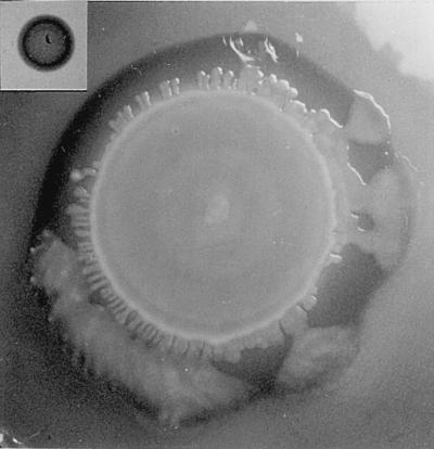
In a first attempt to recover a strain that was luminescent in broth, an Ampr Lux+ colony was inoculated into BHI-ampicillin broth. These cells were diluted to an end-point cell number, and 60 cultures begun from single cells were obtained. All of these cultures were ampicillin resistant but negative for lux activity after 16 h of growth with moderate aeration. This result indicated that a simple approach for separating the two populations would not work and that a different selection strategy was required. To begin selection, five Ampr Lux+ colonies were passaged three times to HEA–100 mM glucose agar, followed by three passages in early log phase in BHI-ampicillin broth, as described in Materials and Methods. This strategy was chosen for two reasons. First, HEA–100 mM glucose agar supports colony growth of serovar Enteritidis, so this agar was used to grow colonies to a large size (>5 mm after 16 h). Secondly, successive passage in broth during early-log-phase growth dilutes out late-log-phase populations and maintains early-log-phase cells under conditions that limit fewer metabolites. The last culture in broth was allowed to grow to stationary phase (for 16 h with moderate aeration). Luminescence that was 10 times greater than background could then be detected from three of the five passaged cultures transformed with pSB402 after 8 h of growth. Central inoculation of HEA–100 mM glucose agar showed that colony morphology of Ampr Lux+ and wild-type cultures with or without pBR322 differed and that cells were luminescent in broth were more likely to migrate across agar surfaces and produce larger colonies (Fig. 1). These cells gave a positive reaction for serovar D1 O-chain. In addition, immunoreactivity for these cells with factor 12 antiserum, which reacts with α1,4-glucosylated galactose of O-chain, was as strong as the reaction with factor 9, which reacts with tyvelose. These results correlate positively with cells producing a large amount of glucosylated HMW O-chain.
FIG. 1.

Colony morphology of SE6-HCD on inducing agar medium after serial passage. A 5-μl volume of cells of serovar Enteritidis maintained for two passages in early log phase and allowed to grow to stationary phase during the third passage was centrally inoculated on HEA–100 mM glucose. Plates were incubated for 16 h at 37°C. The diameter of the large terraced colony obtained from SE-HCD at its widest point is 62 mm. (Inset) Colony morphology of SE6-E21 transformed with pBR322 after serial passage (diameter, 8 mm), which is a phenotype similar to that of wild type lacking pBR322.
Growth characteristics of SE-HCD.
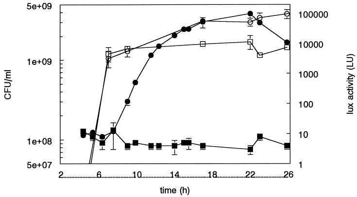
One luminescent colony was chosen for further analysis and designated SE-HCD. SE-HCD grew to an HCD than wild type (3.2 × 10⁹ ± 0.25 × 10⁸ CFU/ml [mean ± standard deviation] versus 1.5 × 10⁹ ± 0.02 × 10⁸ CFU/ml, moderate aeration) or wild type transformed with pBR322 (1.0 × 10⁹ ± 0.05 × 10⁸ CFU/ml) (Fig. 2). SE-HCD was visibly luminescent in broth culture when cell density reached 2.5 × 10⁹ CFU/ml and lux activity exceeded 150,000 LU. Therefore, selection for high cell density by assay of luminescence had restored SE6 to a growth potential of >3.0 × 10⁹ CFU/ml in BHI broth under conditions of moderate aeration. Examination of logarithmic growth revealed no differences between wild type, wild type transformed with pSB402, and wild type transformed with pBR322 (data not shown).
FIG. 2.
Correlation of luminescence and cell density in stationary phase. Three colonies of SE6-HCD and wild-type SE6 were analyzed; error bars indicate average deviations between readings. Solid squares, lux activity of wild-type SE6; solid circles, lux activity of SE-HCD; open squares, wild type SE6 (CFU/ml); open circles, SE-HCD (CFU/ml). Incubation was at 37°C with moderate aeration for the amount of time indicated.
Further correlation of CFU with opacity revealed that with vigorous aeration, the cell density of SE-HCD could reach a peak of 1.45 × 10¹¹ ± 5.36 × 10⁹ CFU/ml at a 1/10 OD reading of 0.48 ± 0.04, whereas wild type peaked at previously observed values of 2.2 × 10⁹ CFU/ml. To establish that such readings were repeatable, growth curves of SE-HCD with vigorous high aeration were plotted in triplicate. Swarming migration and the highest cell counts that surpassed 1011 CFU/ml were encountered between 0.43 and 0.53 1/10 OD. However, such high cell densities were followed by a decrease in cell numbers as opacity continued to increase past 0.6 1/10 OD, so that peak 1/10 OD of 0.8 correlated with 3.7 × 10⁹ CFU/ml. Swarming migration could not be detected past 0.6 1/10 OD. Thus, induction of swarm cell migration and attainment of a very high cell density by serovar Enteritidis was dependent upon the presence of vigorous aeration, attainment of a 1/10 OD between 0.43 and 0.53, and the continued production of O-chain during growth to high cell density. Cultures grown with moderate aeration never surpassed a 1/10 OD of 0.35 and were never observed to undergo swarming migration.
Mathematical modeling of high-cell-density growth of SE-HCD.
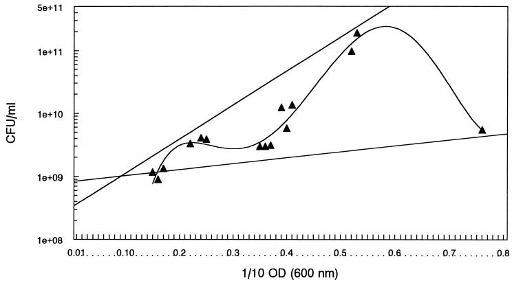
The correlation between opacity and cell count at peak 1/10 OD indicated that the highest opacities did not correlate with the highest cell densities. To understand this departure from convention, three growth curves were examined, beginning with cultures grown without aeration to a 1/10 OD of 0.1, which is an opacity reached just prior to detection of any luminescence and which corresponds to a cell count of 109. Cultures were then aerated vigorously until opacity peaked. Cell numbers were determined by plating serial 10-fold dilutions collected every 2 h. The readings that had the three highest and lowest ratios of CFU/ml to 1/10 OD were plotted, with opacity on the x axis and cell density on the y axis. A linear regression line was generated for the two sets of numbers, and each line was extrapolated back to 109 CFU/ml. The resulting graph defined a boundary that encompassed all observed values of CFU/ml versus 1/10 OD readings generated during experiments (Fig. 3). Examination of previous growth curves indicated that data collected over the course of several months fit within these boundaries. This finding suggested that it was possible to describe a mathematical relationship between opacity and cell density, even though the relationship was not linear, logarithmic, or exponential.
FIG. 3.
High-cell-density growth characteristics of SE-HCD. Straight lines, linear regression lines indicate observed highest and lowest boundaries for opacity versus cell density values after cultures attained a 0.1 1/10 opacity and 109 CFU/ml. Biphasic curvilinear line, data points generated during growth curve analysis conducted in triplicate were analyzed by curvilinear analysis (fifth polynomial). Closed triangles, opacity of cells diluted 10-fold (1/10 OD). Swarming migration can be detected by passaging cells to agar at a 1/10 OD of 0.5.
To generate information about the mathematical relationship between cell density and opacity, another growth curve was examined in triplicate, again beginning with cells grown for 8 h without aeration that had reached a beginning density of 109 CFU/ml. At the times indicated (Fig. 3), plate counts and 1/10 OD readings were obtained. These data points were superimposed on the graph depicting expected data boundaries defined by linear regression (Fig. 3). Curvilinear analysis (fifth polynomial) was then applied to the new growth curve data set (Fig. 3). Results indicated that the relationship between opacity and cell number for cultures that grow to high cell density is a function of time in growth phase and degree of aeration. The growth curve of serovar Enteritidis that grows to high cell density is biphasic, with the first phase comprising ratios typically encountered during moderate aeration, whereas the second phase peaks at cell densities 50 times greater when aeration is vigorous (Fig. 3). Thus, growth curve analysis of SE-HCD indicated that with moderate aeration, cell density is expected to be in the range of 3 × 10⁹ to 4 × 10⁹ CFU/ml but that vigorous aeration enables cells to divide and eventually reach a brief peak density of nearly 1.5 × 10¹¹ CFU/ml. Increasing aeration above 8 did not yield higher cell densities; instead, it appeared to hasten loss of cells at the very highest opacity readings. Curvilinear analysis suggests that some opacity/cell density ratios would be unlikely; for example, a 1/10 OD of 0.3 would be unlikely to yield a cell count of 1010 CFU/ml.
Cell shape changes during high-cell-density growth of SE-HCD.
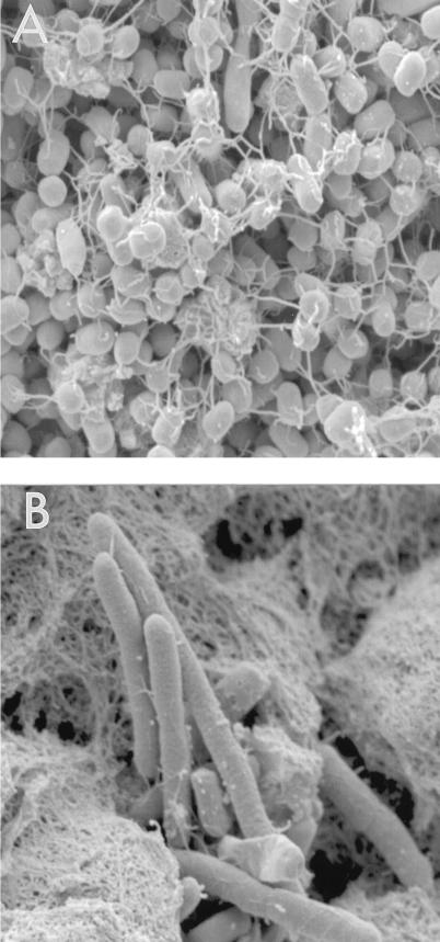
The reason why cell loss occurred when opacity was at its highest was investigated, because this result suggested that common assumptions made about the relationship between opacity and cell numbers were not true at peak opacity. SEM indicated that at least two types of cells could be detected in different ratios as opacity increased (Fig. 4). One type of cell, a hyperflagellated coccobacillus (Fig. 4A), was predominant at peak cell density, which occurred around 0.5 1/10 OD. These cells underwent swarming migration when they were passaged to agar. The second cell shape was elongated as much as 50-fold compared to the coccobacillus. Lacking discernible surface appendages such as flagella (Fig. 4B), this cell type eventually increased in cultures as opacity increased, so that at the very highest 1/10 ODs of >0.7, it was predominant. In addition, the accumulation of a large amount of cellular debris at higher ODs occurred concomitantly with the loss of the coccobacillary cell type. Thus, the replacement of a coccobacillus by a hyperelongated cell type and the accumulation of cellular debris were involved in generating the relationship between opacity and cell density once opacity surpassed 0.6 1/10 OD. These results strongly suggest that SE-HCD is capable of undergoing additional stages of cellular differentiation en masse compared to strains that do not have the potential to grow to high cell density. However, the observation of these changes is dependent upon the degree of aeration encountered during growth.
FIG. 4.
SEM of SE-HCD. (A) Cells at peak cell density (>1011 CFU/ml). A hyperflagellated coccobacillary cell was the predominant cell type. (B) Cells at peak opacity (1/10 OD = 0.8). An elongated cell lacking surface appendages was the only type observed, and it was surrounded by much cellular debris. Magnification, ×10,000.
Detection of different patterns of luminescence between isogenic variants of parental SE-HCD.
During the initial growth curve analyses of SE-HCD, a point of variation was observed to occur between isogenic colonies at a 1/10 OD of 0.2. To further examine the degree of variation present within the SE-HCD phenotype at this opacity, 12 isolated colonies of SE-HCD were used to start cultures for assay of cell density and lux activity. Analysis of variance by F test indicated that variation at 0.2 1/10 OD was significantly different from readings taken earlier or later at a 1/10 OD of 0.18 or 0.3 (P value, <0.01). Assay of several colonies was required to establish that variation existed at a 1/10 OD of 0.2, because outliers in the group contributed to the significance of the variation. A 1/10 OD of 0.2 reached at 17 h after growth was initiated with moderate aeration (Fig. 2). Results indicated that most colonies yield an average lux reading of 314,000 LU at this point, but an occasional colony, perhaps 1 in 10, is expected to have a reading that is very high or very low. Since it was not known whether this variation indicated that different pathotypes existed, virulence assays in hens and chicks were performed with clonal variants SE-HCD1 and SE-HCD2. SE-HCD1 had a peak of 616,600 LU at 0.2 1/10 OD, which declined to 10,000 at a 1/10 OD of 0.3. SE-HCD2 lux activity was 340,200 at 0.2 1/10 OD, which increased slightly to 373,700 LU at a 1/10 OD of 0.3. Thus, SE-HCD1 had a high peak that declined rapidly as growth continued, whereas SE-HCD2 maintained a less-intense peak longer. Challenge experiments with isolates producing low readings at 0.2 1/10 OD were not investigated at this time. Both variants used in the following challenge experiments reacted with antisera specific for group D1 O-chain as did parental SE-HCD.
Challenge of 6-day-old chicks with two clonal variants of SE-HCD.
Subcutaneous challenge of chicks with 5 × 10⁷ SE-HCD1 was lethal, killing 70% within 3 days. Conversely, subcutaneous challenge with 7 × 10⁷ SE-HCD2 did not kill any chicks. For birds that survived challenge with SE-HCD1, the average number of organisms recovered from spleen suspension (diluted 100-fold) was greater than 2,000 CFU, whereas an average of 162 CFU were recovered from chick spleens after challenge with SE6-HCD2. Subcutaneous challenge of 10 chicks with wild-type SE6 or ampicillin-resistant SE6 killed no chicks, and average yields of organisms from a 100-fold dilution of spleen suspension for these two control strains were 114 and 3.9 CFU, respectively. These results indicated that transformation with pBR322 resulted in the attenuation of strains. Because wild-type SE6 lacking ampicillin resistance yielded more CFU/spleen, it was used as the control isolate for further examination of serovar Enteritidis in hens.
Challenge of hens and contamination of eggs with two clonal variants of SE-HCD.
Uninfected hens between 25 and 45 weeks of age had a daily average egg production of 59.24%, with a standard deviation of 15.22%, as determined by analysis of egg production for 4 days from five different flocks prior to the challenge studies. Hens challenged with wild-type serovar Enteritidis by oral, intravenous, and subcutaneous routes had an average daily egg production of 72.5, 47.8, and 75%, respectively, of prechallenge egg production. Of eggs collected 21 days postchallenge from hens infected by the three different routes of exposure, 1.3, 0.5, and 0.5% were contaminated, respectively (Table 1). These results indicated that egg production was not altered significantly by infection with wild-type serovar Enteritidis, although it may have been marginally derepressed by intravenous challenge. Most contaminated eggs were detected within a few days of challenge in this experiment, which resulted in 4.3, 8, and 0% of eggs from the first day after challenge being contaminated with wild-type strain after oral, intravenous, and subcutaneous challenge, respectively (Table 1).
Hens challenged subcutaneously with 108 CFU of SE-HCD1, which was more lethal to chicks than SE-HCD2, had a daily average egg production of 62.1% of prechallenge egg production. Of 284 eggs collected, 2 were contaminated (0.7%) and both isolates were Ampr Lux+ (Table 1). These results indicated that SE-HCD1 resembled wild type SE in regard to its ability to contaminate eggs, even though it was more virulent in chicks. Results also indicated that maintenance of the reporter plasmid and the Ampr Lux+ phenotype was stable after challenge of birds and eventual recovery of SE-HCD from eggs. Hens challenged by intravenous and subcutaneous routes with SE-HCD2 produced contaminated eggs 22 and 3.5% of the time, with 67 and 100% of eggs collected the first day postchallenge positive for Ampr Lux+ serovar Enteritidis, respectively (Table 1). Oral challenge with SE-HCD2 produced no contaminated eggs. Egg production for these birds increased significantly during a fourth week of collection compared to egg production prechallenge and egg production the first 3 weeks postchallenge (P value, <0.005). However, the egg production of hens challenged intravenously with 108 CFU of SE-HCD2 decreased to 9.52% of the prechallenge egg production (Table 1).
Hens challenged subcutaneously or orally with SE-HCD2 had no overt symptoms of salmonellosis. In contrast, hens challenged intravenously with SE-HCD2 developed signs of gallinarum disease. They became depressed, and most noticeably, every bird exhibited pallor of the wattles and mucous membranes and a slow capillary refill time. These signs suggested that the birds were anemic. Although most birds challenged with SE-HCD2 continued to eat, one of the intravenously challenged birds became moribund and stopped eating 1 week after infection. After euthanasia for humane reasons, necropsy revealed that the bird had developed ascites, and nearly 200 ml of sterile straw-colored peritoneal fluid was recovered. Ascites can be a sequela to severe anemia. The flock was held an additional 2 weeks to see how the illness would resolve. Hens recovered during the fifth week postchallenge, as measured by the return of prechallenge egg production levels and normal mucous membrane and wattle coloration. One bird died from egg yolk peritonitis the final week of the experiment, but this death was not directly due to infection with serovar Enteritidis.
DISCUSSION
Previous research had shown that it was possible to isolate serovar Enteritidis that could grow to a high cell density of >3.0 × 10⁹ CFU/ml, but attenuation occurred because O-chain biosynthesis stopped (12). To overcome this problem of the loss of an important virulence attribute, a reporter plasmid was used to detect production of autoinducers of LuxR homologs and to obtain a smooth strain that could grow to high cell density. These results do not conclusively indicate that O-chain biosynthesis is coordinately regulated with LuxR-regulated homologs, only that this selection strategy is biased toward isolation of strains that grow to high cell density and remain smooth. Thus, the cell density sensor plasmid pSB402 is useful for obtaining virulent serovar Enteritidis. The isolation requirement of a selection strategy that recovered luminescent cells from a negative population may help explain why it has been difficult to detect autoinducible luminescence within the salmonellae (7, 8, 26). At the very least, recognizing that high-cell-density growth is not always associated with virulence within the salmonellae due to the unwanted generation of LPS chemotype conversion emphasizes that challenge experiments are best conducted with characterized strains.
Medium has a profound effect on luminescence. For example, chelation of iron halts high-cell-density growth greater than 109 CFU/ml and abolishes luminescence. It is possible that BHI broth is not ideal for growing SE-HCD, especially since correcting growth conditions for serovar Pullorum, another group D1 serovar that contaminates eggs, changes the shapes of cells from coccobacilli to rods (10). Finding that a heavily flagellated coccobacillary cell type is associated with swarming migration in the salmonellae was surprising because investigations of promiscuously swarming bacteria such as Proteus mirabilis indicate that hyperflagellated elongated cells are associated with cell migration (1, 28). It is possible that passage to agar induces a final change in cell shape to a hyperflagellated elongated structure, but SEM analysis of swarming SE-HCD cells on agar confirms only the presence of the two cell types in broth (10). Regardless of why the swarm cell of SE-HCD is smaller than expected, BHI broth is an acceptable rich complex basal medium for initial investigation of the cell signals and gene expression profiles that accompany these changes in cell shape.
Noticeably lacking from these results is the isolation of a variant that kills some chicks, is recovered in high numbers from chick spleens, and efficiently contaminates eggs. It is possible that plasmids bearing ampicillin resistance interfere with recovery of a strain with such mixed characteristics, which were observed during initial characterization of virulent serovar Enteritidis. Indeed, others have found that plasmid-borne ampicillin resistance is associated with attenuation of serovar Enteritidis (4, 22). Transformation with the low-copy-number pSB402 is stable, although laboratory manipulations such as aging of culture can lead to spontaneous plasmid curing. None of the isolates recovered from animals in this study were cured. When curing occurs, ampicillin resistance and luminescence are lost concurrently (10a), suggesting that plasmid genes are not crossing over into the chromosome frequently. SE-HCD2 appears to be the best variant for assessing the ability of vaccines to prevent egg contamination, because 100% of eggs on day 1 after subcutaneous challenge were contaminated without the suppression of egg production. SE-HCD1 and SE-HCD2 were not particularly orally invasive. This finding is not surprising in view of research that indicates some serovar Enteritidis strains are best adapted to the parenteral environment, whereas others are more associated with oral colonization (12, 20).
The differences in outcomes following challenge of chicks and mature birds with SE-HCD clonal variants were striking. The SE-HCD variants were more virulent than wild-type SE6 by one of the assays used but not by both. In addition, intravenous challenge with SE-HCD2 caused significant illness in mature birds in comparison to illness caused by intravenous challenge with wild type. These results give a first indication that the SE-HCD phenotype can target specific age groups as well as organs and eggs. Two other egg-contaminating group D1 salmonellae, S. pullorum and S. gallinarum, cause illness in birds of different ages (19, 23). Whereas pullorum disease is associated with high mortality in chicks, gallinarum disease (also known as fowl typhoid and infectious leukemia) causes acute salmonellosis in mature birds. Fowl typhoid also causes anemia, which is a clinical sign detected during these investigations (5, 19, 23). Thus, precedence exists for egg-contaminating salmonellae preferentially causing disease in different age groups. The data generated here with serovar Enteritidis indicate that it may be possible to understand how age adaptation occurs. A first indication that these processes require an organism to achieve more of its genetic potential and to thus undergo a broader range of cellular differentiation than that usually observed is suggested by these challenge experiments with smooth serovar Enteritidis that grows to high cell density.
ACKNOWLEDGMENTS
I thank M. Winson, Aberystwyth, Wales, for supplying pSB402 and advice and C. Hughes and colleagues, Cambridge, England, for providing technical guidance during the development of strain SE-HCD. J. Jacks and K. Asokan, SEPRL, coordinated collection of growth curve data at night, and I thank them for their dedication to this project.
Maine Biological Laboratories generously supported this project through CRADA 58-3K95-5-403. Additional support for animal experimentation and characterization of bacterial cells was provided by USDA CRIS 6612-32000-014-OOD.
REFERENCES
- 1.Allison P, Emody L, Coleman N, Hughes C. The role of swarm cell differentiation and multicellular migration in the uropathogenicity of Proteus mirabilis. J Infect Dis. 1994;169:1155–1158. doi: 10.1093/infdis/169.5.1155. [DOI] [PubMed] [Google Scholar]
- 2.Ausubel F M, Brent R, Kingston R E, Moore D D, Seidman J G, Smith J A, Struhl L. Current protocols in molecular biology. New York, N.Y: Greene Publishing/Wiley Interscience; 1988. [Google Scholar]
- 3.Bainton N J, Bycroft B W, Chhabra S R, Stead P, Gledhill L, Hill P J, Rees C E D, Winson M K, Salmond G P C, Stewart G S A B, Williams P. A general role for the lux autoinducer in bacterial cell signalling: control of antibiotic biosynthesis in Erwinia. Gene. 1992;116:87–91. doi: 10.1016/0378-1119(92)90633-z. [DOI] [PubMed] [Google Scholar]
- 4.Chart H, Threlfall E J, Powell N G, Rowe B. Serum survival and plasmid possession by strains of Salmonella enteritidis, S. typhimurium and S. virchow. J Appl Microbiol. 1996;80:31–36. doi: 10.1111/j.1365-2672.1996.tb03186.x. [DOI] [PubMed] [Google Scholar]
- 5.Christensen J P, Barrow P A, Poulsen J E, Poulsen J S D, Bisgaard M. Correlation between viable counts of Salmonella gallinarum in spleen and liver and the development of anaemia in chickens as seen in experimental fowl typhoid. Avian Pathol. 1996;25:769–783. doi: 10.1080/03079459608419180. [DOI] [PubMed] [Google Scholar]
- 6.Cole G. Preparation of microfungi for SEM. In: Aldrich H C, Todd W J, editors. Ultrastructure techniques for microorganisms. New York, N.Y: Plenum Press; 1986. pp. 1–44. [Google Scholar]
- 7.Eberl L, Winson M K, Sternberg C, Stewart G S A B, Christiansen G, Chhabra S R, Bycroft B, Williams P, Molin S, Givskov M. Involvement of N-acyl-l-homoserine lactone autoinducers in controlling multicellular behavior of Serratia liquefaciens. Mol Microbiol. 1996;20:127–136. doi: 10.1111/j.1365-2958.1996.tb02495.x. [DOI] [PubMed] [Google Scholar]
- 8.Fuqua W C, Winans S C, Greenburg E P. Census and concensus in bacterial ecosystems: the LuxR-LuxI family of quorum-sensing transcriptional regulators. Annu Rev Microbiol. 1996;50:727–751. doi: 10.1146/annurev.micro.50.1.727. [DOI] [PubMed] [Google Scholar]
- 9.Gast R K. Recovery of Salmonella enteritidis from inoculated pools of egg contents. J Food Prot. 1993;56:21–24. doi: 10.4315/0362-028X-56.1.21. [DOI] [PubMed] [Google Scholar]
- 10.Guard-Petter J. Induction of flagellation and a novel agar-penetrating flagellar structure in Salmonella enterica grown on solid media: possible consequences for serological identification. FEMS Microbiol Lett. 1997;149:173–180. doi: 10.1111/j.1574-6968.1997.tb10325.x. [DOI] [PubMed] [Google Scholar]
- 10a.Guard-Petter, J. Unpublished data.
- 11.Guard-Petter J, Henzler D J, Rahman M M, Carlson R W. On-farm monitoring of mouse-invasive Salmonella enterica serovar Enteritidis and a model for its association with the production of contaminated eggs. Appl Environ Microbiol. 1997;63:1588–1593. doi: 10.1128/aem.63.4.1588-1593.1997. [DOI] [PMC free article] [PubMed] [Google Scholar]
- 12.Guard-Petter J, Keller L H, Rahman M M, Carlson R W, Silvers S. A novel relationship between O-antigen variation, matrix formation, and invasiveness of Salmonella enteritidis. Epidemiol Infect. 1996;117:219–231. doi: 10.1017/s0950268800001394. [DOI] [PMC free article] [PubMed] [Google Scholar]
- 13.Guard-Petter J, Lakshmi B, Carlson R, Ingram K. Characterization of lipopolysaccharide heterogeneity in Salmonella enteritidis by an improved gel electrophoresis method. Appl Environ Microbiol. 1995;61:2845–2851. doi: 10.1128/aem.61.8.2845-2851.1995. [DOI] [PMC free article] [PubMed] [Google Scholar]
- 14.Jimenez-Lucho V E, Leive L L, Joiner K A. Role of the O-antigen of lipopolysaccharide in Salmonella in protection against complement action. In: Gunsalus I C, Sokatch J R, Ornston L N, Iglewski B H, Clark V L, editors. The bacteria. Vol. 11. New York, N.Y: Academic Press; 1990. pp. 339–354. [Google Scholar]
- 15.Keen N T, Tamaki S, Kobayashi D, Trollinger D. Improved broad-host range plasmids for DNA cloning in Gram-negative bacteria. Gene. 1988;70:191–197. doi: 10.1016/0378-1119(88)90117-5. [DOI] [PubMed] [Google Scholar]
- 16.Meighen E A, Szittner R B. Multiple repetitive elements and organization of the lux operons of luminescent terrestrial bacteria. J Bacteriol. 1992;174:5371–5381. doi: 10.1128/jb.174.16.5371-5381.1992. [DOI] [PMC free article] [PubMed] [Google Scholar]
- 17.Mishu B, Koehler J, Lee L A, Rodriquez D, Brenner F H, Tauxe R V. Outbreaks of Salmonella enteritidis in the contents of naturally contaminated hens’ eggs. Epidemiol Infect. 1991;106:489–496. doi: 10.1017/s0950268800067546. [DOI] [PMC free article] [PubMed] [Google Scholar]
- 18.Petter J G. Detection of two smooth colony phenotypes in a Salmonella enteritidis isolate which vary in their ability to contaminate eggs. Appl Environ Microbiol. 1993;59:2884–2890. doi: 10.1128/aem.59.9.2884-2890.1993. [DOI] [PMC free article] [PubMed] [Google Scholar]
- 19.Pomeroy B S, Nagaraja K V. Fowl typhoid. In: Calnek B W, editor. Diseases of poultry. 9th ed. Ames: Iowa State University Press; 1991. pp. 87–99. [Google Scholar]
- 20.Porter S B, Curtiss R., III Effect of inv mutations on salmonella virulence and colonization in 1-day-old white leghorn chicks. Avian Dis. 1997;41:45–57. [PubMed] [Google Scholar]
- 21.Rahman M M, Guard-Petter J, Carlson R W. A virulent isolate of Salmonella enteritidis produces a Salmonella typhi-like lipopolysaccharide. J Bacteriol. 1997;179:2126–2131. doi: 10.1128/jb.179.7.2126-2131.1997. [DOI] [PMC free article] [PubMed] [Google Scholar]
- 22.Ridley A M, Punia P, Ward L R, Rowe B. Plasmid characterization and pulsed-field electrophoretic analysis demonstrate that ampicillin-resistant strains of Salmonella enteritidis phage type 6a are derived from S. enteritidis phage type 4. J Appl Bacteriol. 1996;81:613–618. doi: 10.1111/j.1365-2672.1996.tb03555.x. [DOI] [PubMed] [Google Scholar]
- 23.Snoeyenbos G H. Pullorum disease. In: Calnek B W, editor. Diseases of poultry. 9th ed. Ames: Iowa State University Press; 1991. pp. 73–86. [Google Scholar]
- 24.Stewart G S A B, Lubinsky-Mink S, Jackson C G, Cassel A, Kuhn J. pHG165: a pBR322 copy number derivative of pUC8 for cloning and expression. Plasmid. 1986;15:172–181. doi: 10.1016/0147-619x(86)90035-1. [DOI] [PubMed] [Google Scholar]
- 25.St. Louise M E, Morse D L, Demelfi T M, Guzewuch J J, Tauxe R V, Blake P A. The emergence of grade A eggs as a major source of Salmonella enteritidis infections: new implications for the control of salmonellosis. JAMA. 1988;259:2103–2107. [PubMed] [Google Scholar]
- 26.Swift S, Bainton N J, Winson M K. Gram-negative bacterial communication by N-acyl homoserine lactones: a universal language? Trends Microbiol. 1994;2:193–197. doi: 10.1016/0966-842x(94)90110-q. [DOI] [PubMed] [Google Scholar]
- 27.Swift S, Winson M K, Chan P F, Bainton N J, Birdsall M, Reeves P J, Rees C E D, Chhabra S R, Hill P J, et al. A novel strategy for the isolation of luxI homologues: evidence for the widespread distribution of a LuxR:LuxI superfamily in enteric bacteria. Mol Microbiol. 1993;10:511–520. doi: 10.1111/j.1365-2958.1993.tb00923.x. [DOI] [PubMed] [Google Scholar]
- 28.Wilkerson M L, Niederhoffer E C. Swarming characteristics of Proteus mirabilis under anaerobic and aerobic conditions. Anaerobe. 1995;1:345–350. doi: 10.1006/anae.1995.1037. [DOI] [PubMed] [Google Scholar]
- 29.Winson, M. K., S. Swift, B. W. Bycroft, P. Williams, and G. A. B. Stewart. Engineering the luxCDABE genes from Photorhabdus luminescens to provide a bioluminescent reporter for constitutive and promoter probe plasmids and mini-Tn5 constructs. FEMS Microbiol. Lett., in press. [DOI] [PubMed]
- 30.Winson, M., S. Swift, L. Fish, J. P. Throup, F. Jorgensen, S. R. Chhabra, B. W. Bycroft, P. Williams, and G. S. A. B. Stewart. Construction and analysis of luxCDABE-based plasmid sensors for investigating N-acyl homoserine lactone-mediated quorum sensing. FEMS Microbiol. Lett., in press. [DOI] [PubMed]


