

Abstract
Background
The differential diagnosis of lymphadenopathies in children including benign and malignant conditions is often difficult to identify by ultrasound (US). The lymphadenopathies in children are frequent and mostly benign, therefore it is essential to decide what patients should undergo further studies.
Objective
To describe the potential usefulness of a new suspicious ultrasound sign on pediatric lymphadenopathies that can orient the diagnosis of malignancy.
Materials and methods
We retrospectively reviewed all pediatric cases with lymphadenopathy suspicious of lymphoma or lymphoproliferative syndrome on soft tissue ultrasound from 2014 to 2021. Two expert ultrasound radiologists reviewed ultrasound images of these patients, associating the internal structure of infiltrated adenopathy with the internal structure of the truffles.
Results
On ultrasound, twelve cases presented enlarged lymphadenopathy with loss of internal structure, without hilum; mostly hypoechoic parenchyma, with some fine echogenic serpiginous linear surrounding hypoechoic pseudo nodular images, resembling the inner structure of black truffles. This US pattern looked suspicious and histological study was recommended. In 9 cases a lymphomatous infiltrated adenopathy was confirmed on biopsy.
Conclusion
The truffle sign is a new potential suspicious ultrasound sign, that can suggest malignant lymphadenopathy in children. This ultrasound pattern can have some probable usefulness to the radiologist in order to recommend further studies, including histological study, that need to be validated by a larger sample. It is important to recognize easily and early the lymphomatous compromise in a lymph node.
Keywords: Lymphadenopathy, Lymphomatous adenopathies, Malignant lymph nodes, Truffle sign, Ultrasound
Background
Observation, description, and interpretation are processes that we radiologists perform every day. One of the techniques we develop in these processes is pattern recognition. Some diseases may show patterns in different imaging techniques that are repeated in most cases. [1]
Certain diseases show classic radiological signs that the radiologist can recognize, allowing an accurate diagnosis.
In other cases, the radiological signs allow the differential diagnosis to be narrowed down.
There are several radiological signs where the imaging pattern evokes or represents a fruit or vegetable, for example, the apple-crown sign, which corresponds to constriction of the lumen of the colon by a stenosing annular colorectal carcinoma and which can be seen in barium enema, computed tomography or magnetic resonance imaging [1, 2].
The differential diagnosis of lymphadenopathies in children including benign and malignant condition, is often difficult to identify by ultrasound (US). The lymphadenopathies in children are frequent, mostly benign, and it is very important to decide what patients should undergo further studies. In the malignant group, acute lymphoblastic leukemia (the most common malignancy in children) [3] and the lymphomatous process can present with adenopathies, among others [4].
Objective
To describe “Truffle sign” a new suspicious ultrasound sign on pediatric lymphadenopathies that can orient malignancy.
Materials and methods
An 8-year retrospective study of the small part ultrasound database, accepted by the Institutional Review Board. A waiver for the informed consent was approved. The inclusion criteria were to have ultrasound images archived in the computational system and the pathology biopsy results.
A series of 12 cases of pediatric patients (younger than 15 years old) that underwent mostly a cervical ultrasound, with the clinical diagnosis of soft tissue mass, enlarged volume, nodule or suspected adenopathy.
All US examinations were performed before the diagnosis or histological results and had images recorded in the computational system.
The images features were analyzed independently blinded by two expert radiologists on ultrasound. The internal structure of infiltrated lymphadenopathy was associated with internal structure of the black truffles. When the internal structure matched, it was called the positive truffle sign.
Results
The universe corresponded to 12 pediatric cases, 3 girls and 9 boys. The age ranged between 4 and 14 years old with a mean age of 9.5 years old.
They presented on ultrasound enlarged lymphadenopathy with loss of internal structure, without echogenic hilum. All of them presented with hypoechoic parenchyma, with some fine echogenic serpiginous linear surrounding hypoechoic pseudo nodular images, resembling the inner structure of black truffles. Figures 1, 2
Fig. 1.
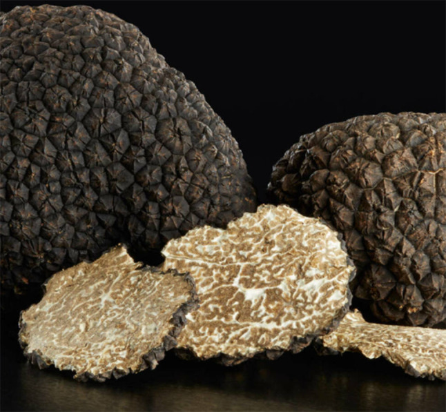
Truffles are fructifications of underground fungi, whose internal structure is serpiginous and somewhat erratic, composed of the dark-colored gleba, where the spores are produced, and crossed by white veinlets, which give a reticulated appearance
Fig. 2.

On US, the relationship between the hypoechoic pseudonodular formations (long arrows) and the echogenic fibrous bands (short arrows) give the reticular pattern to the internal structure of the adenopathy. On histology lymphomatous adenopathies lose their normal internal architecture, they present fibrous bands (short arrows) and pseudonodular formations composed of pathological lymphocytes (long arrows).The truffle internal structure is serpiginous and somewhat erratic, composed of the dark-colored gleba (long arrows), where the spores are produced, crossed by white veinlets (short arrows), which give a reticulated appearance, reminiscent of the structure internal lymphomatous adenopathies
This ultrasound pattern looked suspicious and histological study was recommended. Figures 3, 4, 5, 6.
Fig. 3.

14-year-old boy with right submandibular volumen increase of 1 month of evolution (a, b) Grayscale US shows an enlarged adenopathy, with altered internal structure, loss of the echogenic hilum, and Truffle sign: internal reticular structure with serpingine hypoechoic images and fine irregular echogenic linear tracts. c disorganized vascularization on Doppler US. Biopsy results revealed Large B-cell Non- Hogkin`s lymphoma. PET-FDG shows no distant spread, confirming mononodal involvement
Fig. 4.

9-year-old boy with right cervical swelling of 2 weeks evolution. a US shows adenopathy with a disorganized structure, with loss of the hilum (b) and increased vascularity on Doppler study. Truffle sign: this adenopathy presents a micronodular reticular pattern. Biopsy results revealed Hodgkin's Lymphoma. c FDG-PET/CT: shows a conglomerate of hypermetabolic right cervical adenopathies at level V (max SUV of 7.1) without evidence of disseminated disease
Fig. 5.

10-year-old girl with bilateral cervical swelling. a US shows multiple adenopathies with loss of the hilum and presents with a trufoid-pattern structure b and abnormal vascularity entering from the capsule. Truffle sign: They present hypoechoic serpingine areas delimited by an echogenic linear pattern. c FDG PET/CT: hypermetabolic tumor mass in the anterior mediastinum associated with bilateral cervical, mediastinal, and hilar hypermetabolic lymphadenopathy. Biopsy results revealed a Classic Hodgkin lymphoma (nodular sclerosis variant)
Fig. 6.

On Grayscale and Doppler color US an enlarged lymph node with truffle US pattern, with hipoechogenic pseudonodular and echogenic septa
In nine cases a lymphomatous infiltrated adenopathy was confirmed on biopsy. The “truffle sign” was found to be present in 9 patients with lymphoma. Five patients with Hodgkin's lymphoma, 3 patients with biopsy-diagnosed non-Hodgkin’s lymphoma and 1 lymphoblastic lymphoma. In four cases, the detection was at the mononodal stage or in a single lymph node group, which led to early treatment and thus to a better prognosis.
In the other 3 patients the histological results correspond to: 1 case of leukemia, 1 case of post-transplant lymphoproliferative disorder, and 1 case of reactive lymphoid hyperplasia.
Discussion
The internal structure of lymphomatous adenopathies has been described as heterogeneous reticular or micronodular, given the interfollicular and paracortical proliferation, obliterating the normal follicles and medullary cords of the lymph node. [4–7]
Truffles are fructifications of subterranean fungi, whose internal structure is serpiginous and somewhat erratic, composed of the dark colored gleba, where the spores are produced, and furrowed by white veins, which give a reticulated appearance, reminiscent of the internal structure of lymphomatous lymphadenopathies.
Lymphomatous lymphadenopathies lose their normal internal architecture. Histologically they present fibrous bands (white arrows) and pseudo nodular formations composed of pathological lymphocytes (black arrows). On US, the relationship between the pseudo nodular formations and the fibrous bands give the reticular pattern to the internal structure of the lymphadenopathy.
Because of the importance of high ultrasound suspicion to distinguish between a normal lymph node versus a lymphomatous lymphadenopathy, we propose this new sign that can help the radiologist to remember it easily.
This is even more important in children, whose prognosis depends on the earliest possible treatment.
The most frequent manifestation of lymphoma is the presence of palpable superficial adenopathies, and the most frequently involved groups are the cervical ones, most frequently those of the internal jugular chains and the posterior triangles [8].
There are many ultrasound signs that lead us to suspect lymphomatous adenopathy, but the one that has caught our attention is the altered internal ultrasound structure, evaluated with high-frequency transducers, whose structure evokes the image of the internal structure of the truffle.
In this series of cases with the majority of patients with histological confirmed lymphoma, The “truffle sign” was found to be present both in patients with Hodgkin's lymphoma and in patients with biopsy-diagnosed non-Hodgkin’s lymphoma. In four of our cases, the detection was at the mononodal stage or in a single lymph node group, which led to early treatment and thus to a better prognosis. The FDG PET study is essential for staging.
Conclusion
The purpose of using a radiological sign is to facilitate the identification of the different pathologies associated with it, which is even more important if we are dealing with malignancy.
The altered internal lymph nodes structure as a truffle pattern impresses to be suspicious for a malignancy, especially for lymphomatous infiltration, and that is why we present this preliminary sign, that needs to be validated by a larger sample.
With this new sign, we hope to help remind the radiologist performing ultrasound when to suspect a lymphoproliferative disease, for an early referral for a histological study.
Funding
We have no funding.
Declarations
Conflict of interest
The authors have no conflict of interest to declare.
Footnotes
Publisher's Note
Springer Nature remains neutral with regard to jurisdictional claims in published maps and institutional affiliations.
References
- 1.Roche CJ, O’Keeffe DP, et al. Selections from the Buffet of Food Signs in Radiology. Radiographics. 2002;22(6):1369–1384. doi: 10.1148/rg.226025521. [DOI] [PubMed] [Google Scholar]
- 2.Cortés C. Coronta de manzana: Signo en radiología gastrointestinal. Rev Chil Radiol. 2009;15(2):92–94. [Google Scholar]
- 3.Brillantino E, Rossi DB, et al. An unusual onset of pediatric acute lymphoblastic leukemia. J Ultrasound. 2021;24(4):555–560. doi: 10.1007/s40477-020-00461-y. [DOI] [PMC free article] [PubMed] [Google Scholar]
- 4.Ahuja AT, Ying M, Yuen HY, Metreweli C. ‘Pseudocystic’ appearance of non-Hodgkin’s lymphomatous nodes: an infrequent finding with high-resolution transducers. Clin Radiol. 2001;56(2):111–115. doi: 10.1053/crad.2000.0642. [DOI] [PubMed] [Google Scholar]
- 5.Ahuja A, Ying M. Sonography of neck lymph nodes. Part II: abnormal lymph nodes. Clin Radiol. 2003;58(5):359–366. doi: 10.1016/S0009-9260(02)00585-8. [DOI] [PubMed] [Google Scholar]
- 6.Ahuja AT, Ying M, Ho SY, Antonio G, Lee YP, King AD, Wong KT. Ultrasound of malignant cervical lymph nodes. Cancer Imaging. 2008;8(1):48–56. doi: 10.1102/1470-7330.2008.0006. [DOI] [PMC free article] [PubMed] [Google Scholar]
- 7.Ying M, Bhatia KS, Lee YP, Yuen HY, Ahuja AT. Review of ultrasonography of malignant neck nodes: greyscale, Doppler, contrast enhancement and elastography. Cancer Imaging. 2014;13(4):658–669. doi: 10.1102/1470-7330.2013.0056. [DOI] [PMC free article] [PubMed] [Google Scholar]
- 8.Dogan S, Yildirim A, Erbakirci R, Okur A, Cagli S, Mavili E, Yuce I, Guney E, Ozturk M. The role of ultrasonography for differentiating and management of malignant cervical lymph nodes. Eur J Gen Med. 2016;13(1):7–15. [Google Scholar]