Abstract
Objective
This case report describes the successful treatment of a patient with Crouzon syndrome with severe midfacial deficiency and malocclusion, including reverse overjet.
Materials and Methods
In Phase I treatment, maxillary lateral expansion and protraction were performed. In Phase II treatment, after lateral expansion of the maxilla and leveling of the maxillary and mandibular dentition, an orthognathic approach including simultaneous Le Fort I and III osteotomies with distraction osteogenesis (DO) was used to improve the midfacial deficiency.
Results
After DO, 12.0 mm of the medial maxillary buttress and 9.0 mm of maxillary (point A) advancement were achieved, which resulted in a favorable facial profile and stable occlusion.
Conclusion
Even after 8 years of retention, the patient's profile and occlusion were preserved without any significant relapse.
Keywords: Crouzon syndrome, Craniofacial surgery, Distraction osteogenesis, Rigid external distractor system, Long term follow-up
INTRODUCTION
Crouzon syndrome is an autosomal dominant disorder characterized by premature fusion of multiple craniofacial sutures that causes secondary alteration in facial bones and facial appearance. It is a rare entity that occurs in 1 in 60,000 newborns.1 Partial or complete premature fusion of cranial and/or facial sutures, as well as the synchondrosis, causes typical clinical features of this syndrome, such as a risk of developing raised intracranial pressure, which has the potential to impair both vision and neurocognitive development. Crouzon syndrome also results in a characteristic facial appearance including hypertelorism, exophthalmos, external strabismus, parrot-beaked nose, short upper lip, and mid-facial deficiency with a hypoplastic maxilla. Early prophylactic cranial vault expansion is advised to alleviate the pathological symptoms associated with increased intracranial pressure, such as ocular complications, including optic atrophy or potential impairment of neurocognitive development.2 Additionally, patients with Crouzon syndrome often present with Class III malocclusion, anterior crossbite, and midface concavity due to maxillary deficiency, which requires orthognathic treatment. Le Fort III osteotomy is frequently used to successfully treat craniofacial deformities.3 Additionally, distraction osteogenesis (DO) in combination with Le Fort III osteotomy is an alternative treatment for craniofacial problems in Crouzon syndrome. Both approaches are effective in providing favorable craniofacial function and esthetics in Crouzon syndrome.4–7 However, further clinical evidence is necessary to select a treatment protocol for correcting craniofacial features and malocclusion in patients with Crouzon syndrome.8
Studies have reported successful treatment of patients with midfacial hypoplasia in Crouzon syndrome by Le Fort III DO using a rigid external distractor system (RED) in the mixed dentition.9–11 However, a limited number of studies have explored the long-term stability of these treatments, especially the treatment outcomes of Le Fort III DO. In this report, a patient with Crouzon syndrome was treated through an interdisciplinary approach combining Le Fort I and III DO with orthodontic treatment, leading to a favorable facial appearance and occlusal stability in the long term.
Diagnosis and Etiology
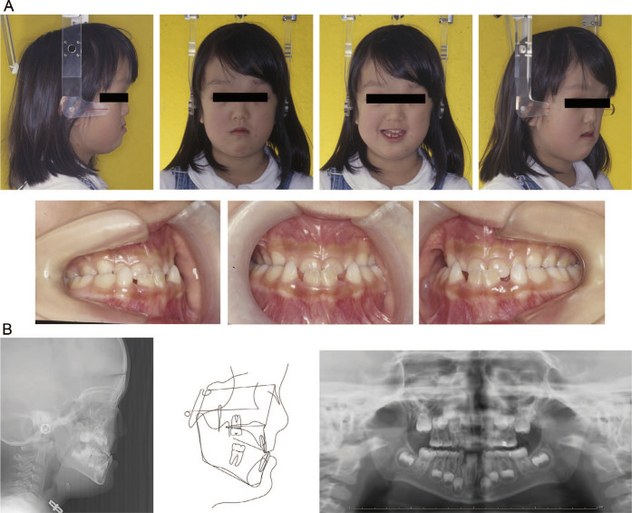
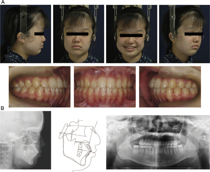
A 5-year-old girl with Crouzon syndrome was referred to the clinic because of midface deficiency and an anterior crossbite (Figures 1 and 2). Before visiting the hospital, fronto-orbital advancement with Le Fort III osteotomy and strabismus surgery was performed at the age of 4 years to improve her intracranial pressure and exorbitism.12 At the time of the first visit to the hospital, the chief complaint was a concave facial profile and an anterior reverse overjet. The patient exhibited severe midfacial deficiency with skeletal Class III malocclusion and total crossbite. During the first phase of orthodontic treatment at 5 years of age, maxillary lateral expansion and protraction using a reverse headgear were performed to improve midfacial deficiency for 4 years. However, limited forward advancement of the maxilla was observed on the superimposition of lateral cephalograms (Supplemental Figure 1).
Figure 1.
Initial records (age, 5 years, 8 months): (A) Facial and intraoral photographs; (B) Radiographs and cephalometric tracing.
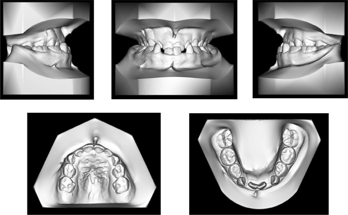
Figure 2.
Initial dental models.
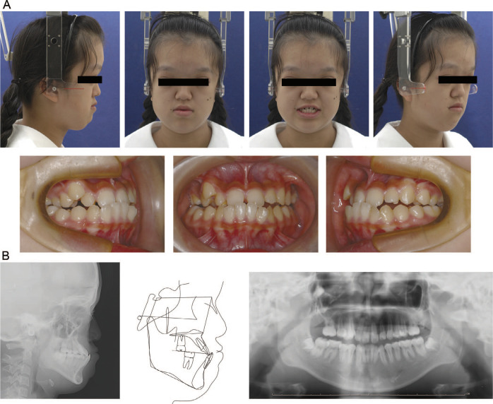
Before beginning Phase II treatment, an extraoral examination of the patient at the age of 14 years and 10 months revealed severe midfacial deficiency, moderate exorbitism, and a concave facial profile with a protruding forehead. The occlusion consisted of anterior and posterior crossbites. The occlusion was classified as Class III dental relationships on both sides, with an overjet of –3.8 mm and an overbite of 3.9 mm. The maxillary dental arch showed lateral constriction and severe crowding, with a labially blocked right canine, whereas the mandibular dental arch exhibited moderate crowding. Dental tubercles were observed on the palatal surface of the maxillary lateral incisors. The maxillary and mandibular skeletal and dental midlines coincided with the facial midline. Additionally, the patient had no symptoms of sleep-disordered breathing. Panoramic radiography revealed congenitally missing second and third molars bilaterally in the maxillary arch. Cephalometric analysis showed a skeletal Class III relationship (ANB, –8.9°) with a retrusive maxilla (SNA, 74.0°). The maxillary incisors were proclined (U1-FH, 129.1°) and the mandibular incisors showed normal inclination (L1-MP, 90.5°) (Figure 3; Table). Neither the maxilla nor the mandible showed further growth from the end of Phase I treatment, which allowed initiation of Phase II treatment at this time (Supplemental Figure 2).
Figure 3.
Pretreatment records (age, 14 years, 10 months): (A) Facial and intraoral photographs; (B) Radiographs and cephalometric tracing.
Treatment Objectives
The treatment objective was to improve the midfacial deficiency with a concave-type facial profile associated with a skeletal Class III jaw deformity. Lateral expansion of the maxillary dentition was required to harmonize the maxillary and mandibular dental arches. Additionally, rotation of the maxillary first molars and crowding in both arches required correction during preoperative orthodontic treatment. The following treatment plan was proposed: (1) maxillary lateral expansion with a quad-helix appliance, (2) placement of preadjusted edgewise appliances in both dental arches to level and align the dentition, (3) simultaneous Le Fort I and III osteotomies with DO, (4) obtaining ideal occlusion by detailing, and (5) retention. A plan was made to move the upper and lower halves of the midface by 12.0 mm and 10.0 mm, respectively.
Treatment Alternatives
Several alternative treatment options were available. These included: (1) extraction of the maxillary and mandibular first premolars and Le Fort III DO. This could be combined with mandibular bilateral sagittal split osteotomy (BSSO) advancement to potentially improve the airway. The maxillary premolars were retained due to the congenital absence of the maxillary second molar. However, maxillary incisor proclination would persist until the end of treatment. Maxillary premolars could be extracted or temporary anchorage devices could be used to retrocline the maxillary incisors. (2) Le Fort III DO or osteotomy for acute midface advancement was considered if there was no need to differentiate between the advancement of the orbital rim and maxilla. In this case, the objective was to improve the patient's exorbitism and midfacial deficiency, while less advancement was necessary for the proclined maxillary incisors. (3) Orthodontic camouflage would provide positive overjet and retain skeletal discrepancies.
Recently, maxillary anterior segment distraction osteogenesis (MASDO) was developed to advance the anterior maxillary segments and improve the retrusion of the maxilla without worsening velopharyngeal function.13,14 However, the effects of improving exorbitism have been limited.
Treatment Progress
At 5 years of age, reverse headgear was used to protract the maxilla to correct a skeletal discrepancy and midfacial deficiency for 4 years.
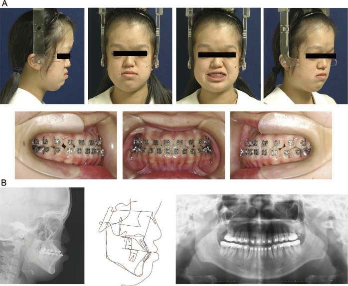
Phase II orthodontic treatment was initiated at 14 years and 10 months of age by lateral expansion of the maxilla using a quadhelical appliance. The intermolar width was increased 3.0 mm by improving the mesial rotation of the maxillary first molars. The mandibular third molars were removed and the dental tubercles on the palatal side of the maxillary lateral incisors were reduced. Subsequently, 0.022-inch pre-adjusted fixed appliances were placed on the maxillary and mandibular teeth for leveling and alignment (Figure 4). The maxillary incisors showed proclination prior to orthognathic surgery.
Figure 4.
Sagittal split ramus osteotomy preoperative records (age, 15 years, 9 months): (A) Facial and intraoral photographs; (B) Radiographs and cephalometric tracing.
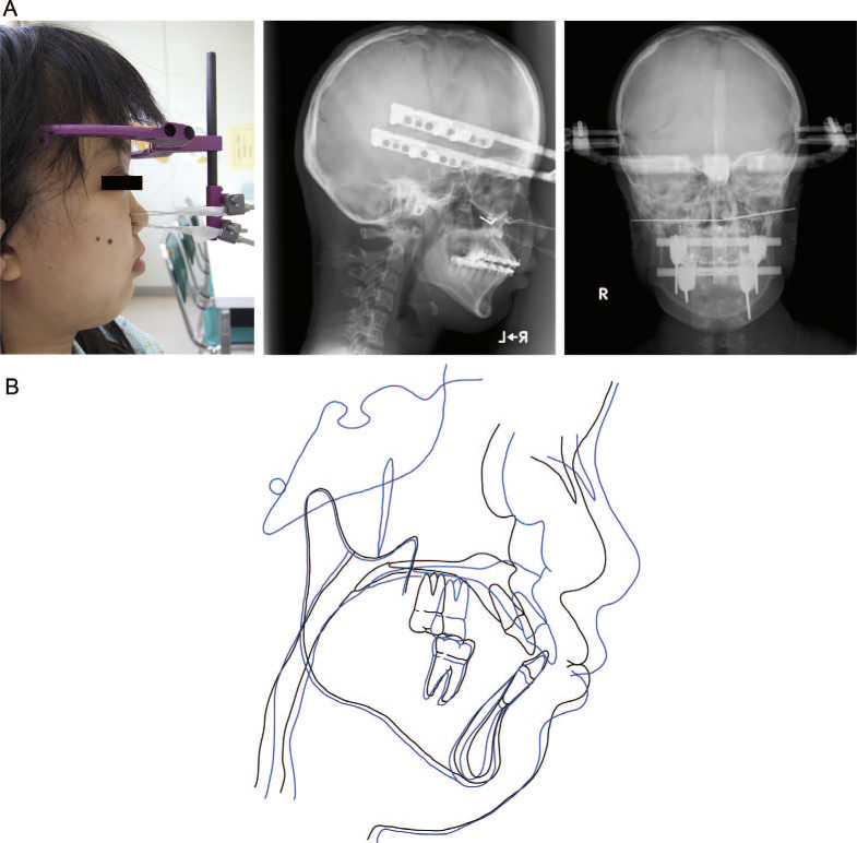
After 1 year of presurgical orthodontic preparation, combined Le Fort I and III DO was performed at the age of 15 years and 10 months to improve exorbitism by forward movement of the orbital rim, while limiting forward movement of the maxillary incisor (Figure 5). Distraction was performed at two levels to produce different advancements in the orbital rim and maxilla. Both segments were distracted 1.0 mm/d. After 12 days, the lower half of the midface reached its planned position with a positive overjet. Four days later, the upper half of the midface reached the planned position, resulting in the preferred facial profile with improved exorbitism and midfacial deficiency. With periodic assessment of facial and intraoral occlusions, the position of the device was adjusted to change the vector of bone movement. Use of extraoral devices may cause significant discomfort; however, they greatly facilitate the manipulation of the vector direction. The intermaxillary elasticity can also be used to change the direction of bone movement. After active distraction, intermaxillary consolidation with an occlusal splint was performed for 1 week.
Figure 5.
Simultaneous Le Fort I and III DO postoperative records (age, 15 years, 10 months): (A) Profile photograph, lateral and posteroanterior cephalograms; (B) superimposed cephalometric tracings: presurgery (black), postsurgery (blue).
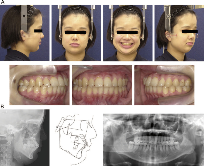
A slight enlargement of the upper airway was observed upon superimposition of the presurgical and postsurgical cephalograms (Figure 5). After 2 years of postoperative orthodontic treatment, all appliances were removed and replaced with Begg-type retainers in both arches. No obvious root resorption was detected on the panoramic radiographs (Figure 6). The facial profile and occlusion did not undergo significant relapse and maintained a favorable status even 8 years after DO surgery (Figures 7 and 8).
Figure 6.
Posttreatment records (age, 17 years, 8 months): (A) Facial and intraoral photographs; (B) Radiographs and cephalometric tracing.
Figure 7.
Postretention records (age, 26 years, 0 months): (A) Facial and intraoral photographs; (B) Radiographs and cephalometric tracing.
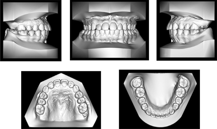
Figure 8.
Postretention dental models.
Treatment Results
The concave facial profile and midfacial deficiency showed substantial improvement with anterior movement of the midfacial bones and combined Le Fort I and III DO. In the present report, combined Le Fort I and III DO resulted in forward movement of the medial maxillary buttress and point A by 12.0 mm and 9.0 mm, respectively, which substantially improved the facial profile and occlusion. Post-treatment facial photographs revealed a straight facial profile. Intraoral photographs showed normal overjet and overbite with favorable occlusion. The molar relationship was Class I on both sides. Maxillary and mandibular crowding were eliminated to achieve proclination of the incisors. Post-treatment cephalometric analysis revealed a skeletal Class I relationship with an ANB angle of –0.7°. The interincisal angle (110.8°) was smaller than the ideal value at the end of the treatment (Figure 9, Table 1). Postsurgical computed tomography was not performed to reduce the radiation dose.
Figure 9.
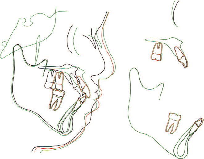
Superimposed cephalometric tracings: pretreatment (black), posttreatment (red), postretention (green).
Table 1. .
Lateral Cephalometric Measurementsa
|
Measurements |
Pretreatment (5 y, 8 mo) |
Before Phase II (14 y, 10 mo) |
Posttreatment (17 y, 8 mo) |
8 y, 4 mo Retention (26 y, 0 mo) |
| Angular (°) | ||||
| SNA | 70.6 | 74.0 | 70.6 | 78.9 |
| SNB | 77.1 | 83.0 | 79.5 | 79.8 |
| ANB | –6.5 | –8.9 | -0.7 | –1.0 |
| Mp-FH | 29.5 | 22.7 | 21.4 | 20.5 |
| Gonial angle | 126.8 | 121.0 | 121.9 | 121.0 |
| U1-FH | 100.4 | 129.1 | 129.4 | 131.7 |
| L1-FH | 63.4 | 66.8 | 60.2 | 57.7 |
| L1-Mp | 87.1 | 90.5 | 98.4 | 101.8 |
| IIA | 117.7 | 117.7 | 110.8 | 106.0 |
| OPA | 6.4 | 6.4 | 8.2 | 8.3 |
| Linear (mm) | ||||
| S-N | 66.9 | 68.5 | 72.2 | 72.2 |
| N-Me | 100.7 | 116.1 | 118.1 | 117.7 |
| N/PP | 48.0 | 43.6 | 48.1 | 48.1 |
| Me/PP | 57.7 | 69.3 | 67.4 | 67.3 |
| PTM-A/PP | 43.8 | 42.6 | 53.0 | 53.0 |
| Go-Me | 52.4 | 63.4 | 62.2 | 63.2 |
| Ar-Go | 39.9 | 52.2 | 53.7 | 53.6 |
| Ar-Me | 82.7 | 101.1 | 101.1 | 101.8 |
| Overjet | –3.1 | –3.8 | 3.7 | 3.8 |
| Overbite | 2.9 | 3.9 | 1.6 | 1.2 |
| A-N perp | –12.5 | –10.0 | –4.2 | –4.2 |
| Wits | –15.2 | –7.4 | 1.1 | 0.4 |
A-N perp indicates A-point to N-perpendicular; ANB, point A-N-point B angle; Ar-Go, distance between articulare (Ar) and Go; Ar-Me, distance Ar and Me; Go-Me, distance between gonion (Go) and Me; L1, lower incisor; MP-FH, angle between Frankfort horizontal (FH) plane and mandibular plane (MP); Me-PP, distance between Me and PP; N-Me, distance between N and Menton (Me); N-PP, distance between N and palatal plane (PP); OP, occlusal plane angle; Ptm-A/PP, distance between point pterygomaxillary fissure (Ptm) and point A angle projected on a palatal plane (PP); S-N, distance between S and N; SNA, sella (S)-nasion (N)-point A angle; SNB, S-N-point B angle; IIA, interincisal angle; U1, upper incisor.
DISCUSSION
Crouzon syndrome is associated with a wide range of craniofacial deformities and severe malocclusion resulting from premature fusion of cranial sutures and synchondroses. Advancement of the frontal bone at an early age is frequently performed to prevent or improve the intracranial pressure.15 Furthermore, Crouzon syndrome can result in skeletal hypoplasia of the midface and severe malocclusion, such as reverse overjet.1 Orthodontic treatment was performed in different phases to correct skeletal and dental discrepancies. In this case, reverse headgear was used to improve midfacial deficiency, which resulted in limited forward movement of the maxilla. A recent study revealed that patients with syndromic craniosynostosis, including those with Crouzon syndrome, exhibit early radiological fusion of the circummaxillary suture.16 These results indicate that an orthopedic approach for correcting the maxilla in patients with syndromic craniosynostosis should be considered with caution. Additionally, meticulous assessment of follow-up radiographic examinations is highly recommended to evaluate the efficacy of treatment and avoid unnecessary intervention. When severe skeletal deficiency persists even after adolescence, surgical intervention is required to achieve normal occlusion.
Due to severe midfacial deficiency and exophthalmos, surgical intervention, including Le Fort III osteotomy, is frequently used in the treatment of malocclusion. This case report demonstrates the long-term results of comprehensive orthodontic treatment in a patient with Crouzon syndrome treated with a combination of Le Fort I and III DO using a RED system.
Le Fort III osteotomy was first described by Gillies in 1950 and was successfully performed by Tessier in 1971.4,17 Ortiz-Monasterio et al.18 introduced the craniofacial monobloc Le Fort III osteotomy as an improved surgical procedure for the treatment of patients with craniosynostosis. DO with Le Fort III osteotomy has also been used as a treatment protocol for patients with severe midfacial deficiencies and exophthalmos.19,20 Several case series have shown an average forward movement of the midface after Le Fort III DO of 12–20 mm21 or 12–22 mm.20 In some studies, Le Fort III DO has been shown to be stable for more than 5 years.22–24
In contrast, combined Le Fort I and Le Fort III osteotomies have been used in cases requiring differential correction of the orbital rim and maxillary component.25,26 Le Fort I and Le Fort III distraction osteotomies have been successfully performed in patients with syndromic craniosynostosis; however, their long-term stability remains largely elusive.27–30
In the present case, 8 years after distraction, minimal relapse of the maxillary advancement was observed. Patients with syndromic craniosynostosis, including Crouzon syndrome, show a higher frequency of sleep apnea due to midface or mandibular hypoplasia.31 Surgical mandibular advancement should be considered in such cases. In the present case, no sleep disorders were detected and the size of the mandible was within the normal range; thus, the patient was treated without mandibular advancement. This case report, along with existing evidence, suggests the advantage of Le Fort I and III DO as an option for orthognathic treatment of malocclusion and for achieving long-term stability in patients with Crouzon syndrome and severe midfacial deficiency.
CONCLUSIONS
A patient diagnosed with Crouzon syndrome was effectively treated using a rigid external distractor system during concurrent Le Fort I and III DO procedures. The patient's exorbitism and midfacial deficiency were notably ameliorated, and her Class III malocclusion was successfully corrected. Eight years after DO, there was minimal relapse of the maxillary advancement.
Considering the present clinical results, simultaneous Le Fort I and III DO is suggested as an efficient treatment with good long-term stability in patients with Crouzon syndrome, which requires different advancements between the orbital rim and maxilla.
SUPPLEMENTAL DATA
Supplemental Figures 1 and 2 are available online.
Supplemental Figure 1. Superimposed cephalometric tracings: start of Phase I treatment (black) and end of Phase I (gray).
Supplemental Figure 2. Superimposed cephalometric tracings: end of Phase I treatment (black) and start of Phase II (gray).
REFERENCES
- 1.Mathijssen IM. Guideline for care of patients with the diagnoses of craniosynostosis: working group on craniosynostosis J Craniofac Surg 2015. 26 1735–1807. [DOI] [PMC free article] [PubMed] [Google Scholar]
- 2.Foster KA, Frim DM, McKinnon M. Recurrence of synostosis following surgical repair of craniosynostosis Plast Reconstr Surg 2008. 121 70e–76e. [DOI] [PubMed] [Google Scholar]
- 3.Meazzini MC, Mazzoleni F, Caronni E, Bozzetti A. Le Fort III advancement osteotomy in the growing child affected by Crouzon's and Apert's syndromes: presurgical and postsurgical growth J Craniofac Surg 2005. 16 369–377. [DOI] [PubMed] [Google Scholar]
- 4.Tessier P. The definitive plastic surgical treatment of the severe facial deformities of craniofacial dysostosis. Crouzon's and Apert's diseases Plast Reconstr Surg 1971. 48 419–442. [DOI] [PubMed] [Google Scholar]
- 5.Kreiborg S, Aduss H. Pre- and postsurgical facial growth in patients with Crouzon's and Apert's syndromes Cleft Palate J 1986. 23 Suppl 1 78–90. [PubMed] [Google Scholar]
- 6.Hopper RA, Sandercoe G, Woo A, et al. Computed tomographic analysis of temporal maxillary stability and pterygomaxillary generate formation following pediatric Le Fort III distraction advancement Plast Reconstr Surg 2010. 126 1665–1674. [DOI] [PubMed] [Google Scholar]
- 7.Tong H, Liu L, Tang X, et al. Midface distraction osteogenesis using a modified external device with elastic distraction for Crouzon syndrome J Craniofac Surg 2017. 28 1573–1577. [DOI] [PubMed] [Google Scholar]
- 8.Saltaji H, Altalibi M, Major MP, et al. Le Fort III distraction osteogenesis versus conventional Le Fort III osteotomy in correction of syndromic midfacial hypoplasia: a systematic review J Oral Maxillofac Surg 2014. 72 959–972. [DOI] [PubMed] [Google Scholar]
- 9.Cedars MG, Linck DL, Chin M, Toth BA. Advancement of the midface using distraction techniques Plast Reconstr Surg 1999. 103 429–441. [DOI] [PubMed] [Google Scholar]
- 10.Fearon JA. Halo distraction of the Le Fort III in syndromic craniosynostosis: a long-term assessment Plast Reconstr Surg 2005. 115 1524–1536. [DOI] [PubMed] [Google Scholar]
- 11.Kuroda S, Watanabe K, Ishimoto K, Nakanishi H, Moriyama K, Tanaka E. Long-term stability of LeFort III distraction osteogenesis with a rigid external distraction device in a patient with Crouzon syndrome Am J Orthod Dentofacial Orthop 2011. 140 550–561. [DOI] [PubMed] [Google Scholar]
- 12.Britto JA, Greig A, Abela C, Hearst D, Dunaway DJ, Evans RD. Frontofacial surgery in children and adolescents: techniques, indications, outcomes Semin Plast Surg 2014. 28 121–129. [DOI] [PMC free article] [PubMed] [Google Scholar]
- 13.Wang XX, Wang X, Li ZL, et al. Anterior maxillary segmental distraction for correction of maxillary hypoplasia and dental crowding in cleft palate patients: a preliminary report Int J Oral Maxillofac Surg 2009. 38 1237–1243. [DOI] [PubMed] [Google Scholar]
- 14.Aikawa T, Haraguchi S, Tanaka S, et al. Rotational movement of the anterior maxillary segment by hybrid distractor in patients with cleft lip and palate Oral Surg Oral Med Oral Pathol Oral Radiol Endod 2010. 110 292–300. [DOI] [PubMed] [Google Scholar]
- 15.Kreiborg S, Marsh JL, Cohen MM, et al. Comparative three-dimensional analysis of CT-scans of the calvaria and cranial base in Apert and Crouzon syndromes J Craniomaxillofac Surg 1993. 21 181–188. [DOI] [PubMed] [Google Scholar]
- 16.Meazzini MC, Corradi F, Mazzoleni F, et al. Circummaxillary sutures in patients with Apert, Crouzon, and Pfeiffer syndromes compared to nonsyndromic children: growth, orthodontic, and surgical implications. Cleft Palate Craniofac J 2021. 58 299–305. [DOI] [PubMed] [Google Scholar]
- 17.Gillies H, Harrison SH. Operative correction by osteotomy of recessed malar maxillary compound in a case of oxycephaly Br J Plast Surg 1950. 3 123–127. [DOI] [PubMed] [Google Scholar]
- 18.Ortiz-Monasterio F, del Campo AF, Carrillo A. Advancement of the orbits and the midface in one piece, combined with frontal repositioning, for the correction of Crouzon's deformities Plast Reconstr Surg 1978. 61 507–516. [DOI] [PubMed] [Google Scholar]
- 19.Polley JW, Figueroa AA, Charbel FT, Berkowitz R, Reisberg D, Cohen M. Monobloc craniomaxillofacial distraction osteogenesis in a newborn with severe craniofacial synostosis: a preliminary report J Craniofac Surg 1995. 6 421–423. [DOI] [PubMed] [Google Scholar]
- 20.Witherow H, Dunaway D, Evans R, et al. Functional outcomes in monobloc advancement by distraction using the rigid external distractor device Plast Reconstr Surg 2008. 121 1311–1322. [DOI] [PubMed] [Google Scholar]
- 21.Safi AF, Kreppel M, Kauke M, Grandoch A, Nickenig HJ, Zöller J. Rigid external distractor-aided advancement after simultaneously performed LeFort-III osteotomy and fronto-orbital advancement J Craniofac Surg 2018. 29 170–174. [DOI] [PubMed] [Google Scholar]
- 22.Meazzini MC, Allevia F, Mazzoleni F, et al. Long-term follow-up of syndromic craniosynostosis after Le Fort III halo distraction: a cephalometric and CT evaluation J Plast Reconstr Aesthet Surg 2012. 65 464–472. [DOI] [PubMed] [Google Scholar]
- 23.Hirjak D, Reyneke JP, Janec J, Beno M, Kupcova I. Long-term results of maxillary distraction osteogenesis in nongrowing cleft: 5-years experience using internal device Bratisl Lek Listy 2016. 117 685–690. [DOI] [PubMed] [Google Scholar]
- 24.Patel PA, Shetye PR, Warren SM, Grayson BH, McCarthy JG. Five-year follow-up of midface distraction in growing children with syndromic craniosynostosis Plast Reconstr Surg 2017. 140 794e–803e. [DOI] [PubMed] [Google Scholar]
- 25.Obwegeser HL. Surgical correction of small or retrodisplaced maxillae. The “dish-face” deformity Plast Reconstr Surg 1969. 43 351–365. [DOI] [PubMed] [Google Scholar]
- 26.Chan C, Garg R, Wlodarczyk JR, Yen S, Urata MM. Simultaneous LeFort III and LeFort I osteotomies in craniometaphyseal dysplasia. Cleft Palate Craniofac J 2021. 58 1560–1568. [DOI] [PubMed] [Google Scholar]
- 27.Matsumoto K, Nakanishi H, Koizumi Y, et al. Segmental distraction of the midface in a patient with Crouzon syndrome J Craniofac Surg 2002. 13 273–278. [DOI] [PubMed] [Google Scholar]
- 28.Takashima M, Kitai N, Murakami S, et al. Dual segmental distraction osteogenesis of the midface in a patient with Apert syndrome Cleft Palate Craniofac J 2006. 43 499–506. [DOI] [PubMed] [Google Scholar]
- 29.Satoh K, Mitsukawa N, Hosaka Y. Dual midfacial distraction osteogenesis: Le Fort III minus I and Le Fort I for syndromic craniosynostosis Plast Reconstr Surg 2003. 111 1019–1028. [DOI] [PubMed] [Google Scholar]
- 30.Sakamoto Y, Nakajima H, Tamada I, Sakamoto T, Le Fort IV. + I distraction osteogenesis using an internal device for syndromic craniosynostosis J Oral Maxillofac Surg 2014. 72 788–795. [DOI] [PubMed] [Google Scholar]
- 31.Bannink N, Nout E, Wolvius EB, Hoeve HL, Joosten KF, Mathijssen IM. Obstructive sleep apnea in children with syndromic craniosynostosis: long-term respiratory outcome of midface advancement Int J Oral Maxillofac Surg 2010. 39 115–121. [DOI] [PubMed] [Google Scholar]


