Abstract
Background
Glucocorticoids are the first-line treatment for Pemphigus vulgaris (PV), but its serious side effects can be life-threatening for PV patients. Tacrolimus (FK506) has been reported to have an adjuvant treatment effect against PV. However, the mechanism underlying the inhibitory effect of FK506 on PV-IgG-induced acantholysis is unclear.
Objective
The objective of this study was to explore the effect of FK506 on desmoglein (Dsg) expression and cell adhesion in an immortalized human keratinocyte cell line (HaCaT cells) stimulated with PV sera.
Methods
A cell culture model of PV was established by stimulating HaCaT cells with 5% PV sera with or without FK506 and clobetasol propionate (CP) treatment. The effects of PV sera on intercellular junctions and protein levels of p38 mitogen-activated protein kinase (p38MAPK), heat shock protein 27 (HSP27), and Dsg were assayed using western blot analysis, immunofluorescence staining, and a keratinocyte dissociation assay.
Results
PV sera-induced downregulation of Dsg3 was observed in HaCaT cells and was blocked by FK506 and/or CP. Immunofluorescence staining revealed that linear deposits of Dsg3 on the surface of HaCaT cells in the PV sera group disappeared and were replaced by granular and agglomerated fluorescent particles on the cell surface; however, this effect was reversed by FK506 and/or CP treatment. Furthermore, cell dissociation assays showed that FK506 alone or in combination with CP increased cell adhesion in HaCaT cells and ameliorated loss of cell adhesion induced by PV sera. Additionally, FK506 noticeably decreased the PV serum-induced phosphorylation of HSP 27, but had no effect on p38MAPK phosphorylation.
Conclusion
FK506 reverses PV-IgG induced-Dsg depletion and desmosomal dissociation in HaCaT cells, and this effect may be obtained by inhibiting HSP27 phosphorylation.
Supplementary Information
The online version contains supplementary material available at 10.1186/s12865-023-00582-z.
Keywords: Pemphigus Vulgaris, Desmoglein, Tacrolimus, Acantholysis, Glucocorticoids
Introduction
Pemphigus is a group of rare, chronic, potentially life-threatening, autoimmune bullous diseases. The two main pemphigus variants are pemphigus vulgaris (PV), which often originates with painful oral erosions, and pemphigus foliaceus (PF), which is characterized by exclusive skin lesions [1]. IgG antibodies against epidermal desmosomes, including desmoglein (Dsg) 1 and Dsg3, two major cell-cell adhesive structure proteins, cause loss of intercellular adhesion, acantholysis, and blister formation [2, 3]. Furthermore, both anti-Dsg1 and -Dsg3 antibodies are involved in the activation of signaling pathways including Ca2+, p38 mitogen activated protein kinases (p38MAPK), protein kinase C (PKC), Src, EGFR/Erk, and several others, resulting in modulation of keratinocyte cell adhesion properties [4].
The MAPK family, a group of highly conserved protein kinases that play an important role in signal transduction by regulating gene transcription in the nucleus in response to changes in the cellular environment, includes four distinct cascades, namely, the extracellular signal-related kinases (ERK1/2), ERK5, p38MAPK, and Jun N-terminal kinases (JNK1/2/3) [5, 6]. p38MAPK inhibition blocks loss of cell cohesion in keratinocyte cultures and blister formation in mouse models induced by PV autoantibodies [7]. The pathogenesis of PV may be related to the activation of p38MAPK and subsequent phosphorylation of the heat shock protein 27 (HSP27), which leads to actin cytoskeleton reorganization and keratin retraction [8]. Specific inhibition of this molecule using SB203580, a selective and ATP-competitive p38 MAPK inhibitor, was effective in preventing loss of adhesive contacts in mouse models [9].
The annual incidence of PV ranges between 0.76 and 32.0 cases per million population, and the epidemiology of PV accounts for about 65–95% of pemphigus cases [10]. Traditionally, PV has been treated with a combination of high-dose systemic glucocorticoids and an adjuvant immunosuppressant. An anti-CD20 antibody, rituximab, is recommended as the first-line treatment for moderate-to-severe PV [11]. Because the long-term administration of glucocorticoids can cause adverse side effects, such as secondary infection, osteoporosis, and obesity, a more effective treatment for PV is needed.
Tacrolimus (FK506) is a calcineurin inhibitor that is used to suppress the immune system in a variety of conditions, including treatment of diverse immune disorders, inhibiting the formation of activated T cells and suppressing the synthesis of pro-inflammatory cytokines [12]. Previous studies have investigated the safety and effectiveness of systemic FK506 or topical use in pemphigus diseases [13–17]. In a study with PV model mice, FK506 monotherapy showed satisfactory effects by inhibiting anti-Dsg3 IgG production, the appearance of the PV phenotype, and development of body weight loss [18]. This study aimed to explore the effect of FK506 and the highly potent glucocorticoid—clobetasol propionate (CP)—on the intercellular junctions and Dsg3 expression in HaCaT cells stimulated with sera from PV patients.
Results
PV sera- and AK23-induced loss of keratinocyte adhesion and Dsg3 depletion
We initially characterized changes in Dsg3 expression in HaCaT cells incubated with PV sera over a time course. AK23 is a monoclonal antibody (mAb) obtained by immunizing mice with recombinant mouse Dsg3 ectodomain and is currently widely used to model the pathogenesis of PV [19]. In this study, AK23 was used as a positive control.
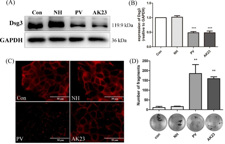
The protein expression of Dsg3 was dramatically decreased after the addition of PV sera at 1 h, and Dsg3 protein levels remained low at 24 h (Supplementary Fig. 1). Dsg3 expression in HaCaT cells was significantly decreased in the PV group and AK23 group when compared with the control group and normal healthy (NH) group (Fig. 1A–B). Fluorescent monoclonal antibody detection showed that Dsg3 was continuously distributed along cellular junctions under control conditions or following treatment with healthy donor sera. In contrast, 5% PV sera or 1 µg/mL AK23 treatment resulted in the disruption of Dsg3 protein expression, the linear fluorescence of Dsg3 on the surface of cells disappeared, and the fluorescent particles were aggregated on the cell surface and cytoplasm membrane (Fig. 1C). However, AK23 showed a weaker effect on the adhesion of HaCaT cells as compared to PV sera.
Fig. 1.
Both pemphigus sera and AK23 caused a decrease in keratinocyte adhesion and Desmoglein (Dsg) 3 internalization in HaCaT cells. (A) Western blot analysis of Dsg3 in total cell lysates of HaCaT cells incubated with 5% PV sera or 1 µg/ml AK23 for 24 h. GAPDH was used as a loading control. Respective controls [normal control group (Con) or normal healthy sera group (NH)] were run in parallel. Both PV sera and AK23 decreased Dsg3 protein levels as compared with the control. (B) Densitometric analysis was derived from the ratio of Dsg3 to GAPDH expression in each group and then normalized to the control group. Statistically significant differences are indicated by *** p < 0.001 vs. Con. Data are represented by mean ± SE (n = 3). (C) Immunostaining revealed that a 24-h incubation with 5% PV sera or 1 µg/ml AK23 induced fragmentation of the Dsg3 staining pattern along the cell borders (scale bar = 50 μm). (D) Dissociation assays in HaCaT cells showed weaker intercellular adhesion consistence with more free cells in cells incubated with 5% PV sera or AK23. ** p < 0.01 vs. Con. Data are shown as mean ± SE (n = 3)
Cell-cell adhesion was also compromised as revealed by the results of the dispase-based dissociation assays. In agreement with the change in Dsg3 expression, cell adhesion was significantly impaired upon the addition of PV sera and AK23 (Fig. 1D). PV sera resulted in a slight increase in the number of fragments in HaCaT cells compared to AK23, however, the comparison was not statistically significant. Taken together, these results demonstrated that HaCaT cells manifest the typical hallmarks of PV when exposed to PV sera or AK23.
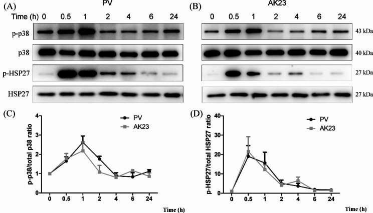
In order to explore which proteins are involved in the pathogenesis of PV, we measured the phosphorylation levels of p38 MAPK and HSP27 in HaCaT cells, using total p38MAPK and total HSP27 as a loading control, respectively. Both PV serum and AK23 induced rapid phosphorylation of p38 MAPK and HSP27, with peak activation at 0.5–2 h (Fig. 2).
Fig. 2.
Time course of p38 mitogen-activated protein kinase (MAPK) and heat shock protein (HSP) 27 activation in HaCaT cells treated with pemphigus vulgaris (PV) sera and AK23 monoclonal antibody. (A, B) (with the exception of 0 h group as blank control ant it is not treated with any reagent), Peaks of phospho-p38 MAPK (p-p38 MAPK) and phospho-HSP27 were detected by western blot. (C, D) Densitometric analysis was derived from the ratios of p-p38 MAPK to p38 MAPK and p-HSP27 to HSP27 expression in each group and normalized to the 0 h group. Peak activation of p38 MAPK and HSP27 by PV sera and AK23 occurred around 0.5–2 h. Levels of total p38 MAPK and HSP27 are shown as loading controls (n = 3)
FK506 inhibited PV sera- and AK23 induced-disruption of cell-cell contact and Dsg3 internalization
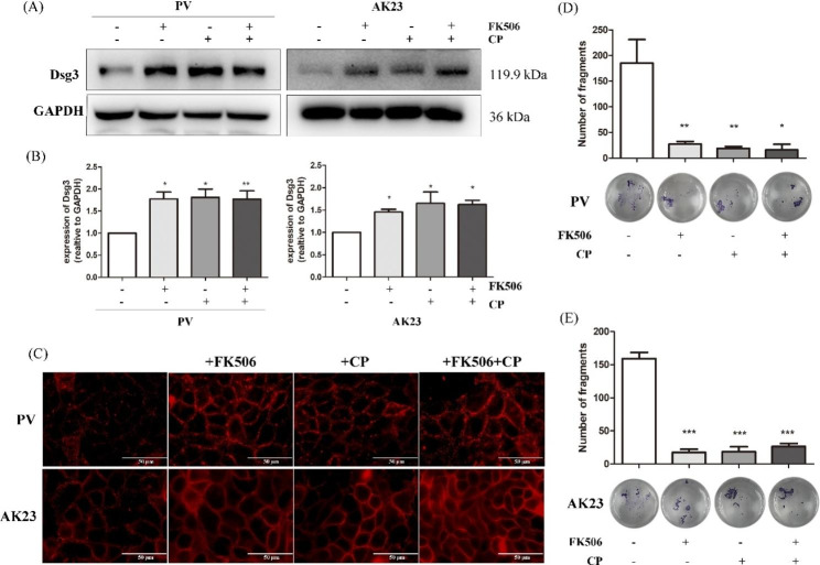
The results of the western blot demonstrated that expression of Dsg3 was dramatically increased when HaCaT cells stimulated with PV sera or AK23 were treated with FK506 and/or CP (Fig. 3A and B). Consistent with these findings, FK506 and/or CP prevented the PV sera-mediated loss of cell surface-localized Dsg3 (Fig. 3C). In the AK23 group, the addition of FK506 and/or CP restored the linear fluorescent distribution of Dsg3 (Fig. 3C). Dispase-based dissociation assays indicated that when cells in the PV sera and AK23 groups were treated with FK506 and/or CP, the number of free cells dramatically decreased (p < 0.05) (Fig. 3D–E).
Fig. 3.
FK506 and clobetasol propionate (CP) rescue disruption of cell-cell contacts and the loss of desmoglein (Dsg) 3 induced by pemphigus vulgaris (PV) sera and AK23. HaCaT cells were co-incubated with 100 nM FK506 and/or 1 µM CP in the present of 5% PV sera or 1 µg/mL AK23 for 24 h prior to western blot and immunostaining. (A) Both PV sera- and AK23-stimulated Dsg3 internalization was blocked by FK506 and CP. (B) Statistically significant differences are indicated by * p < 0.05 and * *p < 0.01 vs. PV group or AK23 group, respectively. Data are represented by mean ± SE. (C) Consistent with western blot, FK506 and/or CP prevented the PV sera- and AK23-mediated loss of cell surface-localized Dsg3. (scale bar = 50 μm). (D and E) Dispase-based dissociation assays indicated that when cells in the PV group and AK23 group were treated with FK506 and/or CP, the number of free cells was dramatically decreased (n = 3). Statistically significant differences are indicated by *** p < 0.001 vs. PV group or AK23 group, respectively
FK506 did not decrease the phosphorylation of p38 MAPK but inhibited the phosphorylation of HSP27 induced by PV sera
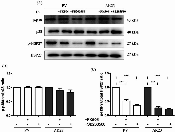
We further analyzed the effect of FK506 on the phosphorylation of both p38 MAPK and HSP27 induced by PV sera. Pretreatment of HaCaT cells with 100 nM FK506 for 2 h, followed by exposure to PV sera or AK23 for 1 h. FK506 significantly blocked phosphorylation of HSP27, but not p38 MAPK (Fig. 4). These results suggested that FK506 may inhibit Dsg3 depletion through inactivation of HSP27.
Fig. 4.
The effect of FK506 on the pemphigus vulgaris (PV) sera-induced phosphorylation of both p38 mitogen-activated protein kinases (MAPK) and heat shock protein (HSP) 27. HaCaT cells were pretreated with 100 nM FK506 or 1 µM SB203580 (p38 MAPK inhibitor) for 2 h, followed by exposure to 5% PV sera or 1 µg/ml AK23 for 1 h. FK506 significantly blocked the phosphorylation of HSP27, but not p38 MAPK. Levels of total p38 MAPK are shown as loading controls (n = 3). (B and C) Data are represented by mean ± SE.
Discussion
The objective of the present work was to investigate the effect of FK506 on the p38 MAPK and HSP27 pathways in an in vitro model of pemphigus [20]. FK506 inhibited pemphigus sera-induced loss of keratinocyte adhesion in vitro. We also found that PV serum and AK23 activated p38 MAPK and HSP27, producing a peak at 0.5–1 h. Activation of p38 MAPK and subsequent phosphorylation of HSP27 seem to be related to the pathogenesis of PV, as both lead to keratin retraction and reorganization of the actin cytoskeleton [21]. Though a protective effect was observed for FK506 on PV sera-induced acantholysis, our results herein showed that the phosphorylation of p38 MAPK was not inhibited by treatment with FK506. Our results indicate that FK506 inhibits acantholysis through a mechanism independent of p38 MAPK phosphorylation (Fig. 5).
Fig. 5.
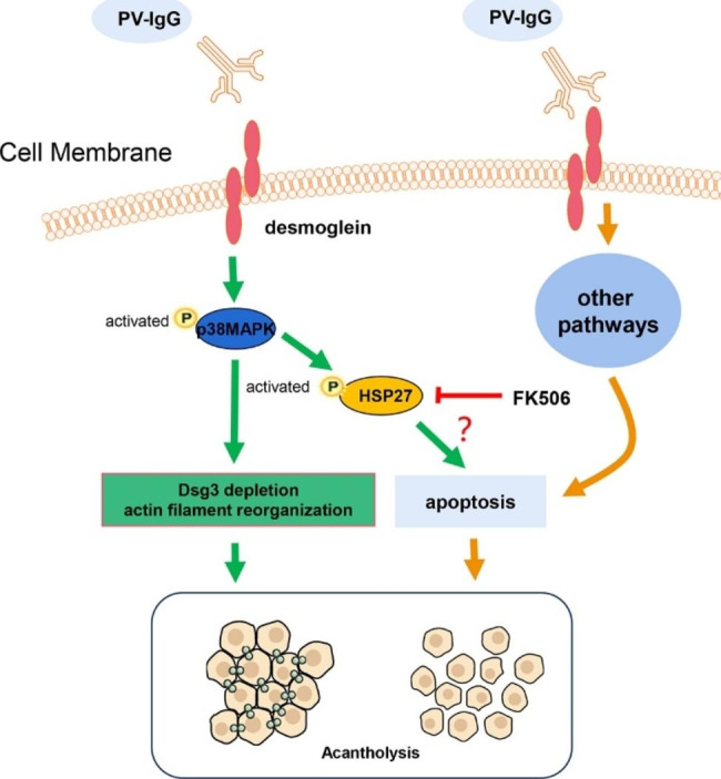
Pictorial presentation of the proposed hypothesis pertaining to apoptosis in the pathogenesis of pemphigus vulgaris. PV-IgG depletes desmoglein (Dsg)3 and causes actin filament reorganization, leading to acantholysis. FK506 blocks heat shock protein (HSP) 27 phosphorylation and prevents changes in Dsg3 depletion and actin filament reorganization
FK506 inhibits calcineurin and the formation of activated T cells, suppresses the synthesis of pro-inflammatory cytokine, and is also associated with the inhibition of nitric oxide synthase activation, cell degranulation, and apoptosis [12, 22] and is widely used as an effective agent to treat autoimmune diseases [23, 24]. Studies have found beneficials results using systemic tacrolimus as an adjuvant treatment in pemphigus [16, 17]. FK506 was reported to have suitable tolerance as a steroid-sparing immunosuppressive agent in the management of PV [16, 17]. However, the mechanism underlying the effectiveness of FK506 for the treatment of PV needs to be further clarified. As far as we know, this is the first study assessing the effectiveness of FK506 for the treatment of pemphigus relating to the p38 MAPK pathway in an in vitro model of pemphigus.
The p38MAPK signaling pathway is an important signaling pathway mediating Dsg3 internalization followed by depletion by endosomes [25]. A previous study reported that the pathogenic activity of polyclonal PV IgG can be attributed to p38 MAPK-dependent clustering and Dsg3 endocytosis and that pathogenic monoclonal Dsg3 antibodies, such AK23, cause loss of adhesion primarily through hindrance, which do not function through p38MAPK pathway [26]. Activation of the p38 MAPK subsequently leads to keratin retraction and reorganization of the actin cytoskeleton, which appears to be involved PV pathogenesis [27]. As previously reported, PV sera induced acantholysis and decreased the total amount of Dsg3 in PV-IgG incubated keratinocytes [28]. Binding of autoantibodies to Dsg3 activated the p38MAPK pathway, resulting in Dsg3 depletion and a collapse in the keratin cytoskeleton [29, 30]. PV IgG causes the internalization of cell-surface Dsg3 into endosomes after binding to keratinocytes as early as 4 h post-exposure, and this phenomenon was able to be blocked by the p38 MAPK inhibitor, SB202190 [27, 30]. In addition, previous observations in tissue culture and animal models suggested a central role for p38 MAPK in the mechanisms underlying acantholysis in PV [29, 31, 32]. In the present study, PV sera and AK23 induced rapid phosphorylation of p38 MAPK, with peak activation at around 1 h (Fig. 2), which was consistent with previous studies [31, 33]. FK506 inhibits desmosomal dissociation of HaCaT cells, but it does not inhibit p38 MAPK phosphorylation pathway (Figs. 3 and 4). We postulated that the phosphorylation of HSP27 may promote the apoptosis pathway, which was inhibited by FK506, however, further studies are needed.
HSP27 belongs to the small molecular weight Hsp family, whose expression is induced in response to a wide variety of physiological and environmental insults [34]. The mammalian small Hsps are rapidly phosphorylated by MAPK-activated proteinase 2/3 (MAPKAP 2/3) at several distinct sites following exposure to a wide range of extracellular stresses [35]. HSP27 is involved in cell death pathways by regulating partner proteins, such as necrosis and apoptosis, and phosphorylated HSP27 regulates actin filaments dynamics and intracellular redox state in cytoskeleton organization during processes such as cell migration or cell stress [34]. A study recently reported a new pathological mechanism of acantholysis in pemphigus, i.e., apoptosis, which emphasizes the involvement of apoptotic enzymes in the occurrence of acantholysis in terms of molecular events and time series [36]. In the present study, the phosphorylation of HSP27 induced by PV sera was inhibited by FK506. However, immunofluorescence assays showed that silencing the HSP27 gene (HSP27-HOMO-877, Gene ID: 3315) did not prevent Dsg3 endocytosis induced by PV sera on the surface of HaCaT cells (Supplementary Figs. 2 and 3). This suggests that HSP27 phosphorylation may be involved in HaCaT apoptosis but not directly involved in endocytosis of Dsg3, as inactivation of HSP27 is a less likely mechanism for acantholysis.
Recent studies have found that calcium (Ca2+) signaling is crucial to a variety of functions of biology. Ca2+ flux-dependent signaling pathways have been reported to play a vital role in the epidermal blistering, and inhibition of Ca2+ signaling blocked keratinocyte adhesion loss in vitro and human skin blistering ex vivo induced by pemphigus Dsg IgG. [2]. Though we are not sure whether FK506 inhibits calcium influx in cell membranes. FK506 has been attributed to have immunodulatory effects on both T cells and non-T cells, including keratinocytes [37]. FK506 may also reduce Dsg3 depletion through the inhibition of a Ca2+ pathway and deserves further investigation.
This study has some limitations. First, anti-Dsg1 antibodies were not used as another positive control. Second, although FK506 inhibits HSP27 phosphorylation and silencing the HSP27 gene does not prevent Dsg3 endocytosis, the question of whether FK506 affects HSP27 expression has not been answered. It remains unclear if FK506 inhibits desmosomal dissociation of keratinocytes through regulating phosphorylation of downstream targets, such as MAPKAP kinase 2/3.
Conclusion
In summary, this study showed that PV sera disrupts desmosome junctions by inducing endocytosis in HaCaT cells, resulting in desmosomal dissociation. Furthermore, FK506 reversed this effect, which was comparable to CP. FK506-dependent inhibition of HSP27 phosphorylation may also contribute to enhanced cell-cell adhesion of the HaCaT monolayer. These findings lay the groundwork for elucidating the mechanism of action of FK506 in the treatment of pemphigus.
Materials and methods
Participants and specimens
Patient blood sera were collected from Guangzhou Institute of Dermatology between October 2019 and October 2020. All six patients were in the active stage prior to treatment, and blood sera from three healthy volunteers were used as healthy control. The characteristics and the titer of anti-Dsg1 and anti-D3 antibodies in the patient’s serum is shown in Table 1. The inclusion and exclusion criteria of PV patients have been reported in the literature [17, 38]. Disease severity was scored using the Pemphigus Disease Area Index (PDAI) score. The mixed serum was prepared by heating at 56 °C for 30 min to inactivate complement. An anti-Dsg antibody was tested by enzyme-linked immunosorbent assay (ELISA) assay. The anti-Dsg antibody concentration in the mixed serum was 169 U/mL for Dsg1 and 117 U/mL for Dsg3. Anti-Dsg antibody titers were negative in the mixed serum from the three healthy volunteers (Dsg1: 1 U/mL; Dsg3: 1 U/mL, 2 females and 1 males, 26–29 years old). This study and all relevant experiments were reviewed and approved by the Guangzhou Institute of Dermatology Research Ethics Committee (NO. 201,802, Guangzhou, Guangdong Province, China).
Table 1.
Characteristics of the six PV patients included in the study
| Factor | Characteristics |
|---|---|
| Gender | 3 male and 3 female |
| Age (mean ± SD) | 52.45 ± 15.20 |
| anti-Dsg3 (U/ml, mean ± SD) | 166.33 ± 52.69 |
| anti-Dsg1(U/ml, mean ± SD) | 20.22 ± 74.45 |
| PDAI score (mean ± SD) | 26.27 ± 8.10 |
Antibodies and reagents
The primary antibodies included mouse anti-Dsg3 antibody (Abcam, Cambridge, UK), rabbit anti-p38 MAPK, rabbit anti-phospho-p38 MAPK (Thr180/Tyr182, Cell signaling Technology, Danvers, MA), mouse anti-HSP27, rabbit anti-phospho-HSP27 (Ser82, Cell signaling Technology), and rabbit anti-GAPDH (Affinity Biosciences, OH, USA). The secondary antibodies included a goat anti-mouse antibody, goat anti-rabbit antibody (Abclonal, Wuhan, China), and Cy3-conjugated goat anti-mouse (Abclonal) antibody.
Other regents included FK506 (Med Chem Express, Shanghai, China), clobetasol propionate (CP) (Macklin, Shanghai, China) and a p38 MAPK inhibitor, SB203580 (Med Chem Express). The pathogenic monoclonal antibody AK23 against Dsg3 was purchased (BML, Tokyo, Japan) and used at a concentration of 1 µg/ml as a positive control.
Cell culture and study design
For all experiment, the human immortalized keratinocyte cell line HaCaT (MissBio Co., Ltd. Guangzhou, China) was used to establish an in vitro model of PV according to a previous study [39, 40]. Cells was maintained in Dulbecco’s modified Eagle’s medium (DMEM) supplemented with 5% fetal bovine serum (FBS) and 1% penicillin–streptomycin solution. HaCat cells were grown in a humidified atmosphere at 37 °C in a 5% CO2 incubator. After the cells reached confluence, the medium was changed, and cell monolayers were treated accordingly.
Four experimental groups were designated in the study: (i) normal control group (Con), which refers to the group that only received DMEM with 5% FBS medium; (ii) normal healthy sera group (NH), refers to the group with the presence of 5% healthy sera in DMEM medium; (iii) PV sera group (PV), refers to the group with the presence of 5% PV sera in DMEM medium; and (iv) AK23 group, refers to 1 µg/mL AK23 in 5% FBS DMEM medium. The HaCaT cells were sub-cultured until reaching 60–70% confluency, and exposed to different conditions as mentioned above for another 24 h. For FK506 studies, cells were incubated in 5% PV sera or AK23 with 100 nM FK506 and/or 1 µM CP.
Dispase-based keratinocytes dissociation assay
The dispase-based keratinocyte dissociation assay is currently the main tool for the analysis of antibody-induced acantholysis in in vitro models of PV [41]. This assay was performed as previously described [2, 42, 43]. Briefly, HaCaT cells were seeded in 24-well plates and grown to 60–70% confluency and exposed to different conditions as mentioned above. After removing the medium and washing with pre-warmed Hank’s Balanced Salt Solution (HBSS, Solarbio, Beijing, China), 2.4 U/ml Dispase II (Solarbio, Beijing, China) in HBSS was incubated for 30 min at 37 °C, and cell monolayers were subsequently released from the well bottom. Each condition was treated for 30 min with thiazolyl blue tetrazolium bromide (namely, methylthiazol tetrazolium, MTT) (Sigma-Aldrich, Taufkirchen, Germany) at a final concentration of 10 µM to better visualize the monolayer sheets. After that, cell monolayers were exposed to mechanical stress by pipetting the monolayers 10 times with a 1-mL electric pipette (Eppendorf, Wesselingberzdorf, Germany) for the production of cell fragments. In each well, the resulting fragments were counted using a binocular microscope (CKX41, Olympus, Tokyo Japan). Each experiment was conducted three times.
Western blotting assay
HaCaT cells were grown in 6-well plates for 18–24 h until the cultures reached 70–80% confluency, and then the medium was replaced with 5% FBS medium for the Con group, 5% healthy sera in DMEM medium for the NH group, 5% PV sera in DMEM medium for the PV sera group, and 1 µg/mL AK23 in 5% FBS DMEM medium for the AK23 group. For FK506 studies, cells were incubated in 5% PV sera or AK23 with 100 nM FK506 and/or 1 µM CP. After which, total cell lysates were prepared from HaCaT cells by extracting the proteins in RIPA buffer (Beyotime, Shanghai, China) with protease and phosphatase inhibitors. Proteins (20 µg) were separated by SDS-polyacrylamide gel electrophoresis (SDS-PAGE) and transferred to polyvinylidene fluoride (PVDF) membranes (Merck Millipore, Darmstadt, Germany). The PVDF membranes were blocked for 2 h with 5% non-fat milk in 0.1% Tris-buffered saline with Tween (TBST) and immunoblotted with primary antibodies (GAPDH 1:10000, Dsg3 1:1000, p38MAPK 1:1000, p-p38MAPK 1:1000, HSP27 1:1000, and p-HSP27 1:1000) at 4 °C for another 12–16 h. For phosphoproteins, the PVDF membranes were blocked with 5% bovine serum albumin (BSA) in 0.1% TBST.
Immunofluorescence assay
The cells were grown to confluence on glass coverslips for 18–24 h in a 24-well plate. After 24 h of incubation with different culture medium as described above, the cells were washed with PBS and subsequently fixed in a 4% paraformaldehyde solution for 15 min then permeabilized with 0.1% Triton X-100 (in PBS) for 10 min. To prevent unspecific binding, a solution of 5% BSA was applied for 1 h at 25 °C before incubation with primary antibody (anti-Dsg3 antibody, 1:250 dilution) at 4 °C for 12–16 h. After three PBS washing steps, a Cy3-conjugated goat anti-mouse antibody was added as the secondary antibody (1:500 dilution) were applied for 1 h at 25 °C. A fluorescence microscope (BX63, Olympus) was used for image acquisition.
Statistical analysis
Western blots were visualized and analyzed using Image J (NIH, Maryland, USA, http:imagej.nih.gov/ij). Statistical analysis was performed using a one-way ANOVA followed by Student-Newman-Keuls correction using GraphPad Prism 5 (GraphPad Software, La Jolla, CA, USA) for comparison of multiple groups. A p value < 0.05 was considered statistically significant. Data are represented as the mean ± SE from three independent experiments.
Electronic supplementary material
Below is the link to the electronic supplementary material.
Acknowledgements
We thank Dr. Liang JY and Dr. Li HP of Guangzhou Institute of Dermatology for their helpful suggestions in the design of the project, and Professor Chen Yuan of Sun Yat-sen University for providing the experimental platform and guiding the research.
Abbreviations
- PV
Pemphigus vulgaris
- Dsg
Desmogleins
- AZA
Azathioprine
- MMF
Mycophenolate mofetil
- CP
Cyclophosphamide
- NH
Normal healthy
- PBS
Phosphate-buffered saline
- MAPK
Mitogen-activated protein kinase
- HSP
Heat shock protein
- DAPI
4′,6-diamidine-2′-phenylindole dihydrochloride
Authors’ contributions
Zhimin Xie performed the experimental investigation formal analysis, and developed the methodology, Zhimin Xie and Xiangnong Dai drafted the manuscript, Qing-qing Li and Sifan Lin reviewed the literature, Xingdong Ye conceptualized the research design and revised the draft. All authors were involved in revising the manuscript and approving the final version.
Funding
This study was financially supported by the Scientific Research Plan (2019) from the Guangzhou Science and Technology Bureau (No. 201904010352) and The School & Institute Joint Fund Project in Basic and Applied Research Areas from the Guangzhou Science and Technology Bureau 2023 (NO.2023A03J0946).
Data Availability
The datasets used and/or analyzed during the current study are available from the corresponding author upon reasonable request.
Declarations
Ethics approval and consent to participate
This study and all relevant experiments were reviewed and approved by Guangzhou Institute of Dermatology Research Ethics Committee (NO.201802). The purpose of the study, type, and number of specimens needed were explained, and written informed consent was obtained from all study participants recruited for this study. The methods were conducted in accordance with approved guidelines and regulations.
Consent for publication
Not applicable.
Competing interests
The authors declare no competing interests.
Footnotes
Publisher’s Note
Springer Nature remains neutral with regard to jurisdictional claims in published maps and institutional affiliations.
Zhimin Xie and Xiangnong Dai are co-first authors.
References
- 1.Schmidt E, Kasperkiewicz M, Joly P. Pemphigus. Lancet. 2019;394(10201):882–94. doi: 10.1016/S0140-6736(19)31778-7. [DOI] [PubMed] [Google Scholar]
- 2.Schmidt MF, Feoktistova M, Panayotova-Dimitrova D, Eichkorn RA, Yazdi AS. Pitfalls in the application of dispase-based keratinocyte dissociation assay for in Vitro Analysis of Pemphigus Vulgaris. Vaccines 2022, 10(2). [DOI] [PMC free article] [PubMed]
- 3.Zhu Y, Su J, Zhang P, Deng M, Wu R, Liu Y, Su Y, Li S. The dysregulation of circulating innate lymphoid cells is related to autoantibodies in pemphigus vulgaris. Int Immunopharmacol. 2023;117:109921. doi: 10.1016/j.intimp.2023.109921. [DOI] [PubMed] [Google Scholar]
- 4.Spindler V, Waschke J. Pemphigus-A Disease of Desmosome Dysfunction caused by multiple mechanisms. Front Immunol. 2018;9:136. doi: 10.3389/fimmu.2018.00136. [DOI] [PMC free article] [PubMed] [Google Scholar]
- 5.Leelahavanichkul K, Amornphimoltham P, Molinolo AA, Basile JR, Koontongkaew S, Gutkind JS. A role for p38 MAPK in Head and Neck cancer cell growth and tumor-induced angiogenesis and lymphangiogenesis. Mol Oncol. 2014;8(1):105–18. doi: 10.1016/j.molonc.2013.10.003. [DOI] [PMC free article] [PubMed] [Google Scholar]
- 6.Sun Y, Liu WZ, Liu T, Feng X, Yang N, Zhou HF. Signaling pathway of MAPK/ERK in cell proliferation, differentiation, migration, senescence and apoptosis. J Recept Signal Transduct Res. 2015;35(6):600–4. doi: 10.3109/10799893.2015.1030412. [DOI] [PubMed] [Google Scholar]
- 7.Egu DT, Walter E, Spindler V, Waschke J. Inhibition of p38MAPK signalling prevents epidermal blistering and alterations of desmosome structure induced by pemphigus autoantibodies in human epidermis. Br J Dermatol. 2017;177(6):1612–8. doi: 10.1111/bjd.15721. [DOI] [PubMed] [Google Scholar]
- 8.Berkowitz P, Hu P, Liu Z, Diaz LA, Enghild JJ, Chua MP, Rubenstein DS. Desmosome signaling. Inhibition of p38MAPK prevents pemphigus vulgaris IgG-induced cytoskeleton reorganization. J Biol Chem. 2005;280(25):23778–84. doi: 10.1074/jbc.M501365200. [DOI] [PubMed] [Google Scholar]
- 9.Berkowitz P, Hu P, Warren S, Liu Z, Diaz LA, Rubenstein DS. p38MAPK inhibition prevents Disease in pemphigus vulgaris mice. Proc Natl Acad Sci U S A. 2006;103(34):12855–60. doi: 10.1073/pnas.0602973103. [DOI] [PMC free article] [PubMed] [Google Scholar]
- 10.Kridin K, Schmidt E. Epidemiology of Pemphigus. JID Innovations: Skin Science from Molecules to Population Health. 2021;1(1):100004. doi: 10.1016/j.xjidi.2021.100004. [DOI] [PMC free article] [PubMed] [Google Scholar]
- 11.Zhao W, Wang J, Zhu H, Pan M. Comparison of guidelines for management of Pemphigus: a review of systemic corticosteroids, Rituximab, and other immunosuppressive therapies. Clin Rev Allergy Immunol. 2021;61(3):351–62. doi: 10.1007/s12016-021-08882-1. [DOI] [PubMed] [Google Scholar]
- 12.Madan V, Griffiths CE. Systemic ciclosporin and tacrolimus in dermatology. Dermatol Ther. 2007;20(4):239–50. doi: 10.1111/j.1529-8019.2007.00137.x. [DOI] [PubMed] [Google Scholar]
- 13.Souaid K, Klejtman T, Kramkimel N, Isnard C, Dupin N, Aractingi S. Topical steroids and topical tacrolimus appear safe regarding the COVID-19 epidemic. Ann Dermatol Venereol. 2021;148(2):122–3. doi: 10.1016/j.annder.2020.12.001. [DOI] [PMC free article] [PubMed] [Google Scholar]
- 14.Hodgson TA, Malik F, Hegarty AM, Porter SR. Topical tacrolimus: a novel therapeutic intervention for recalcitrant labial pemphigus vulgaris. Eur J Dermatol. 2003;13(2):142–4. [PubMed] [Google Scholar]
- 15.Termeer CC, Technau K, Augustin M, Simon JC. Topical tacrolimus (protopic) for the treatment of a localized pemphigus foliaceus. J Eur Acad Dermatol Venereol. 2004;18(5):636–7. doi: 10.1111/j.1468-3083.2004.00980.x. [DOI] [PubMed] [Google Scholar]
- 16.Busing V, Kern JS, Bruckner-Tuderman L, Hofmann SC. Recalcitrant pemphigus vulgaris responding to systemic tacrolimus. Dermatology. 2010;221(2):122–6. doi: 10.1159/000314155. [DOI] [PubMed] [Google Scholar]
- 17.Dastgheib L, Sadati MS, Baghernejhad M. Assessment of the adjuvant effect of tacrolimus in the management of pemphigus vulgaris: a randomized controlled trial. J Dermatolog Treat. 2015;26(1):90–3. doi: 10.3109/09546634.2013.873763. [DOI] [PubMed] [Google Scholar]
- 18.Takae Y, Nishikawa T, Amagai M. Pemphigus mouse model as a tool to evaluate various immunosuppressive therapies. Exp Dermatol. 2009;18(3):252–60. doi: 10.1111/j.1600-0625.2008.00776.x. [DOI] [PubMed] [Google Scholar]
- 19.Emtenani S, Hertl M, Schmidt E, Hudemann C. Mouse models of pemphigus: valuable tools to investigate pathomechanisms and novel therapeutic interventions. Front Immunol. 2023;14:1169947. doi: 10.3389/fimmu.2023.1169947. [DOI] [PMC free article] [PubMed] [Google Scholar]
- 20.Lotti R, Atene CG, Zanfi ED, Bertesi M, Zanocco-Marani T. In Vitro, Ex Vivo, and in vivo models for the study of Pemphigus. Int J Mol Sci. 2022;23(13):7044. doi: 10.3390/ijms23137044. [DOI] [PMC free article] [PubMed] [Google Scholar]
- 21.Yazdanpanah MJ, Taji AR, Javidi Z, Pezeshkpoor F, Rahsepar AA, Tavallaie S, Momenzadeh A, Shojaie-Noori S, Khoddami M, Rahsepar S, et al. High levels of anti-heat shock protein 27 antibody in pemphigus vulgaris. Ann Dermatol. 2013;25(2):266–8. doi: 10.5021/ad.2013.25.2.266. [DOI] [PMC free article] [PubMed] [Google Scholar]
- 22.Patil S, mustaq S, Hosmani J, Khan ZA, Yadalam PK, Ahmed ZH, Bhandi S, Awan KH. Advancement in therapeutic strategies for immune-mediated oral Diseases. Dis Mon. 2023;69(1):101352. doi: 10.1016/j.disamonth.2022.101352. [DOI] [PubMed] [Google Scholar]
- 23.Park YJ, Yoo SA, Kim M, Kim WU. The role of calcium-Calcineurin-NFAT signaling pathway in Health and Autoimmune Diseases. Front Immunol. 2020;11:195. doi: 10.3389/fimmu.2020.00195. [DOI] [PMC free article] [PubMed] [Google Scholar]
- 24.Trindade VC, Carneiro-Sampaio M, Bonfa E, Silva CA. An update on the management of childhood-onset systemic Lupus Erythematosus. Paediatr Drugs. 2021;23(4):331–47. doi: 10.1007/s40272-021-00457-z. [DOI] [PMC free article] [PubMed] [Google Scholar]
- 25.Egu DT, Schmitt T, Waschke J. Mechanisms causing Acantholysis in Pemphigus-lessons from Human skin. Front Immunol. 2022;13:884067. doi: 10.3389/fimmu.2022.884067. [DOI] [PMC free article] [PubMed] [Google Scholar]
- 26.Saito M, Stahley SN, Caughman CY, Mao X, Tucker DK, Payne AS, Amagai M, Kowalczyk AP. Signaling dependent and Independent mechanisms in pemphigus vulgaris blister formation. PLoS ONE. 2012;7(12):e50696. doi: 10.1371/journal.pone.0050696. [DOI] [PMC free article] [PubMed] [Google Scholar]
- 27.Jolly PS, Berkowitz P, Bektas M, Lee HE, Chua M, Diaz LA, Rubenstein DS. p38MAPK signaling and desmoglein-3 internalization are linked events in pemphigus acantholysis. J Biol Chem. 2010;285(12):8936–41. doi: 10.1074/jbc.M109.087999. [DOI] [PMC free article] [PubMed] [Google Scholar]
- 28.Lanza A, Perillo L, Landi C, Femiano F, Gombos F, Cirillo N. Controversial role of antibodies against linear epitopes of desmoglein 3 in pemphigus vulgaris, as revealed by semiquantitative living cell immunofluorescence microscopy and in-cell ELISA. Int J ImmunoPathol Pharmacol. 2010;23(4):1047–55. doi: 10.1177/039463201002300409. [DOI] [PubMed] [Google Scholar]
- 29.Fuchs M, Foresti M, Radeva MY, Kugelmann D, Keil R, Hatzfeld M, Spindler V, Waschke J, Vielmuth F. Plakophilin 1 but not plakophilin 3 regulates desmoglein clustering. Cell Mol Life Sci. 2019;76(17):3465–76. doi: 10.1007/s00018-019-03083-8. [DOI] [PMC free article] [PubMed] [Google Scholar]
- 30.Mao X, Li H, Sano Y, Gaestel M, Mo Park J, Payne AS. MAPKAP kinase 2 (MK2)-dependent and -independent models of blister formation in pemphigus vulgaris. J Invest Dermatol. 2014;134(1):68–76. doi: 10.1038/jid.2013.224. [DOI] [PMC free article] [PubMed] [Google Scholar]
- 31.Berkowitz P, Chua M, Liu Z, Diaz LA, Rubenstein DS. Autoantibodies in the autoimmune Disease pemphigus foliaceus induce blistering via p38 mitogen-activated protein kinase-dependent signaling in the skin. Am J Pathol. 2008;173(6):1628–36. doi: 10.2353/ajpath.2008.080391. [DOI] [PMC free article] [PubMed] [Google Scholar]
- 32.Hartlieb E, Rotzer V, Radeva M, Spindler V, Waschke J. Desmoglein 2 compensates for desmoglein 3 but does not control cell adhesion via regulation of p38 mitogen-activated protein kinase in keratinocytes. J Biol Chem. 2014;289(24):17043–53. doi: 10.1074/jbc.M113.489336. [DOI] [PMC free article] [PubMed] [Google Scholar]
- 33.Lee HE, Berkowitz P, Jolly PS, Diaz LA, Chua MP, Rubenstein DS. Biphasic activation of p38MAPK suggests that apoptosis is a downstream event in Pemphigus Acantholysis. J Biol Chem. 2009;284(18):12524–32. doi: 10.1074/jbc.M808204200. [DOI] [PMC free article] [PubMed] [Google Scholar]
- 34.Acunzo J, Katsogiannou M, Rocchi P. Small heat shock proteins HSP27 (HspB1), αB-crystallin (HspB5) and HSP22 (HspB8) as regulators of cell death. Int J Biochem Cell Biol. 2012;44(10):1622–31. doi: 10.1016/j.biocel.2012.04.002. [DOI] [PubMed] [Google Scholar]
- 35.Rogalla T, Ehrnsperger M, Preville X, Kotlyarov A, Lutsch G, Ducasse C, Paul C, Wieske M, Arrigo AP, Buchner J, et al. Regulation of Hsp27 oligomerization, chaperone function, and protective activity against oxidative stress/tumor necrosis factor alpha by phosphorylation. J Biol Chem. 1999;274(27):18947–56. doi: 10.1074/jbc.274.27.18947. [DOI] [PubMed] [Google Scholar]
- 36.Ramani P, Ravikumar R, Pandiar D, Monica K, Krishnan RP, Ramasubramanian A, Sukumaran G. Apoptolysis: a less understood concept in the pathogenesis of Pemphigus Vulgaris. Apoptosis: An International Journal on Programmed cell Death. 2022;27(5–6):322–8. doi: 10.1007/s10495-022-01726-z. [DOI] [PubMed] [Google Scholar]
- 37.Al-Daraji WI, Afolayan J, Zelger BG, Abdellaoui A, Zelger B. Modulation of NFAT-5, an outlying member of the NFAT family, in human keratinocytes and skin. Am J Translational Res. 2009;1(2):184–202. [PMC free article] [PubMed] [Google Scholar]
- 38.Liang J, Halipu Y, Hu F, Yakeya B, Chen W, Zhang H, Kang X. Naringenin protects keratinocytes from oxidative stress injury via inhibition of the NOD2-mediated NF-κB pathway in pemphigus vulgaris. Biomed Pharmacother. 2017;92:796–801. doi: 10.1016/j.biopha.2017.05.112. [DOI] [PubMed] [Google Scholar]
- 39.Cirillo N, Campisi G, Gombos F, Perillo L, Femiano F, Lanza A. Cleavage of desmoglein 3 can explain its depletion from keratinocytes in pemphigus vulgaris. Exp Dermatol. 2008;17(10):858–63. doi: 10.1111/j.1600-0625.2008.00719.x. [DOI] [PubMed] [Google Scholar]
- 40.Qiao-Lin Pan, Zhi-Min Xie, Xiang-Nong Dai, Yi Zhang, Xu-Cheng Shen, Qing-Qing Li, Xing-Dong Ye. Effects of Pemphigus Vulgaris Serum on the expression of ATP2C1 and PKP3 in HaCaT cells. Int J Dermatology Venereol. 2021;04(04):224–8. doi: 10.1097/JD9.0000000000000181. [DOI] [Google Scholar]
- 41.Schmitt T, Egu DT, Walter E, Sigmund AM, Eichkorn R, Yazdi A, Schmidt E, Sárdy M, Eming R, Goebeler M et al. Ca(2+) signalling is critical for autoantibody-induced blistering of human epidermis in pemphigus. Br J Dermatol 2021. [DOI] [PubMed]
- 42.Ishii K, Harada R, Matsuo I, Shirakata Y, Hashimoto K, Amagai M. In vitro keratinocyte dissociation assay for evaluation of the pathogenicity of anti-desmoglein 3 IgG autoantibodies in pemphigus vulgaris. J Invest Dermatol. 2005;124(5):939–46. doi: 10.1111/j.0022-202X.2005.23714.x. [DOI] [PubMed] [Google Scholar]
- 43.Radeva MY, Walter E, Stach RA, Yazdi AS, Schlegel N, Sarig O, Sprecher E, Waschke J. ST18 enhances PV-IgG-Induced loss of keratinocyte cohesion in parallel to increased ERK activation. Front Immunol. 2019;10:770. doi: 10.3389/fimmu.2019.00770. [DOI] [PMC free article] [PubMed] [Google Scholar]
Associated Data
This section collects any data citations, data availability statements, or supplementary materials included in this article.
Supplementary Materials
Data Availability Statement
The datasets used and/or analyzed during the current study are available from the corresponding author upon reasonable request.


