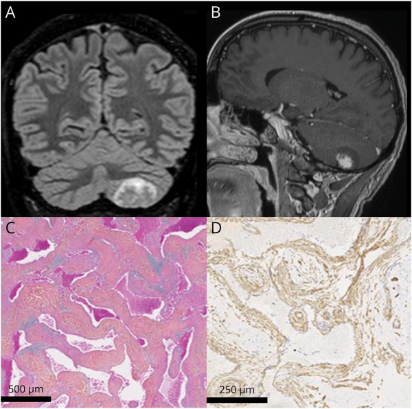
A 47-year-old man with an unremarkable medical history presented with mild dull occipital headaches for 7 months, without other neurologic changes. Structural MRI showed an extraparenchymal, well-delineated left cerebellar lesion with partial postgadolinium enhancement (Figure). Exploration with T1-weighted perfusion (Video 1) revealed a progressive, centrifugal enhancement pattern with slow contrast filling, rapid near the center but slower toward outer edges, suggesting a benign mesenchymal tumor. The absence of mass effect and cerebellar symptoms further supported a slow-growing tumor. A surgical removal was performed. Neuropathologic examination revealed a well-circumscribed lesion with smooth muscle cells and vascular cavities, indicating dural angioleiomyoma (Figure). Dural angioleiomyoma is a rare benign tumor, with <80 cases reported in a recent literature review1 related to soft tissue angioleiomyomas. Partial and flame-like enhancement arising from the tumor base and extending to its periphery seems to be the most typical imaging characteristic,2 and dynamic contrast-enhanced sequence may aid in envisioning diagnosis preoperatively.
Figure. MRI and Neuropathology.
MRI: coronal T2 fluid-attenuated inversion recovery (A) and contrast-enhanced T1-weighted sagittal (B) views. Neuropathology: anfractuous vascular cavities separated by muscular walls (Hematoxylin-Phloxin-Saffron) (C). Immunohistochemical analysis: smooth muscle cells labeled with antiactin antibodies (D).
Dynamic study. Dynamic contrast-enhanced T1-weighted coronal view of a left cerebellar mass, displaying centrifugal enhancement.Download Supplementary Video 1 (2.8MB, mov) via http://dx.doi.org/10.1212/207761_Video_1
Author Contributions
T. Leclerc: drafting/revision of the manuscript for content, including medical writing for content; major role in the acquisition of data; study concept or design; analysis or interpretation of data. C. Oppenheim: drafting/revision of the manuscript for content, including medical writing for content; major role in the acquisition of data; study concept or design; analysis or interpretation of data. A. Tauziede-Espariat: major role in the acquisition of data; analysis or interpretation of data. Hélène Charpentier: major role in the acquisition of data. J. Pallud: major role in the acquisition of data; analysis or interpretation of data. J. Benzakoun: drafting/revision of the manuscript for content, including medical writing for content; major role in the acquisition of data; study concept or design; analysis or interpretation of data.
Study Funding
No targeted funding reported.
Disclosure
The authors report no relevant disclosures. Go to Neurology.org/N for full disclosures.
References
- 1.Tauziède-Espariat A, Pierre T, Wassef M, et al. The dural angioleiomyoma harbors frequent GJA4 mutation and a distinct DNA methylation profile. Acta Neuropathol Commun. 2022;10(1):81. doi: 10.1186/s40478-022-01384-x [DOI] [PMC free article] [PubMed] [Google Scholar]
- 2.Sun L, Zhu Y, Wang H, et al. Angioleiomyoma, a rare intracranial tumor: 3 case report and a literature review. World J Surg Oncol. 2014;12:216. doi: 10.1186/1477-7819-12-216 [DOI] [PMC free article] [PubMed] [Google Scholar]
Associated Data
This section collects any data citations, data availability statements, or supplementary materials included in this article.
Supplementary Materials
Dynamic study. Dynamic contrast-enhanced T1-weighted coronal view of a left cerebellar mass, displaying centrifugal enhancement.Download Supplementary Video 1 (2.8MB, mov) via http://dx.doi.org/10.1212/207761_Video_1


