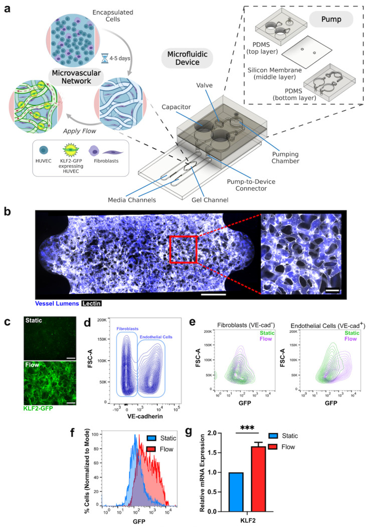
Figure 1. Activation of KLF2-GFP reporter in MVNs with flow.
a Schematic of microfluidic device and attached pump. HUVECs expressing KLF2-GFP and fibroblasts are seeded into the gel channel and self-assemble into MVNs. b Example of MVN stained with lectin (white) and perfused with fluorescent dextran (blue). Scale bar = 500 μm in full device image. Scale bar = 100 μm in close-up image of vasculature. c Image of KLF-GFP (green) expression in MVN after application of flow for 18 hours in MVN after culture in static conditions. Scale bars = 200 μm. d Flow cytometry analysis of cells in MVNs labelled with VE-cadherin. VE-cadherin+ endothelial cells and VE-cadherin− fibroblasts are indicated by bounding rectangles. e Flow cytometry analysis of each population from MVNs cultured under static (green) and flow (purple) conditions. f Percentage of endothelial cells expressing GFP under static and flow conditions. g KLF2 mRNA expression in endothelial cells extracted from MVNs and separated out using flow cytometry as in d (measurement from 5 pooled MVNs). *** indicates p<0.001.

