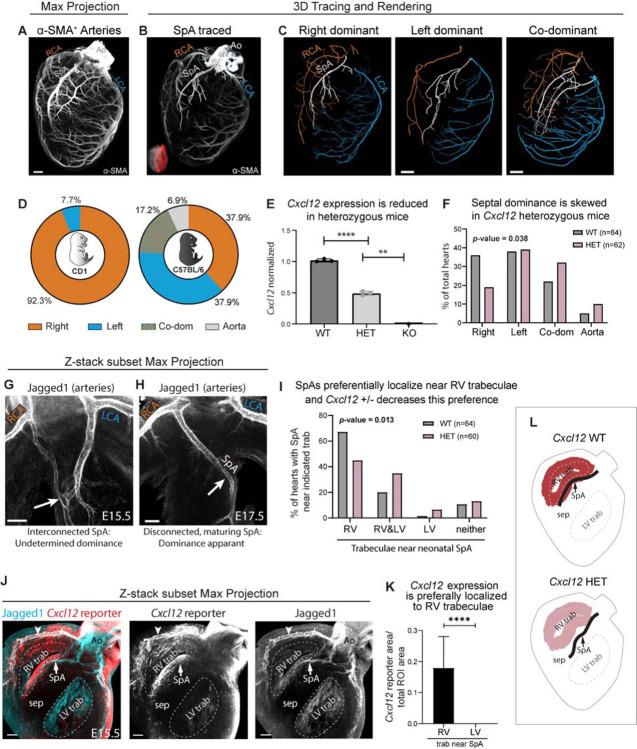
Figure 5. Cxcl12 influences septal artery dominance in mice.
(A) Whole-organ immunolabeling of coronary artery smooth muscle (α-SMA) in a postnatal day 6 mouse heart. (B) Septal artery (SpA) tracing overlayed onto a Z-stack subset max projection of internal coronary arteries (red region in insert). (C) Artery tracings in whole-organ images demonstrating various SpA connections. (D) SpA dominance ratios are strain specific. N=13 CD1 neonatal hearts and N=29 for C56BL/6J. (E) QPCR using whole hearts from embryonic day (E) 16.5. WT, Cxcl12+/+; HET, Cxcl12DsRed/+; KO, Cxcl12DsRed/DsRed. Each dot is the average value for genotypes within single litters normalized to WT littermates. Error bars, mean±st dev: **, p=0.0027, p=0.0016, p=0.0011; ****, p<0.0001 by two-sided Student’s t-test. (F) Blinded scoring of septal artery dominance in WT and HET neonatal mice. P-value by two-tailed Chi-square test. (G and H) Whole-organ immunolabeling of WT embryonic hearts. First, an immature artery network is attached to both sides (G) followed by maturation into a SpA with dominance shortly before birth (H). (I) Blinded scoring of SpA location as related to trabeculae (see Supplementary Figure 8I). P-value by two-tailed Chi-square test. (J) Cxcl12 reporter allele shows that SpA develops in regions of high expression. Arrowhead, expression in artery endothelial cells. (K) Reporter allele fluorescence in standardized regions of interest (ROI) along right or left trabeculae. N=14 hearts. Error bars, mean±st dev: ****, p<0.0001 by two-sided Student’s t-test. (L) Schematic of Cxcl12 bias in trabeculae, and HET’s effect on SpA localization. Ao, aorta; LCA, left coronary artery; LV, left ventricle; RCA, right coronary artery; RV, right ventricle; sep, septum; trab, trabecular cardiomyocytes. Scale bars: A, C 400μm; G - I, 100μm.

