


Nucleic Acids Research, Volume 40, Issue 2, 1 January 2012, Pages 761–774, https://doi.org/10.1093/nar/gkr730
The Editors were alerted in February 2023 about possible errors in the nucleotide sequences given in this article.
While investigating the issue, the Editors have found additional issues with some Figures as detailed below:
Figure 3E, p70S6K1 panel: splice line between Anti-miR-145 and Anti-Control.
Figure 5B, SW1116, p-p70S6K1 panel: splice line between lanes 3 and 4.
Figure 7E, CD31: part of panel 1 and panel 2 appear identical after a 90°clockwise rotation
Figure 8A, top, miR-145: splice line between 8C and 9A.
Figure 8A, bottom, p70S6K1 and β-actin: splice lines between 5C and 6A.
Figure 3E.
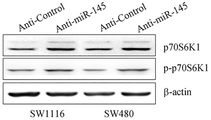
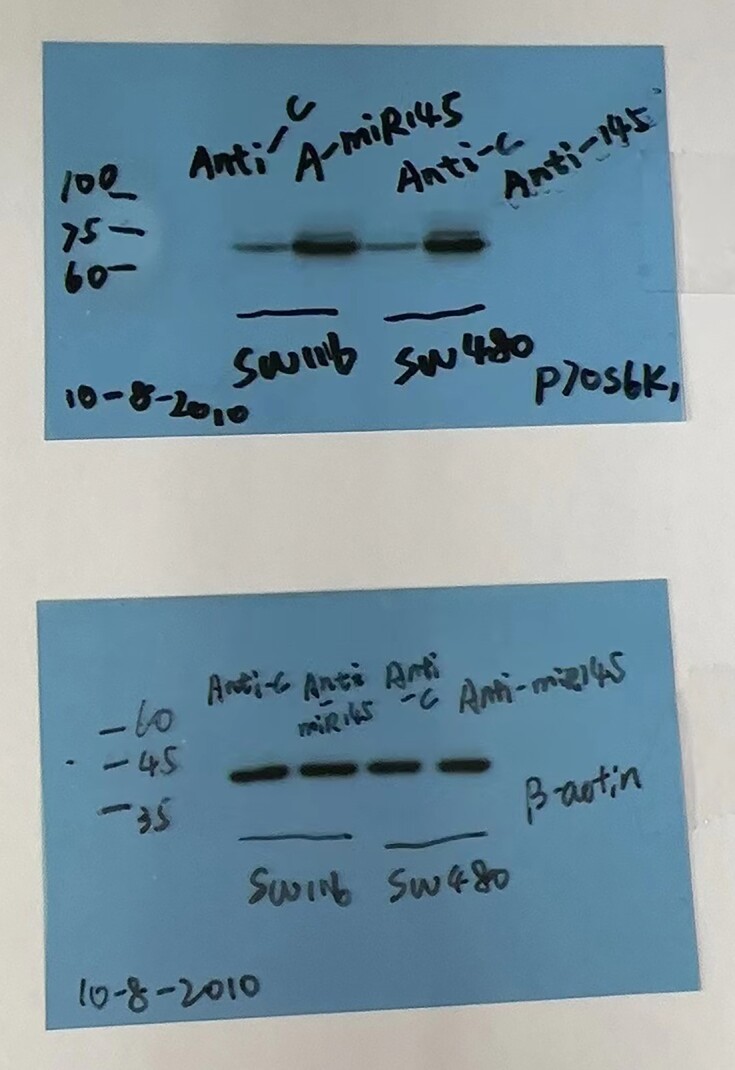
Top, published figure panel. Bottom, replicate experiment showing p70S6K1 in the top film and β-actin in the lower film.
Figure 5B.


Top, published figure panel SW1116. Bottom, replicate experiment showing p-p70S6K1 in the top film and β-actin in the lower film.
Figure 7E.


Top, published figure panel. Bottom, results of two replicate experiments.
Figure 8A.


Top, published figure panel. Bottom, black lines have been inserted where separate blots were spliced into single reconstructed images.
An Expression of Concern was published in March 2023.
Nucleotide sequences: the authors confirm that the p70S6K1 nucleotide sequence presented in the article is correct.
The authors no longer have the original data used for Figures 3, 5 and 7 but have provided the results of contemporaneous replicate experiments, as shown below.
For Figure 8, the authors acknowledge that the results presented in Figure 8A were obtained from two independent experiments as all samples cannot be run on a single gel. The results from both experiments were arranged adjacent to each other. The authors wish to emphasize that all paired normal (A) and cancer (C) samples were run on the same membrane and exposed to the same development time.