Abstract
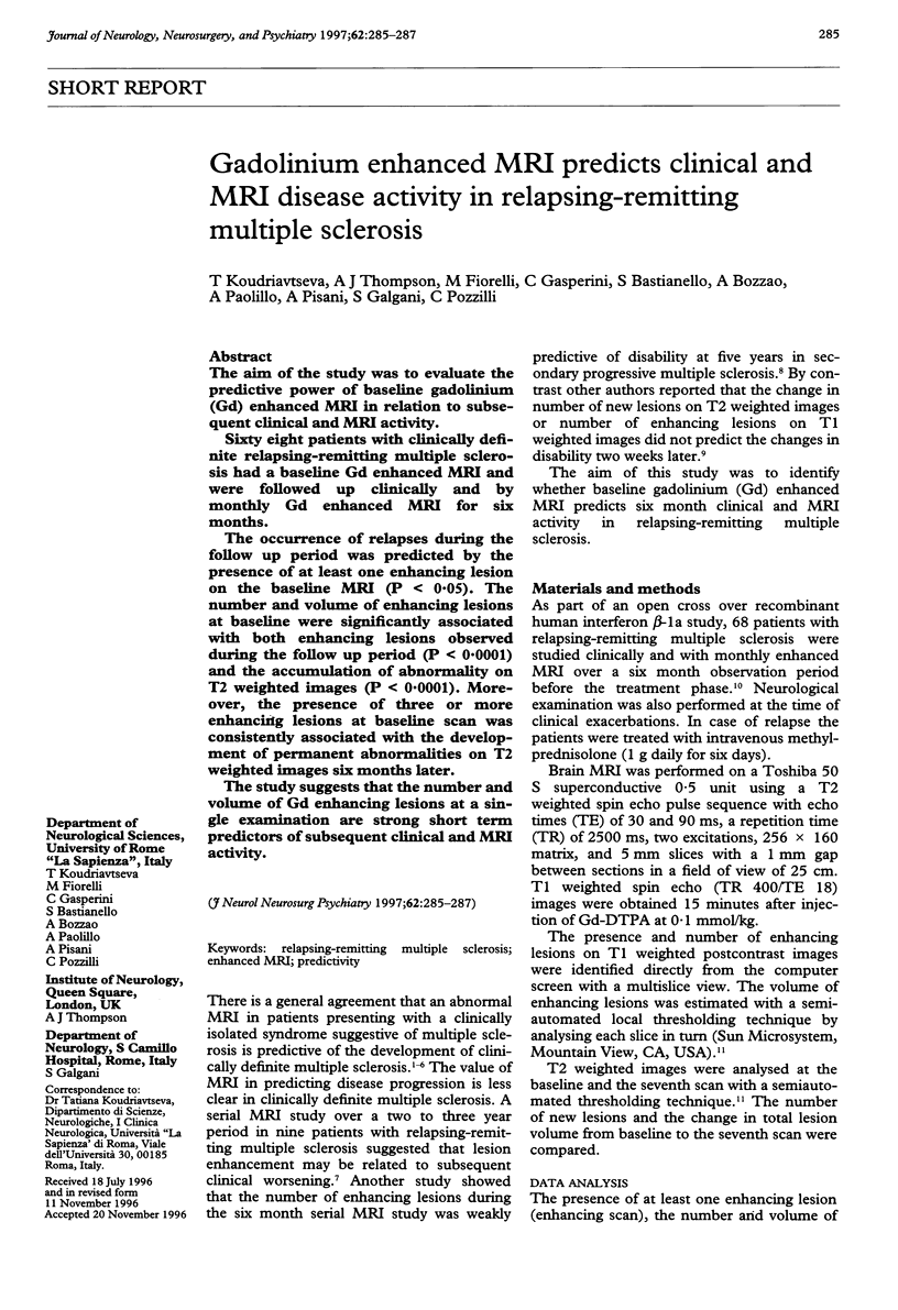
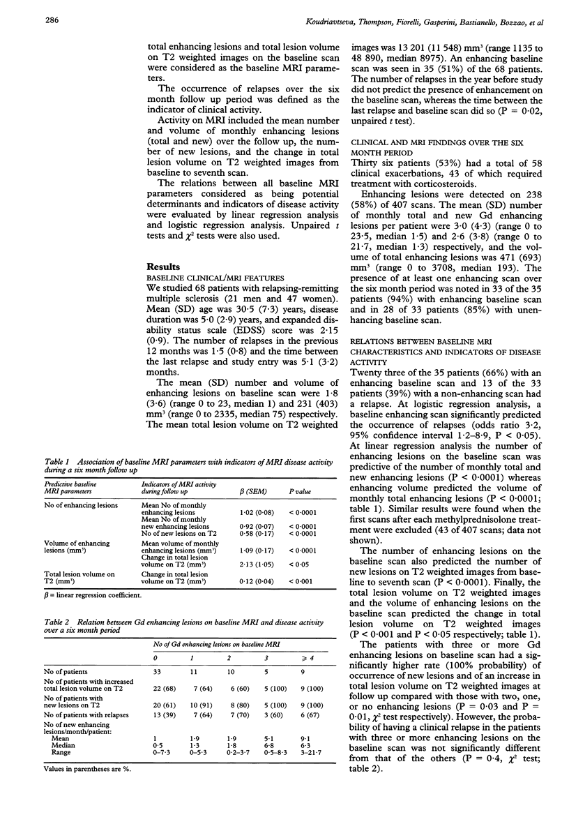
The aim of the study was to evaluate the predictive power of baseline gadolinium (Gd) enhanced MRI in relation to subsequent clinical and MRI activity. Sixty eight patients with clinically definite relapsing-remitting multiple sclerosis had a baseline Gd enhanced MRI and were followed up clinically and by monthly Gd enhanced MRI for six months. The occurrence of relapses during the follow up period was predicted by the presence of at least one enhancing lesion on the baseline MRI (P < 0.05). The number and volume of enhancing lesions at baseline were significantly associated with both enhancing lesions observed during the follow up period (P < 0.0001) and the accumulation of abnormality on T2 weighted images (P < 0.0001). Moreover, the presence of three or more enhancing lesions at baseline scan was consistently associated with the development of permanent abnormalities on T2 weighted images six months later. The study suggests that the number and volume of Gd enhancing lesions at a single examination are strong short term predictors of subsequent clinical and MRI activity.
Full text
PDF


Selected References
These references are in PubMed. This may not be the complete list of references from this article.
- Beck R. W., Cleary P. A., Trobe J. D., Kaufman D. I., Kupersmith M. J., Paty D. W., Brown C. H. The effect of corticosteroids for acute optic neuritis on the subsequent development of multiple sclerosis. The Optic Neuritis Study Group. N Engl J Med. 1993 Dec 9;329(24):1764–1769. doi: 10.1056/NEJM199312093292403. [DOI] [PubMed] [Google Scholar]
- Ellison G. W., Myers L. W., Leake B. D., Mickey M. R., Ke D., Syndulko K., Tourtellotte W. W. Design strategies in multiple sclerosis clinical trials. The Cyclosporine Multiple Sclerosis Study Group. Ann Neurol. 1994;36 (Suppl):S108–S112. doi: 10.1002/ana.410360725. [DOI] [PubMed] [Google Scholar]
- Filippi M., Horsfield M. A., Morrissey S. P., MacManus D. G., Rudge P., McDonald W. I., Miller D. H. Quantitative brain MRI lesion load predicts the course of clinically isolated syndromes suggestive of multiple sclerosis. Neurology. 1994 Apr;44(4):635–641. doi: 10.1212/wnl.44.4.635. [DOI] [PubMed] [Google Scholar]
- Khoury S. J., Guttmann C. R., Orav E. J., Hohol M. J., Ahn S. S., Hsu L., Kikinis R., Mackin G. A., Jolesz F. A., Weiner H. L. Longitudinal MRI in multiple sclerosis: correlation between disability and lesion burden. Neurology. 1994 Nov;44(11):2120–2124. doi: 10.1212/wnl.44.11.2120. [DOI] [PubMed] [Google Scholar]
- Lee K. H., Hashimoto S. A., Hooge J. P., Kastrukoff L. F., Oger J. J., Li D. K., Paty D. W. Magnetic resonance imaging of the head in the diagnosis of multiple sclerosis: a prospective 2-year follow-up with comparison of clinical evaluation, evoked potentials, oligoclonal banding, and CT. Neurology. 1991 May;41(5):657–660. doi: 10.1212/wnl.41.5.657. [DOI] [PubMed] [Google Scholar]
- Losseff N. A., Kingsley D. P., McDonald W. I., Miller D. H., Thompson A. J. Clinical and magnetic resonance imaging predictors of disability in primary and secondary progressive multiple sclerosis. Mult Scler. 1996 Feb;1(4):218–222. [PubMed] [Google Scholar]
- Miller D. H., Barkhof F., Nauta J. J. Gadolinium enhancement increases the sensitivity of MRI in detecting disease activity in multiple sclerosis. Brain. 1993 Oct;116(Pt 5):1077–1094. doi: 10.1093/brain/116.5.1077. [DOI] [PubMed] [Google Scholar]
- Miller D. H., Ormerod I. E., McDonald W. I., MacManus D. G., Kendall B. E., Kingsley D. P., Moseley I. F. The early risk of multiple sclerosis after optic neuritis. J Neurol Neurosurg Psychiatry. 1988 Dec;51(12):1569–1571. doi: 10.1136/jnnp.51.12.1569. [DOI] [PMC free article] [PubMed] [Google Scholar]
- Miller D. H., Ormerod I. E., Rudge P., Kendall B. E., Moseley I. F., McDonald W. I. The early risk of multiple sclerosis following isolated acute syndromes of the brainstem and spinal cord. Ann Neurol. 1989 Nov;26(5):635–639. doi: 10.1002/ana.410260508. [DOI] [PubMed] [Google Scholar]
- Morrissey S. P., Miller D. H., Kendall B. E., Kingsley D. P., Kelly M. A., Francis D. A., MacManus D. G., McDonald W. I. The significance of brain magnetic resonance imaging abnormalities at presentation with clinically isolated syndromes suggestive of multiple sclerosis. A 5-year follow-up study. Brain. 1993 Feb;116(Pt 1):135–146. doi: 10.1093/brain/116.1.135. [DOI] [PubMed] [Google Scholar]
- Pozzilli C., Bastianello S., Koudriavtseva T., Gasperini C., Bozzao A., Millefiorini E., Galgani S., Buttinelli C., Perciaccante G., Piazza G. Magnetic resonance imaging changes with recombinant human interferon-beta-1a: a short term study in relapsing-remitting multiple sclerosis. J Neurol Neurosurg Psychiatry. 1996 Sep;61(3):251–258. doi: 10.1136/jnnp.61.3.251. [DOI] [PMC free article] [PubMed] [Google Scholar]
- Smith M. E., Stone L. A., Albert P. S., Frank J. A., Martin R., Armstrong M., Maloni H., McFarlin D. E., McFarland H. F. Clinical worsening in multiple sclerosis is associated with increased frequency and area of gadopentetate dimeglumine-enhancing magnetic resonance imaging lesions. Ann Neurol. 1993 May;33(5):480–489. doi: 10.1002/ana.410330511. [DOI] [PubMed] [Google Scholar]
- Stone L. A., Frank J. A., Albert P. S., Bash C., Smith M. E., Maloni H., McFarland H. F. The effect of interferon-beta on blood-brain barrier disruptions demonstrated by contrast-enhanced magnetic resonance imaging in relapsing-remitting multiple sclerosis. Ann Neurol. 1995 May;37(5):611–619. doi: 10.1002/ana.410370511. [DOI] [PubMed] [Google Scholar]
- Wicks D. A., Tofts P. S., Miller D. H., du Boulay G. H., Feinstein A., Sacares R. P., Harvey I., Brenner R., McDonald W. I. Volume measurement of multiple sclerosis lesions with magnetic resonance images. A preliminary study. Neuroradiology. 1992;34(6):475–479. doi: 10.1007/BF00598953. [DOI] [PubMed] [Google Scholar]


