Abstract
Background
The identification of vanishing bile duct syndrome (VBDS) is still challenging before liver biopsy. This study tried to explore non-invasive biomarkers for identification of VBDS among children with acute cholestatic hepatitis.
Methods
Between January 2017 and December 2021, 192 children underwent native-liver biopsy for acute cholestatic hepatitis with onset after 6 months of age. VBDS was diagnosed by liver biopsy. Differences of liver biochemical indices were compared between children with and without VBDS. Diagnostic performances for VBDS were tested by receiver operating characteristic (ROC) curve analyses.
Results
Among the 192 patients, 24 (12.5%) were diagnosed with VBDS based on liver biopsy. At biopsy, their levels of total bilirubin (TB), direct bilirubin (DB), γ-glutamyl transpeptidase (GGT), total bile acid, triglyceride, and total cholesterol (TCH) were higher than patients without VBDS (all P<0.05). However, only GGT and TCH could distinguish patients with VBDS from patients without VBDS with an area under ROC curve (AUC) >0.850. Using GGT >446 U/L as a cut-off value, the sensitivity was 87.5%, the specificity was 91.6%, and the AUC was 0.948 (P<0.001). Using TCH >6.4 mmol/L as a cut-off value, the sensitivity was 100.0%, the specificity was 89.8%, and the AUC was 0.983 (P<0.001). A total of 28 patients had both GGT >446 U/L and TCH >6.4 mmol/L, including 21 patients with VBDS and 7 without VBDS (21/28 vs. 3/143, P<0.0001). Three patients with VBDS would be missed for GGT <446 U/L.
Conclusions
Both GGT and TCH can be used as non-invasive biomarkers for identification of VBDS among children with acute cholestatic hepatitis.
Keywords: Bile duct loss, diagnostic yield, hypercholesterolemia
Highlight box.
Key findings
• Both γ-glutamyl transpeptidase (GGT) and total cholesterol (TCH) can be used as non-invasive biomarkers for identification of vanishing bile duct syndrome (VBDS) among children with acute cholestatic hepatitis who develop cholestatic jaundice after 6 months of age.
What is known and what is new?
• VBDS typically arises from acute cholestatic hepatitis. Patients without cholestasis are impossibly diagnosed with VBDS.
• Children with VBDS present cholestasis with elevated levels of GGT and TCH. The diagnosis of VBDS is unlikely if a child presents as cholestasis without high GGT and hypercholesterolemia. GGT and TCH can be used as biomarkers for identification of VBDS, and TCH has a better performance than GGT.
What is the implication, and what should change now?
• Both GGT and TCH should be routinely monitored among children with acute cholestatic hepatitis. VBDS is highly suspected if TCH >6.4 mmol/L and/or GGT >446 U/L are detected among children with acute cholestatic hepatitis.
Introduction
Vanishing bile duct syndrome (VBDS) is a rare acquired condition characterized by progressive loss of intralobular bile ducts, and sometimes can even lead to end stage liver disease requiring transplant (1,2). Known causes of VBDS include drug induced liver injury (DILI), graft versus host disease (GVHD), autoimmune disease, etc. (3). The loss of bile ducts is possibly resulted from an inflammatory response directed at cholangiocytes (1). VBDS is a pathological diagnosis. A diagnosis of classical form of VBDS is established if no bile ducts are detected in >50% of portal tracts in a biopsy with at least ten portal areas (4). Mild or partial form of VBDS is defined as that bile ducts are absent in 25–50% of portal areas (3,4). VBDS typically arises in the setting of acute cholestatic hepatitis (4). Patients with VBDS present as cholestatic jaundice, pruritus, fatigue, hypercholesterolemia, etc.
VBDS often has a poor prognosis. To establish a definite diagnosis, a liver biopsy is needed and should not be deferred when VBDS is suspected in clinical practices (5). However, repetitive biopsies are needed for its diagnosis in some instances because the loss of bile ducts is a gradual process (1). In addition, a non-invasive biomarker for VBDS is still absent at present. Therefore, it is challenging for clinicians to decide when to perform a liver biopsy. VBDS has been reported in adults in a relatively large cohort, and significant differences have been found in liver biochemical indices between adult patients with and without VBDS (3). We wondered whether these differences could be used as biomarkers for identification of VBDS among children with acute cholestatic hepatitis before liver biopsy.
This study enrolled children with acute cholestatic hepatitis who developed jaundice after 6 months of age and underwent native-liver biopsy. We compared the difference of liver biochemical indices between children with VBDS and those without VBDS based on liver biopsy. We aimed to explore non-invasive biomarkers for identification of VBDS among children with acute cholestatic hepatitis. We present this article in accordance with the STARD reporting checklist (available at https://tp.amegroups.com/article/view/10.21037/tp-23-305/rc).
Methods
Patients and definitions
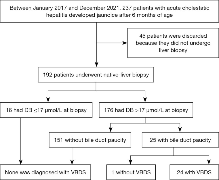
This is a retrospective study from the Center for Pediatric Liver Diseases, Children’s Hospital of Fudan University. Participants were identified by reviewing their medical records. Between January 2017 and December 2021, a total of 237 patients with acute cholestatic hepatitis, who developed cholestatic jaundice after 6 months of age, were admitted into this center. They denied a diagnosis of previous liver disease history. Among them, 45 patients were excluded for further analysis, because they did not undergo liver biopsy (Figure 1). This study enrolled the remaining 192 consecutive patients who performed native-liver biopsy during the course of acute cholestatic hepatitis. Cholestasis was defined as direct bilirubin (DB) >17.1 µmol/L when total bilirubin (TB) <85.5 µmol/L or DB >20% of TB when TB >85.5 µmol/L (6). Based on histologic findings of liver biopsy, the patients were divided into two groups: patients with VBDS and patients without VBDS. The study was approved by the ethics committees of Children’s Hospital of Fudan University (No. 2022-43), and was conducted in full compliance with the Declaration of Helsinki (as revised in 2013). Informed consent was taken from the patients’ parents or legal guardians at admission.
Figure 1.

Flow diagram of patient enrollment. DB, direct bilirubin; VBDS, vanishing bile duct syndrome.
Liver biochemical tests
All the enrolled 192 patients underwent at least one tests of liver biochemical indices, including TB, DB, alanine aminotransferase (ALT), aspartate aminotransferase (AST), total bile acid (TBA), γ-glutamyl transpeptidase (GGT), albumin (ALB), triglyceride (TG), and total cholesterol (TCH). Test results were collected by reviewing their medical records. Tests at biopsy were defined as those carried out within five days before or after liver biopsy. If several tests were done during this period, only the test results obtained closest to liver biopsy were used for analyses.
Differences in liver biochemical indices at biopsy were compared between patients with and without VBDS. This study further tested the diagnostic performances of liver biochemical indices for identification of VBDS among children with acute cholestatic hepatitis, and also explored their cut-off values.
Liver histologic studies
Native-liver tissues were obtained by needle biopsy. Liver histologic studies were performed in the Department of Pathology. Liver tissue sections were read jointly by two pathologists and at least one hepatologist. Using ‘bile duct paucity’ as keywords, preliminary patients were identified by searching liver histologic reports of the enrolled 192 patients. Liver tissue sections of patients with bile duct paucity were further reviewed by a pathologist who was blind to the patients’ clinical information. The loss of bile ducts was assessed in hematoxylin-eosin stained sections, and confirmed in both anti-CK7 and anti-CK19 (GeneTech, Shanghai, China) immunostained sections. Both the numbers of total portal areas and the numbers of portal areas without bile ducts were recorded for calculation of the fraction of portal areas without bile ducts.
The diagnosis of VBDS is established based on paucity of bile ducts in liver biopsy (4). In this study, patients with VBDS included both patients with classical form of VBDS and patients with partial form of VBDS. VBDS was diagnosed if one of the following elements was satisfied: (I) >25% of total portal areas absented bile ducts in a biopsy with at least ten portal areas; (II) at least three portal areas absented bile ducts in a biopsy with <10 portal areas. Although portal areas without bile ducts were detected, patients were still classified into group without VBDS if they did not satisfy any one of the above elements.
Statistical analysis
MedCalc Software (v20.026, https://www.medcalc.org; 2022) was used for statistical analyses. Continuous variables were presented as medians and interquartile ranges, and their differences between patients with and without VBDS were compared by the Mann-Whitney tests. The differences of categorical variables between the two groups were determined by Chi-square or Fisher’s exact test. Diagnostic performances of liver biochemical indices for identification of VBDS among patients with acute cholestatic hepatitis were assessed by the receiver operating characteristic (ROC) curve analysis. Sensitivity, specificity, positive predictive value (PPV), negative predictive value (NPV), and area under the ROC curve (AUC) were calculated. AUC >0.8 meant excellent performance. Patients with missing data were excluded from calculations. P<0.05 was considered significant.
Results
Patients of VBDS
The enrolled 192 patients, including 125 boys and 67 girls, underwent liver biopsy at ages ranging from 7 months to 15 years, with a median age of 5 years. The median time frame from the development of jaundice to biopsy was 27 days (range, 6 to 172 days). By searching liver histologic reports, 25 patients with bile duct paucity were identified. Among them, one case was not diagnosed with VBDS, because he only had two portal areas without bile ducts in a biopsy with nine portal areas. Other 24 patients, including 17 boys and 7 girls, satisfied the diagnostic criteria, and were diagnosed with VBDS (Figure 2). The overall ratio of positive diagnosis of VBDS were 12.5% (24/192). DILI was the most common cause (n=13), and the implicated drugs included piperacillin-tazobactam (n=3), azithromycin (n=3), Chinese herbal medicine (n=3), etc. (Table S1). Sixteen patients had cholestatic jaundice ameliorated (DB <17 µmol/L) but still elevated transaminase at biopsy, and none of them was diagnosed with VBDS. Their levels of ALT, AST, GGT, and TCH were similar to other 152 patients without VBDS (all P>0.05).
Figure 2.
Liver histologic studies of patient 10. Bile ducts absent in portal areas by hematoxylin-eosin (×200, A), CK19 (×200, B), and also CK7 (×200, C) staining. A few hepatocytes are CK7-positive in CK7 staining sections.
At biopsy, all the 24 patients with VBDS presented as cholestasis with high GGT and hypercholesterolemia (Table 1). The median level of DB was 128 µmol/L (range, 20 to 327 µmol/L). The levels of GGT were prominently elevated with a median of 932 U/L (range, 151 to 2,860 U/L), while ALT was only mildly increased (median and range: 205 U/L; 56 to 1,080 U/L). The median of TCH concentrations was 13.9 mmol/L (range, 6.8 to 39.9 mmol/L).
Table 1. Clinical findings of 24 pediatric patients with vanishing bile duct syndrome at biopsy.
| Patient | Gender | Age | TB | DB | ALT | AST | GGT | TBA | TG | TCH | Time frame† (days) | No. of portal areas with/without bile ducts | Etiologic diagnosis | Outcome |
|---|---|---|---|---|---|---|---|---|---|---|---|---|---|---|
| P1 | Male | 7 years | 336 | 188 | 526 | 417 | 1,114 | 518 | 5.2 | 19.6 | 31 | 3/6 | Unknown | Alive |
| P2 | Female | 11 months | 244 | 140 | 190 | 294 | 2,120 | 249 | 3.5 | 27.1 | 37 | 1/10 | DILI | Alive |
| P3 | Female | 7 years | 200 | 122 | 482 | 172 | 796 | 229 | 6.2 | 18.6 | 20 | 2/3 | DILI | Alive |
| P4 | Male | 13 years | 256 | 131 | 122 | 47 | 1,067 | 170 | 1.9 | 13.9 | 71 | 2/7 | Unknown | Alive |
| P5 | Male | 7 years | 106 | 58 | 378 | 381 | 738 | 167 | 1.0 | 6.8 | 28 | 4/8 | Unknown | Alive |
| P6 | Male | 7 months | 126 | 70 | 128 | 207 | 259 | 102 | 2.4 | 7.1 | 20 | 2/8 | DILI | Alive |
| P7 | Male | 3 years | 161 | 125 | 80 | 79 | 194 | 423 | 2.9 | 19.7 | 27 | 2/4 | DILI | Alive |
| P8 | Male | 10 years | 395 | 327 | 214 | 143 | 1,156 | 536 | 3.1 | 21.6 | 38 | 7/8 | Unknown | Alive |
| P9 | Male | 17 months | 35 | 29 | 203 | 157 | 1,111 | 64 | 3.2 | 9.4 | 11 | 1/3 | DILI | Alive |
| P10 | Male | 14 months | 150 | 126 | 358 | 279 | 2,616 | 142 | 7.2 | 22.3 | 19 | 2/6 | DILI | Alive |
| P11 | Female | 15 months | 390 | 323 | 211 | 270 | 1,113 | 305 | 2.8 | 13.9 | 84 | 3/10 | DILI | Death |
| P12 | Male | 15 months | 30 | 20 | 208 | 189 | 470 | 145 | 1.8 | 39.9 | 63 | 3/9 | DILI | Alive |
| P13 | Male | 13 months | 205 | 185 | 56 | 109 | 634 | 53 | 6.4 | 11.4 | 21 | 1/6 | Unknown | Alive |
| P14 | Female | 5 years | 271 | 244 | 174 | 126 | 899 | 429 | 3.2 | 9.4 | 36 | 1/6 | DILI | Loss to follow-up |
| P15 | Male | 12 years | 220 | 197 | 1,080 | 319 | 1,794 | 140 | 2.7 | 11.6 | 25 | 1/5 | Unknown | Alive |
| P16 | Female | 8 years | 96 | 76 | 694 | 305 | 892 | 131 | 2.6 | 11.8 | 33 | 4/6 | DILI | Alive |
| P17 | Male | 12 years | 199 | 104 | 162 | 64 | 151 | 406 | 4.1 | 8.5 | 43 | 2/7 | Unknown | Alive |
| P18 | Male | 5 years | 121 | 113 | 196 | 201 | 517 | 460 | 2.7 | 33.3 | 31 | 4/9 | Unknown | Alive |
| P19 | Male | 8 months | 165 | 132 | 345 | 299 | 2,860 | 368 | 4.4 | 16.8 | 60 | 0/15 | DILI | Loss to follow-up |
| P20 | Male | 2 years | 75 | 65 | 101 | 160 | 1,443 | 143 | 3.8 | 36.8 | 74 | 1/9 | Unknown | Alive |
| P21 | Male | 11 years | 193 | 152 | 639 | 146 | 966 | 26 | 2.6 | 9.1 | 28 | 2/11 | DILI | Alive |
| P22 | Female | 2 years | 251 | 208 | 162 | 369 | 616 | 453 | 1.6 | 9.5 | 27 | 0/13 | DILI | LT |
| P23 | Male | 5 years | 308 | 248 | 317 | 292 | 1,043 | 149 | 3.3 | 11.6 | 29 | 3/3 | GVHD | Alive |
| P24 | Female | 8 years | 102 | 78 | 180 | 110 | 659 | 17 | 2.1 | 15.6 | 32 | 4/5 | Unknown | Alive |
†, Time frame from the development of jaundice to liver biopsy. TB (μmol/L), total bilirubin; DB (μmol/L), direct bilirubin; ALT (U/L), alanine aminotransferase; AST (U/L), aspartate aminotransferase; GGT (U/L), γ-glutamyl transpeptidase; TBA (μmol/L), total bile acid; TG (mmol/L), triglyceride; TCH (mmol/L), total cholesterol; DILI, drug induced liver injury; LT, liver transplantation; GVHD, graft versus host disease.
Difference between patients with and without VBDS
There was no difference on age and gender between patients with and without VBDS (both P>0.05) (Table 2). The levels of TB, DB, TBA, and GGT at biopsy were higher in the group with VBDS than the group without VBDS at biopsy (all P<0.05). However, the levels of ALT, AST, and ALB showed no difference. The patients with VBDS had higher levels of TG and TCH than the patients without VBDS (both P<0.05). Hypercholesterolemia (TCH >5.2 mmol/L) was diagnosed in all the 24 patients with VBDS, but only in 30 (17.9%) of the 168 patients without VBDS (24/24 vs. 30/168, P<0.0001).
Table 2. Differences between patients with and without vanishing bile duct syndrome.
| Characteristics | Patients with VBDS (n=24) | Patients without VBDS (n=168) | P |
|---|---|---|---|
| Age (years) | 5.0 (1.2, 8.0) | 4.5 (2.0, 8.0) | 0.5109 |
| Time frame (days)† | 31.0 (26.0, 40.5) | 24.0 (16.0, 44.5) | 0.1077 |
| Gender (No. of male/female) | 17/7 | 108/60 | 0.6494 |
| Total bilirubin (μmol/L) | 196.0 (113.5, 253.5) | 90.0 (55.5, 176.0) | 0.0017 |
| Direct bilirubin (μmol/L) | 128.5 (77.0, 192.5) | 69.0 (35.5, 131.0) | 0.0031 |
| Alanine aminotransferase (U/L) | 205.5 (162.0, 368.0) | 301.0 (146.0, 599.5) | 0.2303 |
| Aspartate aminotransferase (U/L) | 195.0 (134.5, 296.5) | 227.0 (112.5, 466.0) | 0.2656 |
| γ-glutamyl transpeptidase (U/L) | 932.5 (625.0, 1135.0) | 126.0 (60.0, 222.0) | <0.0001 |
| Total bile acid (μmol/L) | 168.5 (135.5, 414.5) | 92.0 (30.0, 259.7) | 0.0032 |
| Albumin (g/L) | 38.2 (35.5, 39.6) | 38.5 (34.7, 42.0) | 0.5980 |
| Triglyceride (mmol/L) | 3.0 (2.5, 3.9) | 1.9 (1.2, 2.9) | 0.0001 |
| Total cholesterol (mmol/L) | 13.9 (9.5, 20.6) | 4.1 (3.3, 4.8) | <0.0001 |
Data are shown as median (interquartile range). †, time frame from the development of jaundice to biopsy. VBDS, vanishing bile duct syndrome.
Biomarkers for identification of VBDS
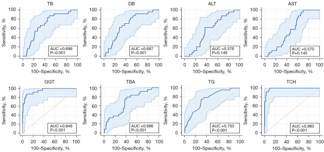
Based on ROC curve analyses, TB, DB, TBA, GGT, TG, and TCH at biopsy shown some diagnostic values to distinguish patients with VBDS from those without (all P<0.05, Table 3). However, only GGT and TCH had AUC >0.850 (Figure 3). Using GGT >446 U/L as a cut-off value, the sensitivity was 87.5%, the specificity was 91.6%, the PPV was 60.0%, the NPV was 98.1%, and the AUC was 0.948 (P<0.001). Using TCH >6.4 mmol/L as a cut-off value, the sensitivity was 100.0%, the specificity was 89.8%, the PPV was 61.5%, the NPV was 100.0%, and the AUC was 0.983 (P<0.001).
Table 3. Performance of liver laboratory indexes on identification of vanishing bile duct syndrome.
| Variables | AUC | P | Cut-off value | Sensitivity (%) | Specificity (%) | PPV (%) | NPV (%) |
|---|---|---|---|---|---|---|---|
| TB (μmol/L) | 0.698 (0.628, 0.762) | 0.0001 | >95 | 87.5 (67.6, 97.3) | 51.2 (43.4, 59.0) | 20.4 (17.1, 24.1) | 96.6 (90.8, 98.8) |
| DB (μmol/L) | 0.687 (0.616, 0.751) | 0.0003 | >64 | 87.5 (67.6, 97.3) | 47.0 (39.3, 54.9) | 19.1 (16.1, 22.5) | 96.3 (90.0, 98.7) |
| ALT (U/L) | 0.576 (0.503, 0.647) | 0.1487 | ≤214 | 62.5 (40.6, 81.2) | 63.1 (55.3, 70.4) | 19.5 (14.3, 25.9) | 92.2 (87.4, 95.2) |
| AST (U/L) | 0.570 (0.497, 0.641) | 0.1454 | ≤417 | 100.0 (85.8, 100.0) | 29.8 (23.0, 37.3) | 16.9 (15.6, 18.3) | 100.0 |
| GGT (U/L) | 0.948 (0.906, 0.975) | <0.0001 | >446 | 87.5 (67.6, 97.3) | 91.6 (86.3, 95.3) | 60.0 (47.1, 71.7) | 98.1 (94.6, 99.3) |
| TBA (μmol/L) | 0.686 (0.615, 0.752) | 0.0008 | >130 | 79.2 (57.8, 92.9) | 58.8 (50.9, 66.4) | 21.8 (17.5, 26.9) | 95.1 (89.8, 97.7) |
| ALB (g/L) | 0.533 (0.460, 0.606) | 0.5588 | ≤40 | 83.3 (62.6, 95.3) | 38.3 (30.9, 46.2) | 16.3 (13.5, 19.4) | 94.1 (86.5, 97.6) |
| TG (mmol/L) | 0.753 (0.681, 0.815) | <0.0001 | >2.5 | 75.0 (53.3, 90.2) | 69.4 (61.3, 76.7) | 28.6 (22.2, 35.9) | 94.4 (89.4, 97.2) |
| TCH (mmol/L) | 0.983 (0.950, 0.997) | <0.0001 | >6.4 | 100.0 (85.8, 100.0) | 89.8 (83.7, 94.2) | 61.5 (49.8, 72.1) | 100.0 |
95% CIs in parentheses. AUC, area under ROC curve; ROC, receiver operating characteristic; PPV, positive predictive value; NPV, negative predictive value; TB, total bilirubin; DB, direct bilirubin; ALT, alanine aminotransferase; AST, aspartate aminotransferase; GGT, γ-glutamyl transpeptidase; TBA, total bile acid; ALB, albumin; TG, triglyceride; TCH, total cholesterol.
Figure 3.
Diagnostic performances of liver biochemical indices for vanishing bile duct syndrome. TB, total bilirubin; AUC, area under ROC curve; ROC, receiver operating characteristic; DB, direct bilirubin; ALT, alanine aminotransferase; AST, aspartate aminotransferase; GGT, γ-glutamyl transpeptidase; TBA, total bile acid; TG, triglyceride; TCH, total cholesterol.
At biopsy, 171 patients simultaneously tested TCH and GGT. Among them, 28 had both GGT >446 U/L and TCH >6.4 mmol/L (Table 4), including 21 patients with VBDS and 7 without VBDS (21/28 vs. 3/143, P<0.0001). Three patients with VBDS would be missed for GGT <446 U/L. The sensitivity was 87.5%, the specificity was 95.2%, the PPV was 75.0%, and the NPV was 97.9%.
Table 4. Performance of GGT and TCH for identification of vanishing bile duct syndrome.
| Indicators | VBDS confirmed by liver biopsy | Total | P | |
|---|---|---|---|---|
| Positive | Negative | |||
| GGT >446 U/L | <0.0001 | |||
| Positive | 21 | 14 | 35 | |
| Negative | 3 | 152 | 155 | |
| TCH >6.4 mmol/L | <0.0001 | |||
| Positive | 24 | 15 | 39 | |
| Negative | 0 | 132 | 132 | |
| GGT >446 U/L and TCH >6.4 mmol/L | <0.0001 | |||
| Positive | 21 | 7 | 28 | |
| Negative | 3 | 140 | 143 | |
GGT, γ-glutamyl transpeptidase; TCH, total cholesterol; VBDS, vanishing bile duct syndrome.
Discussion
VBDS typically arises from acute cholestatic hepatitis (4), and has been reported in children as isolated cases (7-10). In this study, we enrolled 192 children who underwent native-liver biopsy for acute cholestatic hepatitis and a total of 24 children were diagnosed with VBDS. To our best knowledge, this was the largest cohort of pediatric VBDS patients, though it was unclear that 12 of them belonged to partial form of VBDS or classical form of VBDS for specimens had less than 10 portal areas. The overall ratio of positive diagnosis of VBDS was 12.5% in this cohort. It might be overestimated, because patients with rapidly resolved jaundice were more likely to reject a liver biopsy. However, given the adverse outcome for VBDS, we suggest that the occurrence of VBDS should be routinely monitored among patients with acute cholestatic hepatitis.
In this cohort, cholestatic jaundice had already ameliorated at biopsy in 16 children, in whom none was diagnosed with VBDS. It is consistent with the findings from adult DILI, where patients without cholestasis are impossibly diagnosed with VBDS (11). In adult VBDS, the elevation of alkaline phosphatase (ALP) was more obvious than ALT and AST (3). ALP is usually used as an indicator for bile duct injury in adults, while GGT is a preferred indicator for bile duct injury in children (12). Similarly, we found that GGT was markedly elevated, while ALT and AST were only mildly elevated in pediatric VBDS. Apart from high GGT, hypercholesterolemia had also been reported previously in children with VBDS (9,13). In our cohort, all the 24 children with VBDS had hypercholesterolemia and high GGT. Our data demonstrate that high GGT and hypercholesterolemia are common features of children with VBDS. Therefore, we infer that the diagnosis of VBDS is unlikely if a child presents as cholestasis without high GGT and hypercholesterolemia. Due to the small sample size, further researches are needed to confirm our findings.
Identification of VBDS is still a challenge before liver biopsy. This study tried to explore non-invasive biomarkers for identification of VBDS among children with acute cholestatic hepatitis. Similar to adult VBDS (3), significant differences were also found in liver biochemical indices between children with and without VBDS. We further found that both GGT and TCH could be used as non-invasive biomarkers for identification of VBDS with AUC >0.85. Using GGT >446 U/L as a cut-off value, the specificity could be 91.6%, but three children with VBDS would be missed. In addition, 14 out of the 166 patients without VBDS also had GGT >446 U/L. It might be attributed to that elevation of GGT levels could result from many other causes, such as sclerosing cholangitis, bile duct obstruction, etc. (14). Interestingly, TCH had a better performance for identification of VBDS than GGT. Using TCH >6.4 mmol/L as a cut-off value, the specificity could be 89.8%, and none of patients with VBDS was missed. The combination of GGT and TCH did not further improve the diagnostic value. Therefore, both GGT and TCH can be used as non-invasive biomarkers for identification of VBDS among children with acute cholestatic hepatitis. We suggest that both GGT and TCH should be routinely monitored among children with acute cholestatic hepatitis. VBDS is highly suspected if TCH >6.4 mmol/L and/or GGT >446 U/L are detected among children with acute cholestatic hepatitis, and liver biopsy is suggested.
Conclusions
Children with VBDS present as cholestasis with high GGT and hypercholesterolemia. The diagnosis of VBDS is unlikely if a child presents as cholestasis without high GGT and hypercholesterolemia. Both GGT and TCH can be used as non-invasive biomarkers for identification of VBDS among children with acute cholestatic hepatitis, and TCH has a better performance than GGT. These markers are still potential, and additional tests are needed to confirm the diagnosis of VBDS.
Supplementary
The article’s supplementary files as
Acknowledgments
The authors extend their great thanks to the patients and their families for their participation.
Funding: This study was supported by the Key Development Program of Children’s Hospital of Fudan University (No. EK2022ZX05).
Ethical Statement: The authors are accountable for all aspects of the work in ensuring that questions related to the accuracy or integrity of any part of the work are appropriately investigated and resolved. This study was approved by the ethics committees of Children’s Hospital of Fudan University (No. 2022-43) and was conducted in full compliance with the Declaration of Helsinki (as revised in 2013). Informed consent was taken from the patients' parents or legal guardians at admission.
Footnotes
Reporting Checklist: The authors have completed the STARD reporting checklist. Available at https://tp.amegroups.com/article/view/10.21037/tp-23-305/rc
Data Sharing Statement: Available at https://tp.amegroups.com/article/view/10.21037/tp-23-305/dss
Peer Review File: Available at https://tp.amegroups.com/article/view/10.21037/tp-23-305/prf
Conflicts of Interest: All authors have completed the ICMJE uniform disclosure form (available at https://tp.amegroups.com/article/view/10.21037/tp-23-305/coif). The authors have no conflicts of interest to declare.
References
- 1.Eiswerth MJ, Heckroth MA, Ismail A, et al. Infliximab-Induced Vanishing Bile Duct Syndrome. Cureus 2022;14:e21940. 10.7759/cureus.21940 [DOI] [PMC free article] [PubMed] [Google Scholar]
- 2.Gaudel P, Brown P, Byrd K. Vanishing Bile Duct Syndrome in the Presence of Hodgkin Lymphoma. Cureus 2022;14:e26842. 10.7759/cureus.26842 [DOI] [PMC free article] [PubMed] [Google Scholar]
- 3.Bonkovsky HL, Kleiner DE, Gu J, et al. Clinical presentations and outcomes of bile duct loss caused by drugs and herbal and dietary supplements. Hepatology 2017;65:1267-77. 10.1002/hep.28967 [DOI] [PMC free article] [PubMed] [Google Scholar]
- 4.LiverTox: Clinical and research information on drug-induced liver injury. Bethesda (MD): National Institute of Diabetes and Digestive and Kidney Diseases; 2012. [Google Scholar]
- 5.Bakhit M, McCarty TR, Park S, et al. Vanishing bile duct syndrome in Hodgkin's lymphoma: A case report and literature review. World J Gastroenterol 2017;23:366-72. 10.3748/wjg.v23.i2.366 [DOI] [PMC free article] [PubMed] [Google Scholar]
- 6.Wang NL, Lu Y, Gong JY, et al. Molecular findings in children with inherited intrahepatic cholestasis. Pediatr Res 2020;87:112-7. 10.1038/s41390-019-0548-8 [DOI] [PubMed] [Google Scholar]
- 7.Li L, Zheng S, Chen Y. Stevens-Johnson syndrome and acute vanishing bile duct syndrome after the use of amoxicillin and naproxen in a child. J Int Med Res 2019;47:4537-43. 10.1177/0300060519868594 [DOI] [PMC free article] [PubMed] [Google Scholar]
- 8.Takaki Y, Murahashi M, Honda K, et al. L-carbocisteine can cause cholestasis with vanishing bile duct syndrome in children: A case report. Medicine (Baltimore) 2022;101:e31486. 10.1097/MD.0000000000031486 [DOI] [PMC free article] [PubMed] [Google Scholar]
- 9.Palla Velangini S, Boddu D, Balakumar S, et al. Vanishing Bile Duct Syndrome Secondary to Hodgkin Lymphoma in a Child. J Pediatr Hematol Oncol 2022;44:e945-7. 10.1097/MPH.0000000000002505 [DOI] [PubMed] [Google Scholar]
- 10.Maximova N, Sonzogni A, Matarazzo L, et al. Vanishing Bile Ducts in the Long Term after Pediatric Hematopoietic Stem Cell Transplantation. Biol Blood Marrow Transplant 2018;24:2250-8. 10.1016/j.bbmt.2018.07.009 [DOI] [PubMed] [Google Scholar]
- 11.Björnsson ES, Jonasson JG. Idiosyncratic drug-induced liver injury associated with bile duct loss and vanishing bile duct syndrome: Rare but has severe consequences. Hepatology 2017;65:1091-3. 10.1002/hep.29040 [DOI] [PubMed] [Google Scholar]
- 12.Monge-Urrea F, Montijo-Barrios E. Drug-induced Liver Injury in Pediatrics. J Pediatr Gastroenterol Nutr 2022;75:391-5. 10.1097/MPG.0000000000003535 [DOI] [PubMed] [Google Scholar]
- 13.Gökçe S, Durmaz O, Celtik C, et al. Valproic acid-associated vanishing bile duct syndrome. J Child Neurol 2010;25:909-11. 10.1177/0883073809343474 [DOI] [PubMed] [Google Scholar]
- 14.Fawaz R, Baumann U, Ekong U, et al. Guideline for the Evaluation of Cholestatic Jaundice in Infants: Joint Recommendations of the North American Society for Pediatric Gastroenterology, Hepatology, and Nutrition and the European Society for Pediatric Gastroenterology, Hepatology, and Nutrition. J Pediatr Gastroenterol Nutr 2017;64:154-68. 10.1097/MPG.0000000000001334 [DOI] [PubMed] [Google Scholar]
Associated Data
This section collects any data citations, data availability statements, or supplementary materials included in this article.
Supplementary Materials
The article’s supplementary files as


