. 2023 Nov 14;12:RP87147. doi: 10.7554/eLife.87147

Figure 2. CRX E80A binds to wild-type (WT) sites while CRX K88N occupies novel genomic regions enriched for N88 homeodomain (HD) motif in vivo.

(A) Enrichment heatmap depicting CRX ChIP-seq normalized reads centered at all possible CRX peaks ±2 kb, sorted by binding intensity in WT samples. Clusters were defined by hierarchical clustering of CRX-binding intensity matrix from all genotypes (Materials and methods). (B, C) Genome browser representations of ChIP-seq normalized reads for different CRX species in P14 WT and mutant mouse retinas at Rho and Atf2. (D) Enrichment heatmap showing fraction of CRX ChIP-seq peaks fall in different genomic environments. (E) Logo representations of de novo found short HD motifs under CRX ChIP-seq peaks in WT and mutant mouse retinas with DREME E-value on the right.

Figure 2.

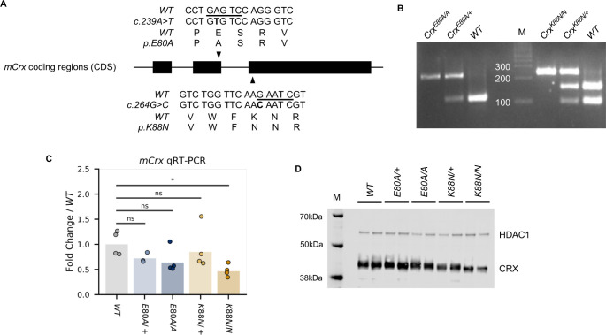
Figure 2—figure supplement 1. Wild-type (WT) and mutation knock-in mouse CRX sequences and genotyping identifications.

Figure 2—figure supplement 1.

(A) Alignment of mCrx cDNA and protein sequences showing the nucleotide substitutions and amino acid changes of CrxE80A (top) and CrxK88N (bottom) alleles. Only coding regions of mCrx exons are shown and the diagram is not to scale. Underlined bases in WT sequences indicate the restriction enzyme HinfI cut sites used in the genotyping PCR. (B) Representative mutation knock-in mouse genotyping gel image. (C) Barchart and stripplot showing mCrx mRNA expression levels in P14 WT and mutant mouse retinas. p-values for one-way analysis of variance (ANOVA) with Turkey honestly significant difference (HSD) test are indicated. (D) Immunoblots of nuclear extracts obtained from P14 WT and mutant mouse retinas showing that full-length CRX protein are produced and localized to the nucleus fraction in all mutant mouse retinas. HDAC1 was used as a loading control.
Figure 2—figure supplement 1—source data 1.