



Abstract
This study aimed to investigate the olfactory fossa anatomy (Keros types) and its relationship with changes in adjacent anatomical structures using cone-beam computed tomography (CBCT). In this descriptive-analytical study, the paranasal CBCT of 120 healthy adults over 18 years of age were evaluated. The olfactory fossa depth on both sides and the degree of asymmetry on both sides were reported. Correlation of olfactory fossa depth with the size of adjacent anatomical structures such as middle concha length, maximum orbital height and distance from ethmoid roof to nasal floor and ethmoid roof height to the palate in the anterior and posterior, length, and lateral angle of the lamella and cribriform plate distance. The lower concha junction was examined. The most common olfactory fossae on both sides of the Keros classification were Type II, Type I, and Type III, respectively. The mean dimensions of adjacent anatomical structures on the right and left did not differ significantly. The length and lateral angle of the lamella and the height of the ethmoid roof to the floor of the nose, and the height of the ethmoid roof to the palate in the back, on the right, and left in Type III were greater. The lateral angle of the left lamella was greater in Type III. The relationship between olfactory fossa depth and changes in anatomical structures were not significantly correlated with increasing olfactory fossa depth.
Supplementary Information
The online version contains supplementary material available at 10.1007/s12070-023-03538-2.
Keywords: Cribriform plate, Ethmoid, Endoscopic sinus surgery, CBCT, Olfactory fossa
Introduction
The frontal bone is dense and thick in the ethmoid roof region. The thicker frontal bone medially becomes the thinner lamella of the ethmoid bone. The lateral lamella of the cribriform plate forms the thinner medial wall of the ethmoid roof. Given that the area is the thinnest part of the ethmoid roof, the risk of intracranial penetration is more during endoscopic surgeries [1]. The shape and form of the olfactory fossa are variable so it may be situated in different levels concerning the anterior cranial fossa. Based on the Keros classification, the depth of the olfactory fossa relative to the ethmoid roof is categorized into three main types; I (1–3 mm), II (4–7 mm), and III (8–16 mm) [2]. Today, endoscopic sinus surgery (ESS) is a well-known method to treat sinonasal diseases. Surgeons should be well aware of all the anatomical features of paranasal sinuses and adhere strictly to the principles of identifying anatomical landmarks and susceptible adjacent structures. The principles are considered the most critical factors affecting the outcomes and complications of the ESS [1–3]. Having enough information about the complex anatomy of the base of the skull and its anatomical connections, including the fovea ethmoidalis, olfactory fossa, and the lateral lamella of the cribriform plate, is crucial to prevent complications in endoscopic sinus surgery. Such variations in anatomy are the most important factors that affect the results of ESS and its degree of complications. Acquiring knowledge specific to the ethmoidal labyrinth is critical to assess possible risks such as bleeding (injury to the anterior ethmoidal artery) or skull base damage (the lateral lamella of olfactory fossa). Therefore, a preoperative radiological evaluation is necessary. Cone-beam computed tomography (CBCT) may be an appropriate alternative to CT scans for studying this region [4]. This method is based on the principles of rotational tomography and is extensively utilized in dentistry. It exhibits advantages such as higher resolution, especially bony structures, less radiation exposure in many cases, and lower costs compared to most CT scans [5]. During the recent few years, the use of CBCT has prevailed in diagnosing ear, nose, and throat diseases [6]. Although the Multidetector computed tomography (MDCT) modality is still the number one choice of many surgeons and use as standard preoperative imaging, CBCT due to mentioned advantages can also be helpful and a useful replacement for CT scans.
The lateral lamella of the cribriform plate in the ethmoidal sulcus is the most dangerous part during the ESS. In addition, it is the most susceptible area to the risk of intracranial penetration across the base of the skull since the anterior ethmoidal artery leaves the ethmoid sinus in this site and proceeds anteriorly in the ethmoidal sulcus of the olfactory fossa [7, 8]. Costa et al. [9] reported no significant difference between the Keros type, age, and gender. They introduced Keros type II as the most common type by examining the nasal cavity based on the CBCT images. Belgin et al. [10] evaluated ethmoid bone using CBCT and found morphological differences in the anatomy of the bone. The ethmoid roof is considered a vital anatomical landmark in the ESS and a common site for the complications of iatrogenic penetration into the skull base [11]. Having sufficient information about complex skull base anatomy and anatomical connections such as the ethmoid roof, as well as the lateral lamella of the cribriform plate is essential for preventing complications in the ESS [7, 12]. Thus, the present study sought to determine ethmoid roof and olfactory fossa anatomy by using CBCT.
Materials and Methods
This descriptive-analytical study was conducted on the right and left sides of the CBCT images archived from 120 patients (58 females and 62 males) aged above 18 years who were referred to the Department of Oral and Maxillofacial Radiology at the University of Isfahan. The images should be of good quality and cover the ethmoid sinus region.
In this regard, a GALILEOS CBCT system (Sirona, Bensheim, Germany) was applied under the exposure conditions of 85 kV and 21–35 mAs at high resolution and source-to-patient distance of approximately 22 mm. Further, the SIDEXSIS software (Sirona Dental Systems GmbH), which provides the tool required for measuring distances, was used to examine the data. After setting the midsagittal plane vertically and the patient’s head in the software, measurements were performed under the supervision of an oral and maxillofacial radiologist on each side in two coronal and sagittal planes with a precision of 0.01 mm. In the sagittal plane, ethmoid roof height was determined in the anterior (The attachment point of frontal sinus posterior wall with cribriform plate) and posterior areas (The attachment point of cribriform plate posterior edge and planum sphenoidal anterior edge). The regions were selected since they are the anterior and posterior endpoints of the cribriform plate and can be easily measured with proper accuracy and repeatability [13].
The measurements in this study are as follows:
Olfactory fossa depth at two sides in the coronal plane, which was determined based on the Keros classification [2].
Lateral lamella length and angulation concerning the horizontal plane in the coronal view (Fig. 1).
Anterior and posterior ethmoid roof height in the sagittal plane (Fig. 2).
Middle turbinate length from the attachment point to the cribriform plate (Fig. 3).
Distance between a cribriform plate with the attachment point of inferior turbinate to sinus wall (Fig. 4).
Maximal vertical orbital height in the coronal plane (Fig. 5).
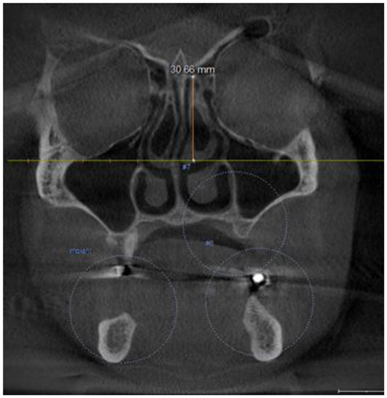
Distance between the ethmoid roof and nasal floor in the coronal plane (Fig. 6).
Fig. 1.

Lateral lamella angle (a) and length (b) in coronal view
Fig. 2.

Ethmoid roof height from anterior and posterior palate in sagittal view
Fig. 3.

Middle turbinate length from the attachment point to the cribriform plate in coronal view
Fig. 4.
Distance between a cribriform plate with the attachment point of inferior turbinate to medial sinus wall in coronal view
Fig. 5.

Maximal vertical orbital height in the coronal view
Fig. 6.

Distance between the ethmoid roof and nasal floor in the coronal view
To determine the depth of olfactory fossa, the standard anatomical points were considered to specify the distances. Then, the vertical distance between the medial ethmoid roof point (MERP) and cribriform plate with the horizontal plane defined by the infraorbital nerve on both sides (MERP and CP height, respectively) were measured. Finally, the CP height was subtracted from the MERP one to obtain the depth of the fossa (Fig. 7) [14, 15].
Fig. 7.

CBCT coronal view of measurements in (a): The horizontal line between infraorbital foramens, (b): MERP, (c): CP
LLCP = MERP-CP
The data were statistically analyzed using one-way ANOVA, Pearson’s correlation, paired t-tests, and SPSS 24 software.
Results
Regarding the type of olfactory fossa, an asymmetry was observed between the right and left sides of 39 individuals (32.5%) in the coronal plane. Additionally, 39.7% of females and 25.8% of males had an asymmetry in the type of fossa, the difference of which was statistically significant (p = 0.049). The asymmetry of 79.2 and 20.8% of cases were > 2 mm, respectively. The height of the right and left sides were equal in 3.3% (4) ones, while 59.2 (71) and 37.5% (45) had greater height on the left and right sides, respectively. Regarding the difference in the fossa depth of more than 2 mm, Keros type III was significantly more than the two others (p=0.03). Based on the Keros classification, type II was detected as the most common olfactory fossa on both sides among males and females. However, no significant difference was found between the two genders concerning the frequency of the types (p < 0.05) (Table 1).
Table 1.
Frequency distribution of the Keros types
| Right Number (%) |
Left Number (%) |
Male Number (%) |
Female Number (%) |
|
|---|---|---|---|---|
| Type I | 40 (33.4%) | 34 (28.3%) | 37 (29.8%) | 37 (31.8%) |
| Type II | 73 (60.8%) | 74 (61.7%) | 82 (66.1%) | 65 (56%) |
| Type III | 7 (5.8%) | 12 (10%) | 5 (4%) | 14 (12%) |
Further, the mean values for adjacent anatomical structures were not significantly different between the right and left sides (p < 0.05). The comparison between the parameters at the right and left sides according to the Keros classification indicated a higher mean distance between the ethmoid roof and nasal floor and posterior ethmoid roof height in types III, II, and I on both sides, respectively. Lateral lamella length and angulation in type III were significantly greater than in types II and I, respectively. However, no significant difference was observed between the mean of other variables in the various types (p < 0.05) (Table 2). As summarized in Table 3, the mean of anterior (p > 0.001) and posterior ethmoid roof height (p = 0.002), the distance between the ethmoid roof and nasal floor (p = 0.01), and the distance between the cribriform plate with the attachment point of inferior turbinate to maxillary sinus medial wall (p = 0.02) are significantly more among males compared to the females. The two genders are not significantly different in terms of the mean of other variables(p > 0.05). Finally, none of the intended parameters was significantly related to age (p < 0.05).
Table 2.
Comparison between the mean of various quantitative variables in the right and left sides based on the Keros classification
| Variable | Side | Type I | Type II | Type III | P-value |
|---|---|---|---|---|---|
| Mean ± SD | Mean ± SD | Mean ± SD | |||
| Middle turbinate length | Right | 24.9 ± 3.6 | 3.6 ± 3.8 | 25.8 ± 3.6 | 0.15 |
| Left | 24.7 ± 3.7 | 26.5 ± 4.4 | 24.8 ± 3.2 | 0.07 | |
| Anterior and posterior ethmoid roof height | Right | 65.3 ± 4.5 | 65.7 ± 4.7 | 67.5 ± 2.2 | 0.49 |
| Left | 65.2 ± 4.2 | 65.9 ± 3.6 | 64.1 ± 3.7 | 0.22 | |
| Posterior ethmoid roof height | Right | 53.3 ± 5.1 | 56.9 ± 4.6 | 58.2 ± 3.01 | < 001 |
| Left | 54.1 ± 5.7 | 56.9 ± 5.2 | 56.6 ± 4.4 | 0.04 | |
| Distance between the ethmoid roof and nasal floor | Right | 49.5 ± 6.3 | 52.9 ± 3.9 | 55.3 ± 5.1 | 0.001 |
| Left | 49.6 ± 4.8 | 51.7 ± 9.5 | 53.3 ± 4.4 | 0.004 | |
| Lateral lamella length | Right | 2.9 ± 1.2 | 5.5 ± 1.2 | 8.6 ± 0.7 | < 0.001 |
| Left | 3.3 ± 1 | 5.5 ± 1.2 | 8.5 ± 1.4 | < 0.001 | |
| Maximal vertical orbital height | Right | 39.3 ± 3.7 | 39.9 ± 2.8 | 41.3 ± 2.2 | 0.27 |
| Left | 40.30 ± 2.6 | 39.9 ± 2.4 | 39.5 ± 2.5 | 0.62 | |
| Distance between cribriform plate with the attachment point of inferior turbinate to sinus medial wall | Right | 26.3 ± 3.1 | 27.3 ± 3.1 | 26.5 ± 4.3 | 0.26 |
| Left | 26.4 ± 2.8 | 27.4 ± 2.9 | 26.8 ± 3.3 | 0.30 | |
| Lateral lamella angulation | Right | 58.4 ± 1.7 | 72.4 ± 7.5 | 79.9 ± 3.6 | < 0.001 |
| Left | 56.2 ± 14.2 | 71.9 ± 8.5 | 68.3 ± 12.5 | < 0.001 |
Table 3.
Comparison between the mean of various quantitative variables (right and left mean) by gender
| Variable | Mean ± SD | Mean ± SD | P-value |
|---|---|---|---|
| Male | Female | ||
| Middle turbinate length | 25.9 ± 3.6 | 25.7 ± 3.7 | 0.71 |
| Anterior ethmoid roof height | 64.3 ± 2.9 | 66.9 ± 4.2 | < 0.001 |
| Posterior ethmoid roof height | 54.6 ± 4.6 | 57.3 ± 5.01 | 0.002 |
| Distance between the ethmoid roof and nasal floor | 50.8 ± 5.2 | 53.1 ± 4.5 | 0.01 |
| Lateral lamella length | 5.1 ± 1.9 | 4.9 ± 1.5 | 0.62 |
| Lateral lamella angulation | 68.6 ± 11.1 | 66.7 ± 9.6 | 0.30 |
| Maximal vertical orbital height | 39.9 ± 9.2 | 39.8 ± 2.6 | 0.86 |
| Distance between cribriform plate with the attachment point of inferior turbinate to sinus medial wall | 26.4 ± 3.1 | 27.6 ± 2.6 | 0.02 |
Discussion
In the present study, anterior and posterior ethmoid roof height, lateral lamella length and angulation, middle turbinate length, maximal vertical orbital height, and distance between a cribriform plate with the attachment point of inferior turbinate were quantitatively analyzed by using CBCT images. However, no study, to the best of the researchers’ knowledge, has evaluated these measurements on CBCT in Isfahan city, Iran. The results revealed no significant difference in the mean of the variables between the right and left sides. In addition, the depth of olfactory fossa on the right and left sides was respectively obtained at 0-9.9 and 1-9.6 mm, whereas Erdem et al. reported 1–15 and 1–12 mm in this regard [16]. Krmpotic-Nemanic et al. and Meloni et al. measured the distance between cribriform plate to the ethmoid roof as 4–7 (70.2% of cases) and 1.3–17 mm (m = 5.94), respectively [17, 18]. Researchers have referred to various orders for the prevalence of types.
Based on the results of the present study, types II, I, and III were respectively common, which is consistent with those of some studies [10, 13, 19, 20]. However, the result is not in line with that obtained by Solares et al. [15], which indicated the prevalence order of types I, III, and II, as well as other researchers, which reflected the order of types II, III, and I [2, 9, 21, 22]. Further, type II was the most prevalent olfactory fossa in males and females in this study, although the difference in the prevalence of each type was insignificant among the two genders. In this regard, Elwany et al. [13] introduced types II, and I like the most common olfactory fossa at two sides among the males and females, respectively. They found that the fossa depth of the two genders significantly differed on both sides. Costa et al. [9]. outlined no difference in various classifications, age, and gender. Along with the depth of the olfactory fossa, the asymmetry of the depth or the lateral lamella length of a cribriform plate between two sides is considered necessary. A greater depth of olfactory fossa increases the possibility of perforation and penetration in the skull base [23]. In the present study, cribriform plate depth was less on the right side than the left one, which agrees with the results of another research [18, 20, 24, 25]. However, Bista et al. [23] reported a higher depth of olfactory fossa on the right side, and Jones et al. [26] pointed out that the two sides were not different. Among 120 cases, 39 (32.5%) had asymmetry in the Keros type at the right and left sides. Kaplanoglu et al. [27] referred to asymmetry of the Keros type in 5.04% of males and 6.87% of females, whereas Elwany et al. [13] detected this issue in 11% of males and 2% of females on the two sides. The difference in the results can be attributed to geography and race. The means of middle turbinate length were not significantly different on the right and left sides, as well as on females and males, which is in line with the results of Meloni et al. [18] Furthermore, no significant relationship was detected between the maximal vertical orbital height at the right side and gender, which is inconsistent with the results of Erdem et al. [16]. They found a greater height among males. Regarding the left side, the distance between the ethmoid roof and nasal floor was significantly more in types II, III, and I, respectively. However, types III, II, and I had a higher value on the right side, respectively. In addition, Keros classification was not significantly related to middle turbinate length and maximal vertical orbital height, which is not in agreement with the results of Erdem et al. [16] Regarding lateral lamella length and angulation, there was no significant difference among males and females, as well as right and left sides. Both parameters were more significant in the types III, II, and I on the left side, respectively, while a greater value was detected in the types III, II, and I on another side, respectively. The results are inconsistent with results obtained by Elwany et al. [13] and Souza et al. [20] which suggested a relationship between asymmetry in the ethmoid roof and lateral lamella angulation. Compared to males, females had lower lateral lamella length and angulation and, consequently, less olfactory fossa depth, which can be considered a reason for the more prevalence of type I among females [3]. The two variables were significantly different on the right and left sides. Further, anterior and posterior ethmoid roof heights at the two sides was not significantly different, although the height was significantly higher among males than females. On the left side, a significantly larger posterior ethmoid roof height was observed in types II and III in comparison with type I. However, type III had a significantly greater value than types II and I on the right side, respectively. Regarding the posterior area, the parameter was not related to the classification of olfactory fossa depth, which is in line with the results of Elwany et al. [13] Finally, the distance between the cribriform plate with the attachment point of the inferior turbinate to medial sinus wall was significantly greater among males than females. In this regard, the results indicated an insignificant difference between the right and left sides, as well as no significant relationship with the Keros types. The results are in agreement with the results of Basak et al. [22]. The differences between the results of the present study and other studies may be attributed to differences in the race of populations under study and the sample sizes.
Conclusion
Based on the results, there was no significant relationship between olfactory fossa depth and changes in anatomical structures. Additionally, lateral lamella length and angulation, and nasal cavity length are enhanced by increasing the depth of the fossa. The CBCT can help examine the changes in anatomical structures.
Electronic Supplementary Material
Below is the link to the electronic supplementary material.
Acknowledgements
Not applicable.
Funding
Not applicable.
Statements and Declarations
The authors also confirmed that the manuscript is original, does not contain any unlawful statements and any material the publication of which would violate any copyright or other personal or proprietary right of any person or entity. They also confirm that have written permission from copyright owners if their work has been used in this manuscript. The authors declare that the following manuscript has not been published elsewhere (in any format or language) and has not been concomitantly sent for another journal for publication consideration. The authors agree that it is their responsibility to pay any fees charged for permissions.
*This manuscript does not contain any studies with human or animal subjects performed by the any of the authors.
Conflicts of Interest/Competing Interests
Not applicable.
Ethics Approval
IR.IAU.KHUISF.REC.1400.221.
Informed Consent
Not applicable.
Footnotes
Publisher’s Note
Springer Nature remains neutral with regard to jurisdictional claims in published maps and institutional affiliations.
References
- 1.Stammberger H, Posawetz W. Functional endoscopic sinus surgery. Eur Arch Otorhinolaryngol. 1990;247:63–76. doi: 10.1007/BF00183169. [DOI] [PubMed] [Google Scholar]
- 2.Keros P. On the practical value of differences in the level of the lamina cribrosa of the ethmoid. Z fur Laryngologie Rhinologie Otologie und ihre Grenzgebiete. 1962;41:809–813. [PubMed] [Google Scholar]
- 3.Ohnishi T, Yanagisawa E. Lateral lamella of the cribriform plate—an important high-risk area in endoscopic sinus surgery. Ear Nose Throat J. 1995;74:688–690. doi: 10.1177/014556139507401006. [DOI] [PubMed] [Google Scholar]
- 4.Güldner C, Diogo I, Windfuhr J, Bien S, Teymoortash A, Werner JA et al (2011) Analysis of the fossa olfactoria using cone beam tomography (CBT). Acta Otolaryngol131:72 – 8. 10.3109/00016489.2010.506653 [DOI] [PubMed]
- 5.Mozzo P, Procacci C, Tacconi A, Tinazzi Martini P, Bergamo Andreis I. A new volumetric CT machine for dental imaging based on the cone-beam technique: preliminary results. Eur Radiol. 1998;8:1558–1564. doi: 10.1007/s003300050586. [DOI] [PubMed] [Google Scholar]
- 6.Güldner C, Ningo A, Voigt J, Diogo I, Heinrichs J, Weber R, et al. Potential of dosage reduction in cone-beam-computed tomography (CBCT) for radiological diagnostics of the paranasal sinuses. Eur Arch Otorhinolaryngol. 2013;270:1307–1315. doi: 10.1007/s00405-012-2177-2. [DOI] [PubMed] [Google Scholar]
- 7.Ohnishi T, Tachibana T, Kaneko Y, Esaki S. High-risk areas in endoscopic sinus surgery and prevention of complications. Laryngoscope. 1993;103:1181–1185. doi: 10.1288/00005537-199310000-00020. [DOI] [PubMed] [Google Scholar]
- 8.Stammberger HR, Kennedy DW. Paranasal sinuses: anatomic terminology and nomenclature. Ann Otol Rhinol Laryngol Suppl. 1995;104:7–16. doi: 10.1177/000348949510410s01. [DOI] [PubMed] [Google Scholar]
- 9.Costa ALF, Paixão AK, Gonçalves BC, Ogawa CM, Martinelli T, Maeda FA, et al. Cone beam computed tomography-based anatomical assessment of the olfactory fossa. Int J Dent. 2019;2019:4134260. doi: 10.1155/2019/4134260. [DOI] [PMC free article] [PubMed] [Google Scholar]
- 10.AKTUNA BELGİN C, BAYRAK, S., ORHAN K (2021) Evaluation of The Ethmoid Bone Using by Cone Beam Computed Tomography In Turkish Subpopulation. Clinical and Experimental Health Sciences. 10.33808/clinexphealthsci.732867
- 11.Zacharek MA, Han JK, Allen R, Weissman JL, Hwang PH. Sagittal and coronal dimensions of the ethmoid roof: a radioanatomic study. Am J Rhinol. 2005;19:348–352. doi: 10.1177/194589240501900405. [DOI] [PubMed] [Google Scholar]
- 12.Schnipper D, Spiegel JH. Management of intracranial complications of sinus surgery. Otolaryngol Clin North Am. 2004;37:453–472. doi: 10.1016/S0030-6665(03)00150-6. [DOI] [PubMed] [Google Scholar]
- 13.Elwany S, Medanni A, Eid M, Aly A, El-Daly A, Ammar S. Radiological observations on the olfactory fossa and ethmoid roof. J Laryngol Otol. 2010;124:1251–1256. doi: 10.1017/S0022215110001313. [DOI] [PubMed] [Google Scholar]
- 14.Alazzawi S, Omar R, Rahmat K, Alli K. Radiological analysis of the ethmoid roof in the malaysian population. Auris Nasus Larynx. 2012;39:393–396. doi: 10.1016/j.anl.2011. [DOI] [PubMed] [Google Scholar]
- 15.Solares CA, Lee WT, Batra PS, Citardi MJ. Lateral lamella of the cribriform plate: software-enabled computed tomographic analysis and its clinical relevance in skull base surgery. Arch Otolaryngol Head Neck Surg. 2008;134:285–289. doi: 10.1001/archotol.134.3.285. [DOI] [PubMed] [Google Scholar]
- 16.Erdem G, Erdem T, Miman MC, Ozturan O. A radiological anatomic study of the cribriform plate compared with constant structures. Rhinology. 2004;42:225–229. [PubMed] [Google Scholar]
- 17.Krmpotić-Nemanić J, Vinter I, Judaš M. Transformation of the shape of the ethmoid bone during the course of life. Eur Arch Otorhinolaryngol. 1997;254:347–349. doi: 10.1007/BF02630727. [DOI] [PubMed] [Google Scholar]
- 18.Meloni F, Mini R, Rovasio S, Stomeo F, Teatini GP. Anatomic variations of surgical importance in ethmoid labyrinth and sphenoid sinus. A study of radiological anatomy. Surg Radiol Anat. 1992;14(1):65–70. doi: 10.1007/bf01628046. [DOI] [PubMed] [Google Scholar]
- 19.Adeel M, Ikram M, Rajput MSA, Arain A, Khattak YJ. Asymmetry of lateral lamella of the cribriform plate: a software-based analysis of coronal computed tomography and its clinical relevance in endoscopic sinus surgery. Surg Radiol Anat. 2013;35:843–847. doi: 10.1007/s00276-013-1106-4. [DOI] [PubMed] [Google Scholar]
- 20.Souza SA, Souza MMAd, Idagawa M, Wolosker ÂMB, Ajzen SA. Computed tomography assessment of the ethmoid roof: a relevant region at risk in endoscopic sinus surgery. Radiologia Brasileira. 2008;41:143–147. doi: 10.1590/S0100-39842008000300003. [DOI] [Google Scholar]
- 21.Anderhuber W, Walch C, Fock C. Configuration of ethmoid roof in children 0–14 years of age. Laryngorhinootologie. 2001;80:509–511. doi: 10.1055/s-2001-17083. [DOI] [PubMed] [Google Scholar]
- 22.Başak S, Akdilli A, Karaman CZ, Kunt T. Assessment of some important anatomical variations and dangerous areas of the paranasal sinuses by computed tomography in children. Int J Pediatr Otorhinolaryngol. 2000;55:81–89. doi: 10.1016/s0165-5876(00)00362-1. [DOI] [PubMed] [Google Scholar]
- 23.Bista M, Maharjan M, Kafle P, Shrestha S (2010) Computed tomographic assessment of lateral lamella of cribriform plate and comparison of depth of olfactory fossa.Journal of the Nepal Medical Association, 49(178) [PubMed]
- 24.Özeren Keşkek C, Aytuğar E. Radiological evaluation of olfactory Fossa with Cone-Beam Computed Tomography. J oral maxillofacial Res. 2021;12(3):e3. doi: 10.5037/jomr.2021.12303. [DOI] [PMC free article] [PubMed] [Google Scholar]
- 25.Lebowitz RA, Terk A, Jacobs JB, Holliday RA. Asymmetry of the ethmoid roof: analysis using coronal computed tomography. Laryngoscope. 2001;111:2122–2124. doi: 10.1097/00005537-200112000-00007. [DOI] [PubMed] [Google Scholar]
- 26.Jones T, Almahdi J, Bhalla R, Lewis-Jones H, Swift A. The radiological anatomy of the anterior skull base. Clin Otolaryngol Allied Sci. 2002;27:101–105. doi: 10.1046/j.1365-2273.2002.00540.x. [DOI] [PubMed] [Google Scholar]
- 27.Kaplanoglu H, Kaplanoglu V, Dilli A, Toprak U, Hekimoğlu B. An analysis of the anatomic variations of the paranasal sinuses and ethmoid roof using computed tomography. Eurasian J Med. 2013;45:115–125. doi: 10.5152/eajm.2013.23. [DOI] [PMC free article] [PubMed] [Google Scholar]
Associated Data
This section collects any data citations, data availability statements, or supplementary materials included in this article.