



Abstract
The authors aimed to develop an extensive preoperative checklist of CT scan findings during endoscopic access to the ventral skull base and implement it in clinical practice. A comprehensive literature review was conducted to identify the radiological landmarks crucial to endoscopic skull base surgery. Four electronic databases were searched: PubMed, MEDLINE, EMBASE, and Google Scholar using search terms/keywords such as “radiological landmarks,” “endoscopic skull base surgery,” “CT scan,” “pituitary surgery,” “anatomical variations,” “internal carotid,” “optic nerve,” “sphenoid sinus,” “pneumatization,” “dehiscence,” and “protrusion”. Inclusion criteria were limited to original articles and systematic reviews published in English, between the years 2000 and 2021, which pertained to the radiological landmarks to be identified during endoscopic skull base surgery. Full-text articles were retrieved and collated into a narrative review focused on a 12-item checklist the authors agreed upon. The mnemonic “O ROAD TO SELLA” was used to represent the checklist and include the following landmarks: Sphenoid Ostium, Sphenoid Rostrum, Onodi cells, Anatomic variations of the sphenoid sinus, Distance between the carotids, Tumor characteristics, Optic nerve dehiscence/protrusion, Septation/insertion of the sphenoid sinus, Entrance to the sellar floor, Lateral recess of the sphenoid sinus, cLinoid process pneumatization, and internal carotid Artery dehiscence/protrusion. The checklist is designed to be used by attending physicians, fellows, and residents and the authors intend to implement it into electronic medical records at the institution’s medical center to monitor the outcomes of EEPS after implementation.
Keywords: Endoscopic sinus surgery, Preoperative checklist, Pituitary surgery, Radiological landmarks, Anatomical variations
Introduction
The origins of pituitary surgery date back to the early 20th century when Sir Victor Horsely performed a temporal craniotomy. This approach was associated with high mortality rates ranging from 50 to 80%. To reduce morbidity, transnasal approaches were developed, with the first being the transethmoidal-transsphenoidal approach by Schloffer in 1907 which was further followed by Cushing's transseptal-transsphenoidal technique that became the gold standard for pituitary surgery over several decades [1]. With advancements in technology, surgical equipment, and techniques, minimally invasive approaches to pituitary surgery evolved. In the late 20th century, Carrau introduced endoscopic endonasal transsphenoidal surgery (EEPS) as a less invasive alternative to traditional pituitary surgery. The EEPS approach accesses the tumor through the nasal cavity, allowing the surgeon to visualize the surgical field with an endoscope with increased accuracy and preservation of critical structures during the procedure [2]. Studies have shown that EEPS has a low complication rate and results in favorable clinical outcomes, such as reduced hospital stays and quicker recovery times compared to traditional approaches, and has become the standard of care for access to the ventral skull base [3].
Endoscopic endonasal pituitary surgery carries potential complications, with an overall incidence ranging from 2 to 20%. Intraoperative and postoperative complications can include CSF (cerebrospinal fluid) leak, vascular injuries, cranial nerve injuries, diabetes insipidus, anterior pituitary dysfunction, bleeding, infections, and visual deficits among others [4–6]. Preoperative BMI (Body Mass Index), age, size of the tumor, and presence of intracranial extension may predispose to these complications [7].
The sphenoid sinus is a complex paranasal sinus in the cranial cavity due to its proximity to critical structures such as the optic nerve, carotid artery, and pituitary gland. Its anatomy includes anterior and posterior walls, roof, floor, and lateral walls, with variability in size, shape, and pneumatization pattern among patients [8]. The roof of the sphenoid sinus is a crucial landmark in endoscopic endonasal transsphenoidal surgery, while its pneumatization pattern can affect surgical access and outcomes [9]. Thus, thorough knowledge of the sphenoid sinus anatomy and anatomic variants is important for safe access and successful endoscopic transsphenoidal surgery.
Surgical checklists, influenced by the aviation sector, have been shown to reduce surgical complications and improve patient outcomes globally, including in the US [10]. Checklists are cost-effective, improve medical-legal situations, and are positively viewed by patients [11]. The WHO Surgical Safety Checklist has been successful in reducing postoperative complication rates and death rates in diverse clinical settings [12]. Since then, several subspecialty checklists have been developed, including those for endoscopic endonasal transsphenoidal surgery, and preoperative CT scan imaging in endoscopic sinus surgery [13–16]. However, none addresses the radiological features of anatomical landmarks crucial for safe endoscopic transsphenoidal pituitary surgery. Recent studies have developed a preoperative CT scan checklist using an 8-item mnemonic “SPHENOID” that focused on the anatomic variations of the sphenoid sinus [17]. Yet the implementation of these checklists has faced many obstacles such as unfamiliarity, staff issues, as well as logistics, and timing constraints [18]. The authors aim to develop a more extensive preoperative checklist of CT scan findings during endoscopic access to the ventral skull base and implement it in clinical practice.
Materials and Methods
A comprehensive literature review was conducted to identify the radiological landmarks crucial to endoscopic skull base surgery. Four electronic databases were searched: PubMed, MEDLINE, EMBASE, and Google Scholar. The search terms used included combinations of the following keywords: “radiological landmarks,” “endoscopic skull base surgery,” “CT scan,” “pituitary surgery,” “anatomical variations,” “internal carotid,” “optic nerve,” “sphenoid sinus,” “pneumatization,” “dehiscence,” and “protrusion.”
Inclusion criteria were limited to original articles and systematic reviews published in English, between the years 2000 and 2021, which pertained to the radiological landmarks to be identified during endoscopic skull base surgery. Exclusion criteria were limited to non-English language articles, case reports, editorials, letters, and duplicate articles.
Two reviewers independently screened the titles and abstracts to assess eligibility. Any discrepancies were resolved through discussion, and full-text articles were retrieved and collated into a narrative review focused on a 12-item checklist the authors agreed upon. The mnemonic “O ROAD TO SELLA” was used to represent the checklist and include the following landmarks to identify preoperatively on CT scan during endoscopic endonasal transsphenoidal surgery to avoid intraoperative and postoperative complications: Sphenoid ostium, Sphenoid rostrum, Onodi cells, anatomic variations of the sphenoid sinus, distance between the carotids, tumor characteristics, optic nerve dehiscence/protrusion, septation/insertion of the sphenoid sinus, entrance to the sellar floor, lateral recess of the sphenoid sinus, clinoid process pneumatization, and internal carotid artery dehiscence/protrusion.
The checklist is designed to be used by attending physicians, fellows, and residents and the authors intend to implement it into the EPIC software for electronic medical records at the institution’s medical center to monitor the outcomes of EEPS after implementation.
Discussion
Multiple anatomical landmarks are reviewed in order to safely navigate endoscopic endonasal pituitary surgery. We will be showcasing many landmarks that constituted our preoperative checklist. Studying these entities will have a great impact on the surgical outcome of pituitary surgery.
Sphenoid Pneumatization/Types of the Sphenoid Sinus
Guldner classified the sphenoid sinus into multiple types based on their pneumatization with respect to the sella: conchal, presellar, sellar, and post-sellar.
“Conchal” (Fig. 1A) type refers to minimal to no pneumatization of the sphenoid sinus. “Pre-sellar” (Fig. 1C) is when the sphenoid sinus pneumatization reaches the anterior wall of the sella. “Sellar” (Figure a-D) type is when the sphenoid sinus pneumatization is at the level of the posterior wall of the sella; whereas “post sellar” (Fig. 1E) is when the pneumatization extends beyond the posterior wall of the sella [19].
Fig. 1.
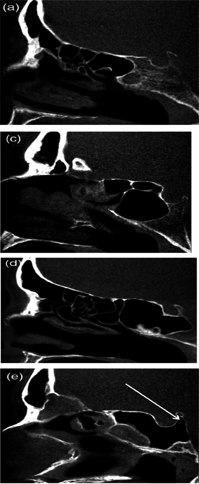
CT scan showing different types of sphenoid sinus pneumatization patterns; a Conchal type; c Pre-cellar; d Sellar; e Post cellar
Localizing the sella on imaging preoperatively is essential in planning safe access to it.
Lateral Recess Pneumatization
It is the pneumatization of the sphenoid sinus beyond an imaginary line connecting the maxillary and vidian nerves [20]. (Fig. 2).
Fig. 2.
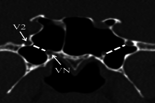
CT scan showing pneumatization of the lateral recess of the sphenoid sinus
With larger lateral sphenoid sinus pneumatization, the incidence of ON and ICA protuberance and dehiscence increase. On another note, lateral recess pneumatization causes an increased risk of vidian and maxillary nerve exposure and injury affecting access to the middle cranial fossa, infratemporal fossa, and petrous apex [17].
Anterior Clinoid Process Pneumatization
Anterior clinoid process pneumatization (Fig. 3) is present in a minority of cases (6–29%) [8]. The more the pneumatization beyond the optic strut is; the higher the likelihood of having a protruded optic nerve or even a free-floating optic nerve into the sphenoid sinus (Figs. 4 and 5).
Fig. 3.
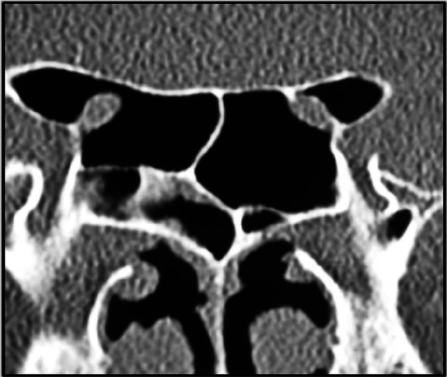
CT scan showing anterior clinoid process pneumatization
Fig. 4.
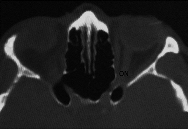
CT scan showing a free-floating optic nerve
Fig. 5.
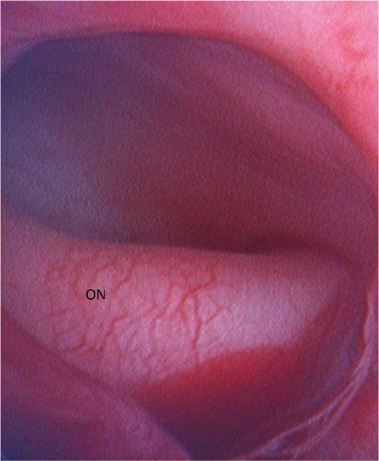
Intra-operative endoscopic demonstration of a free-floating optic nerve
Sphenoid Ostium
The natural sphenoid ostium (Fig. 6) is one of the principal landmarks for accessing safely the sphenoid sinus [21]. The SO is surrounded by multiple structures (Fig. 7). Superiorly is the skull base, inferiorly is the sphenopalatine artery and medially is the nasal septum. Understanding its position with respect to the anterior sphenoid face and skull base is of utmost importance [5]. The sphenoid ostium can also be identified through different references such as the posterior inferior end of the superior turbinate (posterior medial to it) [21], the distance from the nasal spine [22], and the location of the ostium on the vertical axis of the anterior sphenoid face [23]. Moreover, the different pneumatization of the sphenoid sinus itself and any adjacent structure will affect the location of the ostium [24, 25]. Not being able to localize the sphenoid ostium during surgery will lead to dreadful complications since the ostium is near the optic nerve, internal carotid artery, sella, and cavernous sinus [24]. A sphenoid rostrum pneumatization will deflect the ostium laterally making it at the mid-third of the horizontal plane (vs in a non-pneumatized sphenoid rostrum, the ostium is at the medial third) [26]. The lateral wall pneumatization of the sphenoid sinus will also deflect the SO laterally. The distance between the SO and the choana plays a role in influencing the entry point to the sinus. This distance is lengthened with the presence of Onodi cells making the sphenoid ostium higher with regard to the choana. However, it is relevant to note that this distance did not affect the distance between the SO and the skull base [26].
Fig. 6.
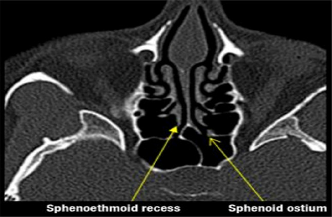
CT scan showing the natural sphenoid ostium and sphenoethmoid recess
Fig. 7.
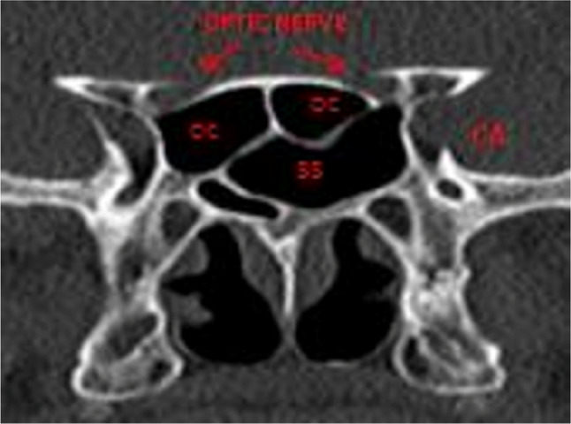
CT scan showing vital structures surrounding the sphenoid ostium such as the optic nerve, internal carotid artery, and the cavernous sinus
Tumor Characteristics
Numerous studies agree that the increased tumor size (defined as macroadenoma if > 1 cm or giant adenoma if > 4 cm; tumor volume > 30cm3; tumor diameter > 3 cm) and tumor extension (defined by a high Knosp grade, cavernous sinus invasion; intraventricular, parasellar, and anterior cranial fossa extension) are significantly associated with increased complication rate after EETS [27, 28]. Every 1 cm increase in the tumor size decreases the likelihood of complete removal by around threefold, and the invasion of the cavernous sinus, determined by a Knosp grade of 3 or 4 is among the factors strongly associated with incomplete tumor resection and hence increased complication rate [29]. Tumor type and biology are not significantly associated with surgical morbidity. However, non-functioning adenomas as compared to secreting adenomas may predispose to increased morbidity and rate of complications [30, 31].
Onodi Cells
These cells are the most posterior ethmoid air cells that pneumatize supero-laterally into the sphenoid sinus adjacent to the optic nerve (Fig. 7). At least 1 Onodi cell will pneumatize into the sphenoid sinus (12%) [17]. This variant carries the risk of injury to the optic nerve if the surgeon dissects through it thinking he/she is in the sphenoid sinus. Recognizing these cells is imperative in evading complications [32]. (Fig. 8).
Fig. 8.
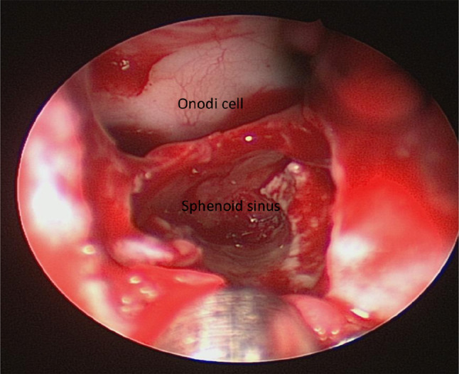
Intra-operative endoscopic view of the sphenoid sinus with an adjacent Onodi cell
Inter-Sphenoid Sinus Septum: Number and Insertion
In the majority of cases, one septum is usually found (63%) while some other sphenoid sinuses might have multiple septa (either complete or incomplete). It is noted that there is a 49% chance for the inter-sphenoid septum to insert into the internal carotid artery (Fig. 9). As the number of sphenoid sinus septa increases, at least one septum will insert into the ICA. No case was found to have the same ICA connecting with more than 1 septum [33]. The inter-sphenoid septum can also insert on the ON. These occurrences are so common that the actual incidence of injury to these neurovascular structures is low [34]. However, training to identify these structures is still beneficial in an attempt to prevent unfavorable outcomes.
Fig. 9.
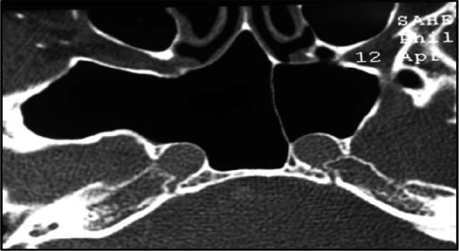
CT scan showing the insertion of the inter-sphenoid sinus septum on the internal carotid artery
Optic Nerve Dehiscence and/or Protrusion
Bony dehiscence over the optic nerve was initially defined as the absence of visible bone density separating the sinus from the course of the nerve, although some may consider the presence of bony thickness of less than 0.5 mm as dehiscence as well [35, 36]. In one study, ON dehiscence occurred in 24% and varied depending on the relationship of the nerve with the posterior paranasal sinuses [37]. Type 2 (ON causes indentation on the lateral wall of SS) and type 3 (ON courses through the sphenoid sinus) had the highest rate of bony dehiscence (82 and 100% respectively) compared to type 1 (ON adjacent to the lateral wall of the sphenoid sinus without indentation) and type 4 (ON lateral to the posterior ethmoid and sphenoid sinuses). In other cohorts, using the same definition, bony dehiscence over the ON was identified in 13.5–17.5%. Types 2 and 3 were most commonly associated with dehiscence, albeit to a lesser degree than in the previous study [38, 39]. All studies agreed that the type 3 ON or those with greater exposure to the sphenoid sinus are at the greatest risk of dehiscence and tend to be associated with protrusion and anterior clinoid process pneumatization [8].
Protrusion of the optic nerve into the ethmoid and the sphenoid sinus was first described by Dessi et al. as a bulging of more than 50% of the optic canal into a pneumatized paranasal sinus and reported a prevalence of 8% protrusion into the sphenoid sinus [34]. However, further studies evaluating the anatomic variations of the sphenoid sinus and its related structures noted a higher prevalence of ON protrusion of around 31% [40, 41]. (Figs. 5, 10 and 11).
Fig. 10.
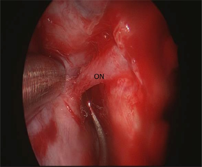
Intra-operative endoscopic image of a free-floating optic nerve
Fig. 11.

Intra-operative endoscopic image of a transected optic nerve
Intercarotid Distance
The intercarotid distance in the setting of no sellar pathology has an average of 18 mm [17]. This distance changes with the type of pituitary pathology. It is usually measured at the level of the mid-cavernous ICA level where it usually protrudes into the sphenoid sinus [42, 43]. With large pituitary tumors, the ICD increases, however with growth hormone tumors there is a narrowing of the ICD, with cases reported as “kissing carotids” [17] (Figs. 12 and 13). Being aware of these variations will prevent dreadful complications to the vasculature.
Fig. 12.
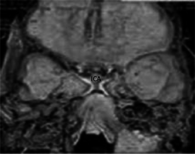
MRI image showing kissing internal carotid artery
Fig. 13.
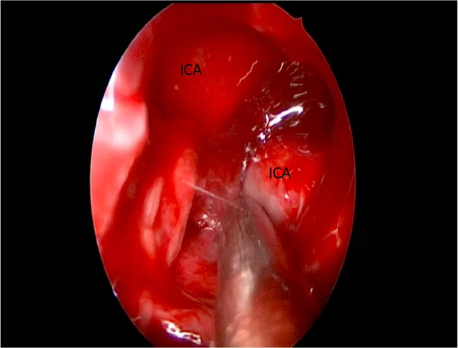
Intra-operative endoscopic view of kissing internal carotid artery
Internal Carotid Artery Dehiscence and/or Protrusion
Similar to ON, ICA dehiscence is defined as a soft carotid bulge without a visible bony coverage or with a submillimetric overlying bone (Fig. 14). The protrusion is defined as a projection of more than 50% of the ICA into the sphenoid sinus. In a cadaveric study on 188 sphenoid sinuses by Kennedy et al., clinical dehiscence over the ICA was identified in 22% of cases [44], which is higher than what was reported in later studies where ICA dehiscence ranged from 1.5 to 14.4% [45–47]. To be more precise in the evaluation of protrusion and dehiscence, it’s important to look at the parasellar and paraclival segments of the ICA. One study involving 90 CT scans reported dehiscence of the pcICA in 3.6% but not of the psICA. Additionally, protrusion of psICA occurred in 26% while that of pcICA occurred in 35% [48]. It’s worth mentioning that the average thickness of bone covering the ICA is around 0.5–1 mm which is higher than that covering the ON, which ranges between 0.28 and 0.5 mm [45]. Therefore, the definition of dehiscence may vary between both structures. The prevalence of ICA protrusion and dehiscence is also affected by the type of pathology. A study by Sasagawa et al. showed that 35.5% of acromegalic patients had ICA protrusion while 22.2% had dehiscence meaning that these vascular anomalies are more frequent in patients with acromegaly [49].
Fig. 14.
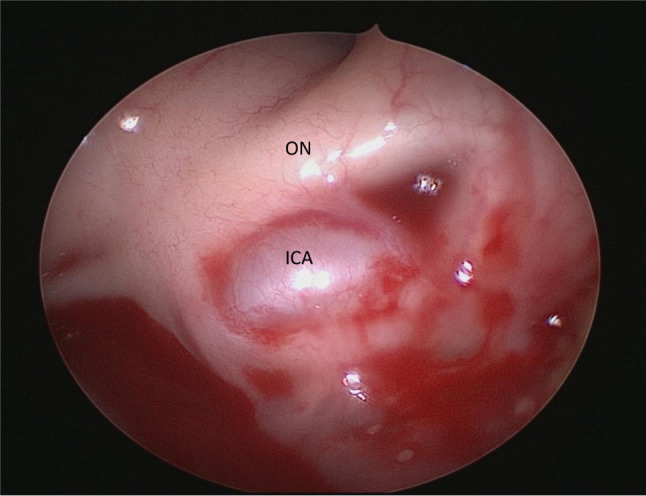
Intra-operative endoscopic view of an internal carotid artery dehiscence
Limitations
The checklist was developed based on a review of literature published in English, which may have limited the inclusion of studies from other languages that could provide additional insights and perspectives on the subject matter. Although the authors conducted a comprehensive literature review, it is possible that some relevant studies were missed, and some important radiological landmarks may have been excluded from the checklist.
The checklist was developed based on the consensus of the authors, and there is a possibility that other experts in the field may have different opinions on which radiological landmarks are most critical for endoscopic skull base surgery.
The study did not assess the practicality or feasibility of implementing the checklist in a clinical setting, and it is possible that some radiological landmarks may be difficult to assess consistently or reliably using CT scans. The study did not evaluate the impact of using the checklist on patient outcomes, and it is unclear whether its implementation leads to improved surgical outcomes or reduced complications.
Conclusion
Following this discussion about how to safely perform an endoscopic endonasal transsphenoidal pituitary surgery; we propose a mnemonic to be utilized as a preoperative checklist to navigate safely during surgery: “O ROAD TO SELLA”.
O–sphenoid Ostium.
R–Rostrum.
O–Onodi cell.
A–Anatomic variation of SS (pneumatization, previous surgery…).
D–Distance between carotids.
T–Tumor pathology.
O–Optic nerve.
S–Septation & Insertion.
E–Entrance to sellar floor.
L–Lateral wall of SS (pneumatization, OC recess…).
L–cLinoid process pneumatization.
A–carotid Artery.
Author Contribution
All authors contributed to the study conception and design. Material preparation, data collection and analysis were performed by NEH, and JH. The first draft of the manuscript was written by UEH and ZK and all authors commented on previous versions of the manuscript. All authors read and approved the final manuscript.
Funding
No funding was received for conducting this study.
Data Availability
The data that support the findings of this study are available from the authors upon reasonable request.
Declarations
Conflict of interest
The authors have no financial or non-financial interests to disclose.
Ethical Approval
This is a review study. The Institutional Review Board at the American University of Beirut Medical Center has confirmed that no ethical approval is required.
Footnotes
Publisher's Note
Springer Nature remains neutral with regard to jurisdictional claims in published maps and institutional affiliations.
References
- 1.Carrau RL, Kassam AB, Snyderman CH. Pituitary surgery. Otolaryngol Clin North Am. 2001;34(6):1143–ix. doi: 10.1016/s0030-6665(05)70371-6. [DOI] [PubMed] [Google Scholar]
- 2.Cappabianca P, Cavallo LM, de Divitiis O, Solari D, Esposito F, Colao A. Endoscopic pituitary surgery. Pituitary. 2008;11:385–90. doi: 10.1007/s11102-008-0087-5. [DOI] [PubMed] [Google Scholar]
- 3.Lucas JW, Zada G. Endoscopic surgery for pituitary tumors. Neurosurg Clin N Am. 2012;23(4):555–569. doi: 10.1016/j.nec.2012.06.008. [DOI] [PubMed] [Google Scholar]
- 4.Vignolles-Jeong J, Kreatsoulas D, Godil S, Otto B, Carrau R, Prevedello D. Complications in endoscopic pituitary surgery. Otolaryngol Clin North Am. 2022;55(2):431–448. doi: 10.1016/j.otc.2021.12.011. [DOI] [PubMed] [Google Scholar]
- 5.Kassam AB, Prevedello DM, Carrau RL, Snyderman CH, Thomas A, Gardner P, Zanation A, Duz B, Stefko ST, Byers K, Horowitz MB. Endoscopic endonasal skull base surgery: analysis of complications in the authors' initial 800 patients. J Neurosurg. 2011;114(6):1544–1568. doi: 10.3171/2010.10.JNS09406. [DOI] [PubMed] [Google Scholar]
- 6.Taghvaei M, Fallah S, Sadaghiani S, Sadrhosseini SM, Tabari A, Fathi M, Zeinalizadeh M. Surgical complications of endoscopic approach to skull base: analysis of 584 consecutive patients. Eur Arch Otorhinolaryngol. 2022;279(6):3189–3199. doi: 10.1007/s00405-022-07256-3. [DOI] [PubMed] [Google Scholar]
- 7.Ivan ME, Iorgulescu JB, El-Sayed I, McDermott MW, Parsa AT, Pletcher SD, Jahangiri A, Wagner J, Aghi MK. Risk factors for postoperative cerebrospinal fluid leak and meningitis after expanded endoscopic endonasal surgery. J Clin Neurosci. 2015;22(1):48–54. doi: 10.1016/j.jocn.2014.08.009. [DOI] [PubMed] [Google Scholar]
- 8.Anusha B, Baharudin A, Philip R, Harvinder S, Shaffie BM. Anatomical variations of the sphenoid sinus and its adjacent structures: a review of existing literature. Surg Radiol Anat. 2014;36(5):419–427. doi: 10.1007/s00276-013-1214-1. [DOI] [PubMed] [Google Scholar]
- 9.Zada G, Agarwalla PK, Mukundan S, Jr, Dunn I, Golby AJ, Laws ER., Jr The neurosurgical anatomy of the sphenoid sinus and sellar floor in endoscopic transsphenoidal surgery. J Neurosurg. 2011;114(5):1319–1330. doi: 10.3171/2010.11.JNS10768. [DOI] [PMC free article] [PubMed] [Google Scholar]
- 10.Treadwell JR, Lucas S, Tsou AY. Surgical checklists: a systematic review of impacts and implementation. BMJ Qual Saf. 2014;23(4):299–318. doi: 10.1136/bmjqs-2012-001797. [DOI] [PMC free article] [PubMed] [Google Scholar]
- 11.Russ SJ, Rout S, Caris J, Moorthy K, Mayer E, Darzi A, Sevdalis N, Vincent C. The WHO surgical safety checklist: survey of patients' views. BMJ Qual Saf. 2014;23(11):939–946. doi: 10.1136/bmjqs-2013-002772. [DOI] [PMC free article] [PubMed] [Google Scholar]
- 12.Haynes AB, Weiser TG, Berry WR, Lipsitz SR, Breizat AH, Dellinger EP, Herbosa T, Joseph S, Kibatala PL, Lapitan MC, Merry AF, Moorthy K, Reznick RK, Taylor B, Gawande AA. A surgical safety checklist to reduce morbidity and mortality in a global population. N Engl J Med. 2009;360(5):491–499. doi: 10.1056/NEJMsa0810119. [DOI] [PubMed] [Google Scholar]
- 13.Christian E, Harris B, Wrobel B, Zada G. Endoscopic endonasal transsphenoidal surgery: implementation of an operative and perioperative checklist. Neurosurg Focus. 2014;37(4):E1. doi: 10.3171/2014.7.FOCUS14360. [DOI] [PubMed] [Google Scholar]
- 14.Laws ER, Wong JM, Smith TR, de Los RK, Aglio LS, Thorne AJ, Cote DJ, Esposito F, Cappabianca P, Gawande A. A checklist for endonasal transsphenoidal anterior skull base surgery. J Neurosurg. 2016;124(6):1634–1639. doi: 10.3171/2015.4.JNS142184. [DOI] [PubMed] [Google Scholar]
- 15.Leong S, Yang N, Ray A, Akbar N, Colley PM, Signore AD, Eloy JA, Govindaraj S, Gudis DA, Helman S, Hsueh W, Iloreta AM, Kacker A, Lieberman S, Pearlman AN, Schaberg MR, Tabaee A, Overdevest JB. Development of an optimized preoperative computed tomography imaging checklist for endoscopic sinus surgery utilizing a systematic review of the literature and the modified Delphi method. Int Forum Allergy Rhinol. 2022 doi: 10.1002/alr.23064. [DOI] [PubMed] [Google Scholar]
- 16.O'Brien WT, Sr, Hamelin S, Weitzel EK. The preoperative sinus CT: avoiding a "CLOSE" call with surgical complications. Radiology. 2016;281(1):10–21. doi: 10.1148/radiol.2016152230. [DOI] [PubMed] [Google Scholar]
- 17.Raseman J, Guryildirim M, Beer-Furlan A, Jhaveri M, Tajudeen BA, Byrne RW, Batra PS. Preoperative computed tomography imaging of the sphenoid sinus: striving towards safe transsphenoidal surgery. J Neurol Surg B Skull Base. 2020;81(3):251–262. doi: 10.1055/s-0039-1691831. [DOI] [PMC free article] [PubMed] [Google Scholar]
- 18.Vats A, Vincent CA, Nagpal K, Davies RW, Darzi A, Moorthy K. Practical challenges of introducing WHO surgical checklist: UK pilot experience. BMJ. 2010;340:b5433. doi: 10.1136/bmj.b5433. [DOI] [PubMed] [Google Scholar]
- 19.Güldner C, Pistorius SM, Diogo I, Bien S, Sesterhenn A, Werner JA. Analysis of pneumatization and neurovascular structures of the sphenoid sinus using cone-beam tomography (CBT) Acta Radiol. 2012;53(02):214–219. doi: 10.1258/ar.2011.110381. [DOI] [PubMed] [Google Scholar]
- 20.Vaezi A, Cardenas E, Pinheiro-Neto C, Paluzzi A, Branstetter BF, 4th, Gardner PA, Snyderman CH, Fernandez-Miranda JC. Classification of sphenoid sinus pneumatization: relevance for endoscopic skull base surgery. Laryngoscope. 2015;125(03):577–581. doi: 10.1002/lary.24989. [DOI] [PubMed] [Google Scholar]
- 21.Kim HU, Kim SS, Kang SS, Chung IH, Lee JG, Yoon JH. Surgical anatomy of the natural ostium of the sphenoid sinus. Laryngoscope. 2001;111(9):1599–1602. doi: 10.1097/00005537-200109000-00020. [DOI] [PubMed] [Google Scholar]
- 22.Yanagisawa E, Yanagisawa K, Christmas DA. Endoscopic localization of the sphenoid sinus ostium. Ear Nose Throat J. 1998;77(2):88–89. doi: 10.1177/014556139807700204. [DOI] [PubMed] [Google Scholar]
- 23.Enatsu K, Takasaki K, Kase K, Jinnouchi S, Kumagami H, Nakamura T, Takahashi H. Surgical anatomy of the sphenoid sinus on the CT using multiplanar reconstruction technique. Otolaryngol Head Neck Surg. 2008;138(2):182–186. doi: 10.1016/j.otohns.2007.10.010. [DOI] [PubMed] [Google Scholar]
- 24.Alrumaih RA, Ashoor MM, Obidan AA, Al-Khater KM, Al-Jubran SA. Radiological sinonasal anatomy. Exploring the Saudi population. Saudi Med J. 2016;37(5):521–526. doi: 10.15537/smj.2016.5.13904. [DOI] [PMC free article] [PubMed] [Google Scholar]
- 25.Wada K, Moriyama H, Edamatsu H, Hama T, Arai C, Kojima H, Otori N, Yanagi K. Identification of Onodi cell and new classification of sphenoid sinus for endoscopic sinus surgery. Int Forum Allergy Rhinol. 2015;5(11):1068–1076. doi: 10.1002/alr.21567. [DOI] [PubMed] [Google Scholar]
- 26.Doubi A, Albathi A, Sukyte-Raube D, Castelnuovo P, Alfawwaz F, AlQahtani A. Location of the sphenoid sinus ostium in relation to adjacent anatomical landmarks. Ear Nose Throat J. 2021 doi: 10.1177/0145561320927907. [DOI] [PubMed] [Google Scholar]
- 27.Hofstetter CP, Nanaszko MJ, Mubita LL, Tsiouris J, Anand VK, Schwartz TH. Volumetric classification of pituitary macroadenomas predicts outcome and morbidity following endoscopic endonasal transsphenoidal surgery. Pituitary. 2012;15(3):450–463. doi: 10.1007/s11102-011-0350-z. [DOI] [PubMed] [Google Scholar]
- 28.Lobatto DJ, de Vries F, Zamanipoor Najafabadi AH, Pereira AM, Peul WC, Vliet Vlieland TPM, Biermasz NR, van Furth WR. Preoperative risk factors for postoperative complications in endoscopic pituitary surgery: a systematic review. Pituitary. 2018;21(1):84–97. doi: 10.1007/s11102-017-0839-1. [DOI] [PMC free article] [PubMed] [Google Scholar]
- 29.Kim JH, Lee JH, Lee JH, Hong AR, Kim YJ, Kim YH. Endoscopic transsphenoidal surgery outcomes in 331 nonfunctioning pituitary adenoma cases after a single surgeon learning curve. World Neurosurg. 2018;109:e409–e416. doi: 10.1016/j.wneu.2017.09.194. [DOI] [PubMed] [Google Scholar]
- 30.Barzaghi LR, Losa M, Giovanelli M, Mortini P. Complications of transsphenoidal surgery in patients with pituitary adenoma: experience at a single centre. Acta Neurochir. 2007;149(9):877–886. doi: 10.1007/s00701-007-1244-8. [DOI] [PubMed] [Google Scholar]
- 31.Jang JH, Kim KH, Lee YM, Kim JS, Kim YZ. Surgical Results of pure endoscopic endonasal transsphenoidal surgery for 331 pituitary adenomas: a 15-year experience from a single institution. World Neurosurg. 2016;96:545–555. doi: 10.1016/j.wneu.2016.09.051. [DOI] [PubMed] [Google Scholar]
- 32.Batra PS, Citardi MJ, Gallivan RP, Roh H-J, Lanza DC. Software-enabled CT analysis of optic nerve position and paranasal sinus pneumatization patterns. Otolaryngol Head Neck Surg. 2004;131(06):940–945. doi: 10.1016/j.otohns.2004.07.013. [DOI] [PubMed] [Google Scholar]
- 33.Dziedzic T, Koczyk K, Gotlib T, Kunert P, Maj E, Marchel A. Sphenoid sinus septations and their interconnections with parasphenoidal internal carotid artery protuberance: radioanatomical study with literature review. Wideochir Inne Tech Maloinwazyjne. 2020;15(1):227–233. doi: 10.5114/wiitm.2019.85837. [DOI] [PMC free article] [PubMed] [Google Scholar]
- 34.Dessi P, Moulin G, Castro F, Chagnaud C, Cannoni M. Protrusion of the optic nerve into the ethmoid and sphenoid sinus: prospective study of 150 CT studies. Neuroradiology. 1994;36(07):515–516. doi: 10.1007/BF00593511. [DOI] [PubMed] [Google Scholar]
- 35.Fujii K, Chambers SM, Rhoton AL., Jr Neurovascular relationships of the sphenoid sinus. A microsurgical study. J Neurosurg. 1979;50(1):31–39. doi: 10.3171/jns.1979.50.1.0031. [DOI] [PubMed] [Google Scholar]
- 36.Kainz J, Stammberger H. Danger areas of the posterior rhinobasis. An endoscopic and anatomical-surgical study. Acta Otolaryngol. 1992;112(5):852–861. doi: 10.3109/00016489209137484. [DOI] [PubMed] [Google Scholar]
- 37.DeLano MC, Fun FY, Zinreich SJ. Relationship of the optic nerve to the posterior paranasal sinuses: a CT anatomic study. AJNR Am J Neuroradiol. 1996;17(4):669–675. [PMC free article] [PubMed] [Google Scholar]
- 38.Itagi RM, Adiga CP, Kalenahalli K, Goolahally L, Gyanchandani M. Optic nerve canal relation to posterior paranasal sinuses in Indian ethnics: review and objective classification. J Clin Diagn Res. 2017 doi: 10.7860/JCDR/2017/23447.9510. [DOI] [PMC free article] [PubMed] [Google Scholar]
- 39.Sapçi T, Derin E, Almaç S, Cumali R, Saydam B, Karavuş M. The relationship between the sphenoid and the posterior ethmoid sinuses and the optic nerves in Turkish patients. Rhinology. 2004;42(1):30–34. [PubMed] [Google Scholar]
- 40.Sirikci A, Bayazit YA, Bayram M, Mumbuç S, Güngör K, Kanlikama M. Variations of sphenoid and related structures. Eur Radiol. 2000;10(5):844–848. doi: 10.1007/s003300051016. [DOI] [PubMed] [Google Scholar]
- 41.Unal B, Bademci G, Bilgili YK, Batay F, Avci E. Risky anatomic variations of sphenoid sinus for surgery. Surg Radiol Anat. 2006;28(2):195–201. doi: 10.1007/s00276-005-0073-9. [DOI] [PubMed] [Google Scholar]
- 42.Hamid O, El Fiky L, Hassan O, Kotb A, El Fiky S. Anatomic variations of the sphenoid sinus and their impact on trans-sphenoid pituitary surgery. Skull Base. 2008;18(01):9–15. doi: 10.1055/s-2007-992764. [DOI] [PMC free article] [PubMed] [Google Scholar]
- 43.Wiebracht ND, Zimmer LA. Complex anatomy of the sphenoid sinus: a radiographic study and literature review. J Neurol Surg B Skull Base. 2014;75(06):378–382. doi: 10.1055/s-0034-1376195. [DOI] [PMC free article] [PubMed] [Google Scholar]
- 44.Kennedy DW, Zinreich SJ, Hassab MH. The internal carotid artery as it relates to endonasal sphenoethmoidectomy. Am J Rhinol. 1990;4(1):7–12. doi: 10.2500/105065890782020962. [DOI] [Google Scholar]
- 45.Kazkayasi M, Karadeniz Y, Arikan OK. Anatomic variations of the sphenoid sinus on computed tomography. Rhinology. 2005;43(2):109–114. [PubMed] [Google Scholar]
- 46.Meloni F, Mini R, Rovasio S, Stomeo F, Teatini GP. Anatomic variations of surgical importance in ethmoid labyrinth and sphenoid sinus. A study of radiological anatomy. Surg Radiol Anat. 1992;14(1):65–70. doi: 10.1007/BF01628046. [DOI] [PubMed] [Google Scholar]
- 47.Johnson DM, Hopkins RJ, Hanafee WN, Fisk JD. The unprotected parasphenoidal carotid artery studied by high-resolution computed tomography. Radiology. 1985;155(1):137–141. doi: 10.1148/radiology.155.1.3975391. [DOI] [PubMed] [Google Scholar]
- 48.Dal Secchi MM, Dolci RLL, Teixeira R, Lazarini PR. An Analysis of anatomic variations of the sphenoid sinus and its relationship to the internal carotid artery. Int Arch Otorhinolaryngol. 2018;22(2):161–166. doi: 10.1055/s-0037-1607336. [DOI] [PMC free article] [PubMed] [Google Scholar]
- 49.Sasagawa Y, Tachibana O, Doai M, Hayashi Y, Tonami H, Iizuka H, Nakada M. Carotid artery protrusion and dehiscence in patients with acromegaly. Pituitary. 2016;19(5):482–487. doi: 10.1007/s11102-016-0728-z. [DOI] [PubMed] [Google Scholar]
Associated Data
This section collects any data citations, data availability statements, or supplementary materials included in this article.
Data Availability Statement
The data that support the findings of this study are available from the authors upon reasonable request.