

Abstract
Biochemists who study single cells have been constrained by the lack of a general methodology of high time resolution and high measurement sensitivity for quantitatively assaying enzyme activities using natural substrates in solution. The methods we describe will remove this limitation. In brief, nanogram tissue samples are dissected from frozen-dried tissue. The samples are `extracted' in microdroplets of assay cocktail. The enzyme activity, indicated fluorometrically by the oxidation/reduction of NAD(P), is followed in real time on a computer display. In the development of this method, we evaluated several parameters required for optimization; the most important of these evaluations, including numerous empirically derived relationships, are reported here and in supplemental material provided with reprints.
With these methods, assays of pyruvate orthophosphate dikinase on samples enriched in bundlesheath cells and mesophyll cells of Flaveria brownii yielded the predictable results. Assays of this enzyme in guard cells dissected from Vicia faba leaflets gave results like those recently reported by another laboratory for protoplasts derived from these cells. The results of assays by this method and by enzymic cycling for NAD(P)triose-P dehydrogenase were comparable. Phosphoenolpyruvate carboxylase, the most extensively studied enzyme activity, was present at high levels in guard cells, which has been demonstrated previously in other reports based on diverse assay approaches.
Full text
PDF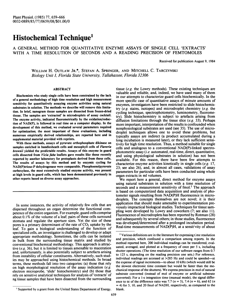
Selected References
These references are in PubMed. This may not be the complete list of references from this article.
- Aoyagi K., Bassham J. A. Pyruvate orthophosphate dikinase of c(3) seeds and leaves as compared to the enzyme from maize. Plant Physiol. 1984 Jun;75(2):387–392. doi: 10.1104/pp.75.2.387. [DOI] [PMC free article] [PubMed] [Google Scholar]
- Chen T. M., Dittrich P., Campbell W. H., Black C. C. Metabolism of epidermal tissues, mesophyll cells, and bundle sheath strands resolved from mature nutsedge leaves. Arch Biochem Biophys. 1974 Jul;163(1):246–262. doi: 10.1016/0003-9861(74)90475-5. [DOI] [PubMed] [Google Scholar]
- Holaday A. S., Chollet R. Photosynthetic/Photorespiratory Carbon Metabolism in the C(3)-C(4) Intermediate Species, Moricandia arvensis and Panicum milioides. Plant Physiol. 1983 Nov;73(3):740–745. doi: 10.1104/pp.73.3.740. [DOI] [PMC free article] [PubMed] [Google Scholar]
- Jones M. G., Outlaw W. H., Lowry O. H. Enzymic assay of 10 to 10 moles of sucrose in plant tissues. Plant Physiol. 1977 Sep;60(3):379–383. doi: 10.1104/pp.60.3.379. [DOI] [PMC free article] [PubMed] [Google Scholar]
- Jongkind J. F., Ploem J. S., Reuser A. J., Galjaard H. Enzyme assays at the single cell level using a new type of microfluorimeter. Histochemistry. 1974;40(3):221–229. doi: 10.1007/BF00501957. [DOI] [PubMed] [Google Scholar]
- Mroz E. A., Lechene C. An NADH-coupled assay for femtogram or nanogram quantities of chymotrypsin. Anal Biochem. 1983 Jan;128(1):181–185. doi: 10.1016/0003-2697(83)90360-3. [DOI] [PubMed] [Google Scholar]
- Mroz E. A., Lechene C. Fluorescence analysis of picoliter samples. Anal Biochem. 1980 Feb;102(1):90–96. doi: 10.1016/0003-2697(80)90322-x. [DOI] [PubMed] [Google Scholar]
- Outlaw W. H., Kennedy J. Enzymic and substrate basis for the anaplerotic step in guard cells. Plant Physiol. 1978 Oct;62(4):648–652. doi: 10.1104/pp.62.4.648. [DOI] [PMC free article] [PubMed] [Google Scholar]
- Outlaw W. H., Lowry O. H. Organic acid and potassium accumulation in guard cells during stomatal opening. Proc Natl Acad Sci U S A. 1977 Oct;74(10):4434–4438. doi: 10.1073/pnas.74.10.4434. [DOI] [PMC free article] [PubMed] [Google Scholar]
- Outlaw W. H., Manchester J., Brown P. H. High Levels of Malic Enzyme Activities in Vicia faba L. Epidermal Tissue. Plant Physiol. 1981 Nov;68(5):1047–1051. doi: 10.1104/pp.68.5.1047. [DOI] [PMC free article] [PubMed] [Google Scholar]
- Outlaw W. H., Manchester J., Dicamelli C. A. Histochemical Approach to Properties of Vicia faba Guard Cell Phosphoenolpyruvate Carboxylase. Plant Physiol. 1979 Aug;64(2):269–272. doi: 10.1104/pp.64.2.269. [DOI] [PMC free article] [PubMed] [Google Scholar]
- Outlaw W. H., Manchester J., Dicamelli C. A., Randall D. D., Rapp B., Veith G. M. Photosynthetic carbon reduction pathway is absent in chloroplasts of Vicia faba guard cells. Proc Natl Acad Sci U S A. 1979 Dec;76(12):6371–6375. doi: 10.1073/pnas.76.12.6371. [DOI] [PMC free article] [PubMed] [Google Scholar]
- Outlaw W. H., Manchester J. Guard cell starch concentration quantitatively related to stomatal aperture. Plant Physiol. 1979 Jul;64(1):79–82. doi: 10.1104/pp.64.1.79. [DOI] [PMC free article] [PubMed] [Google Scholar]
- Outlaw W. H., Manchester J., Zenger V. E. The relationship between protein content and dry weight of guard cells and other single cell samples of Vicia faba L. leaflet. Histochem J. 1981 Mar;13(2):329–334. doi: 10.1007/BF01006886. [DOI] [PubMed] [Google Scholar]
- Outlaw W. H., Mayne B. C., Zenger V. E., Manchester J. Presence of Both Photosystems in Guard Cells of Vicia faba L: IMPLICATIONS FOR ENVIRONMENTAL SIGNAL PROCESSING. Plant Physiol. 1981 Jan;67(1):12–16. doi: 10.1104/pp.67.1.12. [DOI] [PMC free article] [PubMed] [Google Scholar]
- Outlaw W. H., Schmuck C. L., Tolbert N. E. Photosynthetic Carbon Metabolism in the Palisade Parenchyma and Spongy Parenchyma of Vicia faba L. Plant Physiol. 1976 Aug;58(2):186–189. doi: 10.1104/pp.58.2.186. [DOI] [PMC free article] [PubMed] [Google Scholar]
- Outlaw W. H., Tarczynski M. C. Guard Cell Starch Biosynthesis Regulated by Effectors of ADP-Glucose Pyrophosphorylase. Plant Physiol. 1984 Feb;74(2):424–429. doi: 10.1104/pp.74.2.424. [DOI] [PMC free article] [PubMed] [Google Scholar]
- Pette D. Microphotometric measurement of initial maximum reaction rates in quantitative enzyme histochemistry in situ. Histochem J. 1981 Mar;13(2):319–327. doi: 10.1007/BF01006885. [DOI] [PubMed] [Google Scholar]
- ROTMAN B. Measurement of activity of single molecules of beta-D-galactosidase. Proc Natl Acad Sci U S A. 1961 Dec 15;47:1981–1991. doi: 10.1073/pnas.47.12.1981. [DOI] [PMC free article] [PubMed] [Google Scholar]
- Salmon J. M., Kohen E., Viallet P., Hirschberg J. G., Wouters A. W., Kohen C., Thorell B. Microspectrofluorometric approach to the study of free/bound NAD(P)H ratio as metabolic indicator in various cell types. Photochem Photobiol. 1982 Nov;36(5):585–593. doi: 10.1111/j.1751-1097.1982.tb04420.x. [DOI] [PubMed] [Google Scholar]
- Van Kirk C. A., Raschke K. Release of Malate from Epidermal Strips during Stomatal Closure. Plant Physiol. 1978 Mar;61(3):474–475. doi: 10.1104/pp.61.3.474. [DOI] [PMC free article] [PubMed] [Google Scholar]
- Van Noorden C. J., Tas J., Vogels I. M. Cytophotometry of glucose-6-phosphate dehydrogenase activity in individual cells. Histochem J. 1983 Jun;15(6):583–599. doi: 10.1007/BF01954149. [DOI] [PubMed] [Google Scholar]