

Abstract
A herniation is the abnormal protrusion of an organ or other body structure through a defect or natural opening in a covering membrane, muscle, or bone. The parotid gland is the largest of the salivary glands, which is situated below the external acoustic meatus and overlaps the masseter muscle anteriorly. In this rare case report, a 35 years old female patient complained of pain in the cheek region for 3 years and gave a history of pain aggravating while having sour food. On palpation, multiple nodules were found bilaterally in the cheek area which was tender. Ultrasonography of the masseter and parotid region showed herniation of the lower lobe of the parotid gland into the masseter muscle which is 1 cm on the right side and 0.8 cm on the left side. The findings obtained from ultrasound images were further confirmed with Magnetic Resonance Imaging (MRI).
Keywords: Herniation, Parotid gland, Masseter muscle
Introduction
A herniation is an abnormal protrusion of a body structure through an opening that may be natural or a defect caused by trauma. The parotid gland is the largest of the salivary gland which is well encapsulated and overlaps the masseter muscle anteriorly.
Herniation of the parotid gland is very rare and uncommon. In the literature, a case was reported [1] in a 7-months-old boy with the herniation of the parotid gland through the Stenson’s duct following a mechanical blow on his cheek. Another case of herniation of buccal fat pad [2] into the oral cavity was reported in a 2¾-year-old girl after a fall from a sofa and the pad tissue was excised under General Anaesthesia (GA).
Here we report a rare case of herniation of the parotid gland into masseter muscle bilaterally with no history of trauma. To the best of our knowledge, this is the first case reporting the herniation of parotid into the masseter muscle.
Case Report
A 35-year-old female patient visited our outpatient department 2 months ago with a chief complaint of pain in the left and right side of the cheek region for the past 3 years. Her history of presenting illness revealed that the pain is only after mastication and aggravates while having sour food. On eliciting her medical history, the patient had been diagnosed with allergic rhinitis and hypothyroidism for the past 10 years and vertigo for the last 6 months. The patient is on medication for the above said medical conditions. Her personal history revealed that she had a habit of clenching her teeth, especially during stressful conditions. She was diagnosed with Myofascial pain dysfunction syndrome (MPDS) a year ago and she got relieved by wearing occlusal splints. She also reported a relief in pain in the cheek region after having anti-allergic drug, Montelukast levocetirizine which she takes for allergic rhinitis.
On examination, no swelling or redness was present in the cheek area (Fig. 1A). Multiple palpable nodules were felt on cheeks bilaterally, both extra orally and intraorally, which were tender on palpation. Intraorally mucosa appears normal and moist. The Stenson’s duct appeared normal with normal salivary flow and consistency. Provisionally, we reached a diagnosis of myositis ossificans with a differential diagnosis of lymphadenopathy and cysticercosis.
Fig. 1.
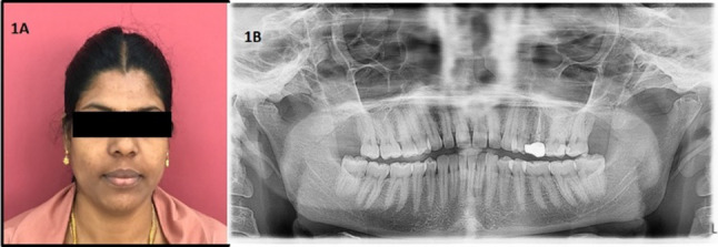
A Profile picture of the patient. B OPG showing flattening of condyles
We advised an Orthopantomogram (OPG), which showed flattening of the condylar head bilaterally (Fig. 1B). As there were no ossifications seen in the OPG, we could rule out myositis ossificans, and hence advised for ultrasonography (USG) was advised. As the patient reported pain from having sour food, we included the parotid region in the USG scan. USG of the cheek and parotid region showed herniation of the lower lobe of the parotid gland into the masseter muscle which is 1 cm on the right side and 0.8 cm on the left side (Fig. 2).
Fig. 2.
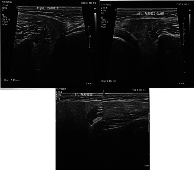
Ultrasonography showing herniation of the lower lobe of the parotid gland into the masseter muscle which is 1 cm on the right side and 0.8 cm on the left side
The patient was referred to an oral and maxillofacial surgeon and subsequently referred for a contrast MRI of the neck and parotid region (Fig. 3). MRI report also confirmed the ultrasound findings. The patient is put under follow-up.
Fig. 3.

A Contrast 3 T MRI performed on Wipro GE Signa HD showing the anterior tongue-like projection of parotid glands extending beyond the anterior margin of masseters bilaterally in the axial section. B Sagittal section
Discussion
This case was a diagnostic challenge for us as the patient came with a complaint of pain in the cheek region after having food which gets aggravated with sour food. Multiple tender nodules were evident on palpation in the cheek region (masseter muscle region) bilaterally, which made us arrive at a provisional diagnosis of myositis ossificans. USG gave an impression of herniation of the parotid gland into masseter muscle bilaterally which was confirmed with MRI.
A herniation is preceded either by trauma or any congenital defect. In the literature, a case was reported with the herniation of buccal fat pad through Stenson’s duct in a toddler due to a fall followed by trauma to the muscle [2]. Another case was reported in a 20 months-old girl [3] with a herniation of the parotid gland and the associated buccal fat pad through a discrete 1 cm laceration in the wall of the mucosa caused by a tin whistle trauma in her mouth.
All these cases were reported in child patients as they are more prone to trauma or injury and the gland was visible in the oral cavity. Fleming suggests that this is because small children like to investigate objects by putting them into their mouths [4]. This is a rare case of herniation of the parotid gland into the masseter muscle in an adult patient without any known history of trauma. The irregularity on the condyle in the OPG suggests her long-standing bruxism habit which may be the cause of herniation. To the best of our knowledge, this is the first case report of herniation of the parotid gland into the masseter muscle.
The parotid gland contains several important structures, including the facial nerve, retromandibular vein, external carotid artery, superficial temporal artery, and branches of the great auricular nerve. Damage to any of these vital structures during parotid surgery can cause significant morbidity, hence the patient is put under follow-up.
Conclusion
A herniation is preceded either by trauma or a congenital defect. This is a rare case of herniation of the parotid gland into masseter muscle which is confirmed by Ultrasound and MRI scans.
Acknowledgements
Not applicable.
Funding
Not applicable.
Declarations
Conflict of interest
No conflict of interest is declared by the authors.
Ethical Approval
Not applicable (as this is a case report ethical clearance is not required).
Informed Consent
Obtained from patient.
Footnotes
Publisher's Note
Springer Nature remains neutral with regard to jurisdictional claims in published maps and institutional affiliations.
References
- 1.Jaber MR. Parotid gland tissue herniation through the opening of the parotid duct after blunt trauma on the cheek. Sch J App Med Sci. 2018;6(4):1603–1605. [Google Scholar]
- 2.Royan SJ. Herniation of the buccal fat pad: a case report. Malaysian J Oral Maxillofac Surg. 2009;7(1):19–20. [Google Scholar]
- 3.Francis EC, Browne KM, Eadie PA. The tin whistle: a rare and serious cause of penetrating oropharyngeal trauma in children. Case Rep Emerg Med. 2014 doi: 10.1155/2014/562418. [DOI] [PMC free article] [PubMed] [Google Scholar]
- 4.Fleming P. Traumatic herniation of buccal fat pad: a report of two cases. Br J Oral Maxillofac Surg. 1986;24:265–268. doi: 10.1016/0266-4356(86)90091-4. [DOI] [PubMed] [Google Scholar]