Abstract
Objective
This study aims to investigate the associations of glymphatic system with the presence, severity and neuroimaging phenotypes of cerebral small vessel disease (CSVD) in a community-based population.
Method
This report included 2219 community-dwelling people aged 50–75 years who participated in the PolyvasculaR Evaluation for Cognitive Impairment and vaScular Events cohort. The diffusivity along perivascular spaces based on diffusion tensor imaging (DTI-ALPS index) was measured to assess glymphatic pathway. The presence and severity of CSVD were estimated using a CSVD score (points from 0 to 4) and a modified CSVD score (points from 0 to 4), which were driven by 4 neuroimaging features of CSVD, including white matter hyperintensity (WMH), enlarged perivascular spaces (EPVS), lacunes, cerebral microbleeds. Brain atrophy (BA) was also evaluated. Binary or ordinal logistic regression analyses were carried out to investigate the relationships of DTI-ALPS index with CSVD.
Result
The mean age was 61.3 (SD 6.6) years, and 1019 (45.9%) participants were men. The average DTI-ALPS index was 1.67±0.14. Individuals in the first quartile (Q1) of the DTI-ALPS index had higher risks of the presence of CSVD (OR 1.77, 95% CI 1.33 to 2.35, p<0.001), modified presence of CSVD (odds ratio (OR) 1.80, 95% CI 1.38 to 2.34, p<0.001), total burden of CSVD (common OR (cOR) 1.89, 95% CI 1.43 to 2.49, p<0.001) and modified total burden of CSVD (cOR 1.95, 95% CI 1.51 to 2.50, p<0.001) compared with those in the fourth quartile (Q4). Additionally, individuals in Q1 of the DTI-ALPS index had increased risks of WMH burden, modified WMH burden, lacunes, basal ganglia-EPVS and BA (all p<0.05).
Conclusion
A lower DTI-ALPS index underlay the presence, severity and typical neuroimaging markers of CSVD, implying that glymphatic impairment may interact with CSVD-related pathology in the general ageing population.
Trial registration number
Keywords: cerebrovascular disorders, cognitive dysfunction, magnetic resonance imaging, stroke, cerebrovascular circulation
WHAT IS ALREADY KNOWN ON THIS TOPIC
Only few clinical reports have paid attention to the potential role of glymphatic transport in cerebral small vessel disease (CSVD), majority of which were conducted using relatively small sample sizes, specific patient populations and incomplete assessments of neuroimaging features of CSVD.
WHAT THIS STUDY ADDS
Impaired glymphatic pathway as denoted by a low diffusivity along perivascular spaces based on diffusion tensor imaging (DTI-ALPS) index was associated with presence, severity and typical neuroimaging markers of CSVD.
Furthermore, adding the DTI-ALPS index to conventional models based on vascular risk factors enhanced predictive ability for CSVD.
HOW THIS STUDY MIGHT AFFECT RESEARCH, PRACTICE OR POLICY
Impaired glymphatic system may underlie CSVD-related neuroimaging injury in the general ageing population.
Further research is needed to illustrate dynamic interactions between glymphatic pathway and physiopathological mechanisms in the CSVD process and implication of glymphatic dysfunction on clinical deficits of CSVD.
Introduction
Cerebral small vessel disease (CSVD) is a whole-brain disorder that mainly damages small brain vessels and is characterised by representative neuroimaging markers, including white matter hyperintensity (WMH), lacunes, enlarged perivascular spaces (EPVS), cerebral microbleeds (CMBs) as well as brain atrophy (BA).1 Although typical MRI features are key to clinical diagnosis of CSVD, several patients experience stroke and cognitive decline, as well as motor, mood and sleep disorders.2 Considering heterogeneity and complexity of clinical and imaging manifestations in CSVD, a comprehensive insight of CSVD is crucial to improve accuracy of diagnosis and prognosis in the early stages.
Recent advances reported a novel cerebrospinal fluid (CSF) circulation and waste clearance pathway, defined as ‘glymphatic system’, which was facilitated by aquaporin 4 in the endfeet of astrocytes.3 In this framework, CSF flowed into the periarterial spaces, exchanged with interstitial fluid in the brain parenchyma and flowed out of the perivenous spaces to get rid of metabolic waste and solute.3 4 An emerging approach based on diffusion tensor imaging (DTI) by calculating diffusivity along perivascular spaces (the DTI-ALPS index) was demonstrated as an alternative proxy for measuring the activity of glymphatic system in vivo.5 Several literatures provided strong evidence of a low level of DTI-ALPS index in ageing,6 and a range of nervous system diseases, such as neurodegenerative disorders and stroke.7 8 However, to our knowledge, only limited clinical studies put attention to the potential implication of glymphatic transport on CSVD, majority of which were conducted using relatively small sample sizes, incomplete neuroimaging assessments and specific patient populations.9 10 The relation of glymphatic transport to sporadic CSVD during the ageing process remains entirely unclear.
Using data from a relatively large community-based population, we aims to identify the associations between glymphatic system as evidenced by the DTI-ALPS index and the presence, severity and different neuroimaging phenotypes of CSVD. In particular, we investigated the pattern and magnitude of the relationships between the DTI-ALPS index and the presence and severity of CSVD. Additionally, we examined the improvement of predictive performance of glymphatic system for CSVD by adding the DTI-ALPS index on basic models based on vascular risk factors. Furthermore, for individuals with CSVD, we explored whether the DTI-ALPS was related to cognitive performance in individuals with CSVD.
Methods
Study design and population
Data used in this report were derived from the PolyvasculaR Evaluation for Cognitive Impairment and vaScular Events (PRECISE) study. Details about the PRECISE cohort have been depicted elsewhere.11 Briefly, the PRECISE study is an ongoing prospective cohort in China to investigate prevalence and progression of multiterritorial atherosclerosis as well as associations between polyvascular lesions and subsequent vascular events and cognitive function. Eligibility for recruitment were community-dwelling people aged 50–75 years who were selected through a cluster sample from six villages and four communities in Lishui City, Zhejiang Province, China. In this study, 3067 residents who conducted baseline assessments between May 2017 and September 2019 were selected. Participants were excluded only when their structural MRI or DTI data were uninterpreted. The trial was registered in ClinicalTrials. gov (unique identifier: NCT03178448). This study complied with the Strengthening the Reporting of Observational Studies in Epidemiology initiative.12
Baseline characteristics collection
The baseline survey was conducted via face-to-face consultation and examination at The Fifth Affiliated Hospital of Wenzhou Medical University by trained researchers. Data collected included demographic characteristics (age and sex), educational attainment, medical histories (such as hypertension, diabetes, hypercholesterolaemia, stroke or transient ischaemic attack, atrial fibrillation and coronary artery disease), smoking and drinking statuses, body mass index, systolic and diastolic blood pressure and drug treatments. Additionally, fasting blood samples were collected during baseline interview and sent to laboratory to assay lipid profile, fasting blood glucose, glycosylated haemoglobin and homocysteine levels. Cognitive function was measured using Montreal Cognitive Assessment (MoCA), and total MoCA scores were used for education correction.13
MRI protocol and collection
MRI was performed by trained investigators at baseline interview at The Fifth Affiliated Hospital of Wenzhou Medical University using the same 3.0 T scanner (Ingenia 3.0T, Philips, Best, The Netherlands) followed the standardised protocol: T1-weighted, T2-weighted, fluid-attenuated inversion recovery (FLAIR), diffusion-weighted imaging, susceptibility-weighted imaging (SWI) as well as DTI. Details about acquisition parameters are provided in online supplemental table S1. All neuroimaging data were scanned at The Fifth Affiliated Hospital of Wenzhou Medical University, stored in Digital Imaging and Communications in Medicine format on discs and processed by Tiantan Neuroimaging Center of Excellence.
svn-2022-002191supp001.pdf (278.5KB, pdf)
Assessment of cerebral small vessel disease
Neuroimaging markers of CSVD, including WMH, lacunes, CMBs, EPVS and BA, were assessed according to the standards for reporting vascular changes on neuroimaging criteria by two bridle-wise neurologists (M Zhou, Y Chen, J Pi, M Zhao and YT), with each neurologist being responsible for two features blinded to the participants’ clinical information.14 When inconsistent results were obtained, one senior neurologist (Y Yang) who knew nothing about the preliminary results decided the final assessment. In brief, WMH was estimated according to Fazekas scale (total score: 0–6; including deep WMH (D-WMH) and periventricular WMH (PV-WMH))15; presence of lacunes was evaluated; CMBs were counted; EPVS was estimated using a previously validated semi-quantitative scale (grade: 0–4; including EPVS in basal ganglia (BG-EPVS) and centrum semiovale (CSO-EPVS))16; BA was evaluated using global cortical atrophy scale.17 The kappa coefficients of CSVD characteristics between estimators were as follows: 0.82, WMH; 0.80, lacune; 0.80, CMBs; 0.90, EPVS and 0.78, BA.18
We assessed the presence and severity of CSVD based on two previously published and validated scales by Wardlaw et al 19 and Rothwell et al.20 In total CSVD burden scale (Wardlaw’s scale; scored from 0 to 4), one point was assigned for WMH burden (D-WMH Fazekas 2–3 or PV-WMH Fazekas 3), presence of lacunes, presence of CMBs or moderate-to-severe BG-EPVS (number >10).19 In modified total CSVD burden scale (Rothwell’s scale; scored from 0 to 6), one point was assigned for modified WMH burden grade 1 (total Fazekas score 3–4), presence of lacunes, CMBs burden grade 1 (numbers from 1 to 4) and frequent-to-severe BG-EPVS (number >20), while two points were allocated for modified WMH burden grade 2 (total Fazekas score 5–6), CMBs burden grade 2 (number >5).20 The CSVD scores of ≥1 point indicated the presence of CSVD.
Assessment of glymphatic system by the DTI-ALPS index
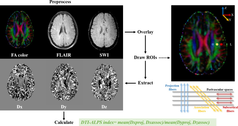
The activity of glymphatic system was evaluated using the DTI-ALPS index.5 This index calculated from diffusivities along perivenous spaces of deep medullary veins on the axial slices at the level of lateral ventricle bodies. At this level, projection fibres (ie, corticofugal corona radiata) ran in the craniocaudal direction (ie, z-axis in the coordinate) adjacent to the lateral ventricle, while association fibres (ie, superior longitudinal fascicle), projected in the anterior-posterior direction (ie, y-axis in the coordinate) and located lateral to the projection fibres. Subcortical fibres ran in x-axis in the coordinate. Since PVSs were mainly perpendicular to both projection and association fibres, diffusivities along PVSs may have been contributed by the significant differences between diffusivity on the x-axis in both fibres (Dxproj and Dxasso for x-axis diffusivity in the projection and association fibres, respectively) and diffusivity which was perpendicular to both the x-axis and major direction of the fibre tracts (Dyproj for the y-axis diffusivity in projection fibres and Dzasso for the z-axis diffusivity in association fibres). To quantify the function of glymphatic transport, the DTI-ALPS index was calculated as follows: DTI-ALPS index=mean (Dxproj, Dxasso)/mean (Dyproj, Dzasoc).5 A low DTI-ALPS index indicated impaired efficiency of glymphatic pathway.
The protocol for determining the DTI-ALPS index is shown in figure 1. Briefly, DTI preprocessing was conducted using FMRIB’s Diffusion Toolbox of the FMRIB Software Library V.6.0 (FSL, https://www.fmrib.ox.ac.uk/fsl/). The command of ‘eddy_correct’ in the FSL was first used to compensate for eddy current-induced and movement-related distortions.21 Diffusion tensor maps and fractional anisotropy (FA) maps were then calculated using the ‘dtifit’ command in the FSL.22 A mask with FA >0.2 was used to avoid the inclusion of CSF voxel.23 Next, FA colour map, FLAIR and SWI images were overlapped with FA map as reference through rigid transformation, minimising the normalised mutual information as cost function using FLIRT function in the FSL.24 Three spherical regions of interest (ROIs) with a diameter of 3 mm were manually drawn by an experienced neurologist (Y Tian) based on the locations of the association (yellow in figure 1), projection (blue in figure 1) and subcortical (red in figure 1) fibres. A recent report indicated that the shape and size of ROIs did not affect the calculation of DTI-ALPS index.25 To ensure that the superior longitudinal fasciculus was thick enough to perfectly maintain perpendicularity with PVSs, ROIs were only placed on the left hemisphere.5 Additionally, FLAIR and SWI images were used to avoid ROIs placed over visible WMH and CMBs lesions.26 Once the ROIs were drawn, diffusivities along the x-axis, y-axis and z-axis were extracted on the diffusion tensor maps for each ROI.5 Finally, the DTI-ALPS index was calculated based on the formula mentioned above.5 To estimate the consistency and reproducibility of the DTI-ALPS index, we randomly selected 100 participants from the list of all enrolled participants. The same neurologist (Y Tian) placed ROIs for the same participants 1 month after the first ROI drawing for estimating intrarater reliability. The intraclass correlation coefficient was 0.88.18
Figure 1.
The assessment of glymphatic system by calculating the DTI-ALPS index. DTI-ALPS, diffusivity along the perivascular spaces based on diffusion tensor imaging; FA, fractional anisotropy; FLAIR, fluid-attenuated inversion recovery; ROI, regions of interest; SWI, susceptibility-weighted imaging.
Statistical analysis
The clinical characteristics were compared among participants with different glymphatic function according to the quartiles of the DTI-ALPS index (quartile (Q)1: <1.57; Q2: 1.57–1.65; Q3: 1.66–1.75; Q4: ≥1.76), and individuals with and without CSVD. Continuous variables are presented as means with SD or medians with IQR and analysed by t-test, Wilcoxon rank-sum statistic, one-way analysis of variance or Kruskal-Wallis test as appropriate. Categorical variables are presented as frequencies with percentages and analysed using χ2 test.
The DTI-ALPS index was assessed for the associations with the presence, severity and different neuroimaging phenotypes of CSVD as a continuous independent variable (per 1-SD decrement) and an ordinal categorical variable according to the quartiles. Binary logistic regression analyses were used to determine the relationships of the DTI-ALPS index with presence and modified presence of CSVD, lacunes, CMBs, WMH burden, EPVS and BA. ORs with 95% CIs were calculated. Ordinal logistic regression analyses were used to identify the relationships of the DTI-ALPS index with total CSVD burden, modified total CSVD burden, modified WMH burden and CMBs burden. Common ORs (cORs) with 95% CIs were calculated. To eliminate the potential bias produced by confounders, three multivariable logistic regression models were performed for each outcome: model 1 was adjusted for age and sex; model 2, vascular risk factors; model 3, current medications.
Moreover, to evaluate the pattern and magnitude of the associations of the DTI-ALPS index with presence and severity of CSVD, binary or ordinal logistic regression models with restricted cubic splines for the DTI-ALPS index (continuous measure) were conducted after adjusting for all potential covariates (model 3). Given the logistic regression results in which the DTI-ALPS index were treated as an ordinal independent variable according to the quartiles, the 75th percentile of DTI-ALPS index was defined as the reference point, and the five knots for the splines were placed at the 5th, 25th, 50th, 75th and 95th percentiles of DTI-ALPS index.27 28
Furthermore, to estimate the predictive power of glymphatic activity for the presence of CSVD, the net reclassification index (NRI) and integrated discrimination improvement (IDI) indices were used to quantify the improvement in predictive performance after addition of the DTI-ALPS index to three basic models based on conventional vascular risk factors.
Additionally, to illustrate the potential implication of glymphatic pathway on clinical deficit (namely cognitive function) in individuals with CSVD, general linear regression analyses were applied to measure the association between the DTI-ALPS index and MoCA scores. Beta coefficient (β) with 95% CI was calculated. Education was adjusted on the basis of the above three multivariable models, and total CSVD burden was added in model 4.
The statistical significance of the result was considered as the two-sided p value of the data analysis <0.05. All statistical analyses were performed using SAS V.9.4 (SAS Institute, Cary, North Carolina, USA).
Results
Baseline characteristics
Among 3067 individuals, 691 participants with unavailable or unqualified DTI data, 156 participants with the DTI-ALPS index processing error and 1 participant without interpretable structural MRI were excluded. We finally enrolled a total of 2219 residents in this study (figure 2). The clinical characteristics of included and excluded individuals are shown in online supplemental table S2. The mean age of all enrolled individuals was 61.3 (SD 6.6) years, and 1019 (45.9%) were men. The mean DTI-ALPS index was 1.67 (SD 0.14). The clinical characteristics of 2219 adults according to the quartiles of the DTI-ALPS index and the presence and modified presence of CSVD are presented in table 1 and online supplemental table S3, respectively.
Figure 2.
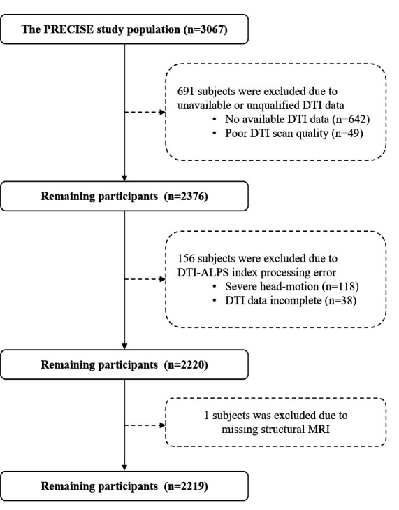
The flow chart of 2219 included individuals. DTI, diffusion tensor imaging; DTI-ALPS, diffusivity along perivascular spaces based on diffusion tensor imaging; PRECISE, PolyvasculaR Evaluation for Cognitive Impairment and vaScular Events.
Table 1.
Clinical characteristics among 2219 individuals according to the quartiles of the DTI-ALPS index
| Characteristic | Quartile 1 (≤1.57) (n=568) |
Quartile 2 (1.57–1.66) (n=534) |
Quartile 3 (1.67–1.76) (n=572) |
Quartile 4 (>1.76) (n=545) |
P value |
| Sociodemographics | |||||
| Age (years), mean±SD | 63.5±6.9 | 61.7±6.6 | 60.7±6.2 | 59.4±5.9 | <0.001 |
| Sex (male), n (%) | 329 (57.9) | 251 (47.0) | 237 (41.4) | 202 (37.1) | <0.001 |
| Education, n (%) | 0.01 | ||||
| Illiteracy | 104 (18.3) | 78 (14.6) | 87 (15.2) | 80 (14.7) | |
| Elementary school | 166 (29.2) | 132 (24.7) | 134 (23.4) | 116 (21.3) | |
| Middle school | 145 (25.5) | 166 (31.1) | 167 (29.2) | 184 (33.8) | |
| High school | 103 (18.1) | 116 (21.7) | 145 (25.4) | 120 (22.0) | |
| University or above | 50 (8.8) | 42 (7.9) | 39 (6.8) | 45 (8.3) | |
| Vascular risk factors | |||||
| Body mass index (kg/m2), mean±SD | 24.1±3.1 | 23.8±2.8 | 23.9±3.1 | 24.1±3.0 | 0.37 |
| Systolic blood pressure (mm Hg), median (IQR) | 131.5 (121.0–143.0) | 128.0 (117.5–139.0) | 128.5 (118.5–138.5) | 128.0 (116.0–139.5) | <0.001 |
| Diastolic blood pressure (mm Hg), median (IQR) | 76.0 (70.0–82.0) | 74.8 (69.0–80.5) | 75.0 (69.0–80.5) | 74.5 (69.0–81.0) | 0.03 |
| Hypertension, n (%) | 293 (51.6) | 223 (41.8) | 237 (41.4) | 223 (40.9) | <0.001 |
| Diabetes mellitus, n (%) | 156 (27.5) | 117 (21.9) | 131 (22.9) | 101 (18.5) | 0.005 |
| Hypercholesterolaemia, n (%) | 109 (19.2) | 113 (21.2) | 150 (26.2) | 122 (22.4) | 0.03 |
| Stroke/Transient ischaemic attack, n (%) | 28 (4.9) | 19 (3.6) | 10 (1.8) | 10 (1.8) | 0.004 |
| Coronary artery disease, n (%) | 0 (0.0) | 2 (0.4) | 5 (0.9) | 3 (0.6) | 0.17 |
| Atrial fibrillation, n (%) | 4 (0.7) | 7 (1.3) | 3 (0.5) | 3 (0.6) | 0.41 |
| Current drinking, n (%) | 129 (22.7) | 101 (18.9) | 100 (17.5) | 67 (12.3) | <0.001 |
| Current smoking, n (%) | 142 (25.0) | 109 (20.4) | 97 (17.0) | 88 (16.2) | <0.001 |
| Laboratory data, median (IQR) | |||||
| Total cholesterol (mmol/L) | 5.3±1.0 | 5.4±1.0 | 5.4±1.0 | 5.4±1.0 | 0.28 |
| Triglycerides (mmol/L) | 1.8±1.2 | 1.8±1.2 | 1.9±1.5 | 1.8±1.2 | 0.18 |
| High-density lipoprotein (mmol/L) | 1.4±0.3 | 1.4±0.4 | 1.4±0.3 | 1.4±0.3 | 0.22 |
| Low-density lipoprotein (mmol/L) | 2.8±0.8 | 2.8±0.8 | 2.8±0.8 | 2.9±0.8 | 0.25 |
| Fasting blood glucose (mmol/L) | 6.2±1.8 | 6.0±1.5 | 6.0±1.4 | 6.0±1.5 | 0.21 |
| Glycosylated haemoglobin (%) | 6.1±1.0 | 6.0±0.9 | 6.0±0.9 | 5.9±0.9 | 0.005 |
| Homocysteine (mmol/L) | 13.2±7.3 | 12.2±6.5 | 11.0±4.2 | 11.3±5.7 | <0.001 |
| Medication, n (%) | |||||
| Antihypertensive | 175 (30.8) | 148 (27.7) | 151 (26.4) | 120 (22.0) | 0.01 |
| Lipid-lowering | 23 (4.1) | 28 (5.2) | 29 (5.1) | 21 (3.9) | 0.6 |
| Antiplatelet | 23 (4.1) | 15 (2.8) | 17 (3.0) | 13 (2.4) | 0.42 |
| Anticoagulant | 2 (0.4) | 0 (0.0) | 1 (0.2) | 0 (0.0) | 0.32 |
| Antidiabetic | 78 (13.7) | 47 (8.8) | 50 (8.7) | 45 (8.3) | 0.006 |
| MoCA adjusted by education, median (IQR) | 21.0 (17.0–25.0) | 22.0 (18.0–25.0) | 23.0 (19.0–25.0) | 23.0 (19.0–25.0) | <0.001 |
CSVD, cerebral small vessel disease; DTI-ALPS, diffusivity along perivascular spaces based on diffusion tensor imaging; MoCA, Montreal Cognitive Assessment.
The DTI-ALPS index in association with the presence and severity of CSVD
As shown in online supplemental figure S1, the prevalence of present CSVD from the first to fourth quartile of the DTI-ALPS index was 43.8%, 30.3%, 26.0% and 21.8%, respectively. Similarly, modified presence of CSVD decreased substantially from the first to fourth quartile of the DTI-ALPS index (57.0%, 40.3%, 35.3%, 33.9%).
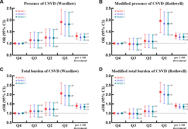
The glymphatic failure as reflected by low DTI-ALPS index was strongly associated with the presence and severity of CSVD (figure 3 and online supplemental table S4). After controlling for all potential confounders in model 3, participants in the lowest quartile of the DTI-ALPS index manifested an increased risk of the presence of CSVD (OR 1.77, 95% CI 1.33 to 2.35, p<0.001), modified presence of CSVD (OR 1.80, 95% CI 1.38 to 2.34, p<0.001), total burden of CSVD (cOR 1.89, 95% CI 1.43 to 2.49, p<0.001) and modified total burden of CSVD (cOR 1.95, 95% CI 1.51 to 2.50, p<0.001) compared with those in the highest quartile of the DTI-ALPS index. Similar results were exhibited when analysed in the continuous models (per 1-SD decrement of the DTI-ALPS index).
Figure 3.
The DTI-ALPS index in association with the presence and severity of CSVD. Forest plots present ORs/cORs for the associations of the DTI-ALPS index with the presence of CSVD (A), modified presence of CSVD (B), total CSVD burden (C) and modified total CSVD burden (D). The colourful lines indicate 95% CIs of ORs/cORs. Model 1 adjusted for age and sex; model 2, vascular risk factors; model 3, current medications. cOR, common OR; CSVD, cerebral small vessel disease; DTI-ALPS, diffusivity along the perivascular spaces based on diffusion tensor imaging.
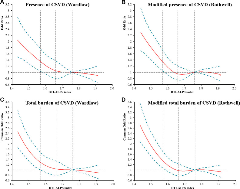
Using multivariable logistic regression models with restricted cubic splines, L-shaped associations of the DTI-ALPS index with the presence and severity of CSVD were found (figure 4).
Figure 4.
Restricted cubic spline plots for the associations of the DTI-ALPS index with the presence and severity of CSVD. Adjusted ORs/cOR for presence of CSVD (A), modified presence of CSVD (B), total CSVD burden (C) and modified total CSVD burden (D) based on restricted cubic spines with 5 knots at 5th, 25th, 50th, 75th and 95th percentiles of the DTI-ALPS index. The solid line indicates adjusted ORs/cORs, and the dashed lines the 95% CI bands. Reference is the first quartile of ALPS index. The vertical dashed lines indicate the 25th, 50th and 75th percentiles of DTI-ALPS index. Data were fitted using a binary/ordinal logistic regression model of restricted cubic spline with five knots (the 5th, 25th, 50th, 75th, 95th percentiles) for the DTI-ALPS index after adjusting for all potential confounders. The lowest 5% and highest 5% of participants are not shown. cOR, common OR; CSVD, cerebral small vessel disease; DTI-ALPS, diffusivity along the perivascular spaces based on diffusion tensor imaging.
The DTI-ALPS index in association with neuroimaging phenotypes of CSVD
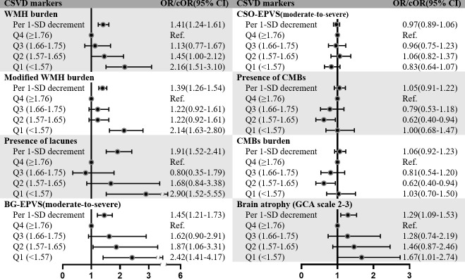
The relationships of the DTI-ALPS index with individual neuroimaging markers of CSVD are presented in figure 5 and online supplemental table S5. Individuals in the first quartile of the DTI-ALPS index were at increased risks of WMH burden (OR 2.16, 95% CI 1.51 to 3.10, p<0.001), modified WMH burden (cOR 2.14, 95% CI 1.63 to 2.80, p<0.001), lacunes (OR 2.90, 95% CI 1.52 to 5.55, p=0.001), moderate-to severe BG-EPVS (OR 2.42, 95% CI 1.41 to 4.17, p=0.001) and BA (OR 1.67, 95% CI 1.01 to 2.74, p=0.04) after adjusting all potential confounders in model 3, compared with those in the fourth quartile. Multivariable logistic regression models revealed no any statistical significances for the relationships of the DTI-ALPS index with the presence of CMBs, CMBs burden or moderate-to-severe CSO-EPVS. Logistic regression analyses according to per 1-SD decrement of the DTI-ALPS index indicated similar links.
Figure 5.
The DTI-ALPS index in association with neuroimaging phenotypes of CSVD. Forest plots present ORs/cORs for the associations of the DTI-ALPS index with different neuroimaging marker of CSVD. The black lines indicate 95% CIs of ORs/cORs. Multivariable logistic regression model adjusted for all potential confounders. BG-EPVS, basal ganglia-EPVS; CSO-EPVS, centrum semiovale-EPVS; CMBs, cerebral microbleeds; cOR, common OR; CSVD, cerebral small vessel disease; DTI-ALPS, diffusivity along the perivascular spaces based on diffusion tensor imaging; EPVS, enlarged perivascular space; GCA, global cortical atrophy scale; WMH, whiter matter hyperintensity.
Predictive value of the DTI-ALPS index for CSVD
We further estimated the incremental value of adding the DTI-ALPS index beyond several traditional vascular risk factors for the prediction of the presence of CSVD (table 2). The addition of the DTI-ALPS index to basic models observably increased the predictive property as verified by NRI and IDI indices (all p<0.001).
Table 2.
Predictive value of the DTI-ALPS index for CSVD
| NRI | IDI | |||
| Estimate (95% CI), % | P value | Estimate (95% CI), % | P value | |
| Presence of CSVD | ||||
| Basic model 1+ALPS index | 21.95 (12.99 to 30.90) | <0.001 | 1.23 (0.76 to 1.70) | <0.001 |
| Basic model 2+ALPS index | 20.45 (11.49 to 29.41) | <0.001 | 0.95 (0.54 to 13.60) | <0.001 |
| Basic model 3+ALPS index | 20.65 (11.69 to 29.61) | <0.001 | 0.91 (0.51 to 1.32) | <0.001 |
| Modified presence of CSVD | ||||
| Basic model 1+ALPS index | 22.81 (14.43 to 31.18) | <0.001 | 1.38 (0.89 to 1.87) | <0.001 |
| Basic model 2+ALPS index | 21.85 (13.47 to 30.23) | <0.001 | 1.03 (0.6 to 1.45) | <0.001 |
| Basic model 3+ALPS index | 21.66 (13.29 to 30.04) | <0.001 | 0.97 (0.56 to 1.39) | <0.001 |
Basic model 1: including age and sex.
Basic model 2: including age, sex, body mass index, stroke/transient ischaemic attack, hypertension, diabetes, hypercholesterolaemia, coronary artery disease, atrial fibrillation, current drinker and current smoker.
Basic model 3: including age, sex, body mass index, stroke/transient ischaemic attack, hypertension, diabetes, hypercholesterolaemia, coronary artery disease, atrial fibrillation, current drinker, current smoker, antihypertensive medication, lipid-lowering medication, antiplatelet medication, anticoagulant medication and antidiabetic medication.
CSVD, cerebral small vessel disease; DTI-ALPS, diffusivity along the perivascular spaces based on diffusion tensor imaging; IDI, integrated discrimination improvement; NRI, net reclassification index.
The DTI-ALPS index in association with cognitive function in CSVD
Multivariable linear regression analyses showed the DTI-ALPS index was strongly associated with MoCA scores in individuals with CSVD after adjusting for potential confounders (all p<0.001, online supplemental table S6).
Discussion
In the current study, we analysed the associations of glymphatic pathway with CSVD and its neuroimaging markers using the DTI-ALPS approach, thereby providing a comprehensive perspective of the vital role of glymphatic transport in pathogenesis of CSVD. We showed that impaired glymphatic transport as denoted by low DTI-ALPS index was forcefully related to the presence and severity of CSVD. Moreover, our results indicated that the DTI-ALPS index had substantial relationships with WMH, lacunes, BG-EPVS and BA. Furthermore, the addition of glymphatic activity to basic models driven by several vascular risk factors exhibited better predictive performances for CSVD. In addition, glymphatic pathway was suggestively related to cognitive deficit in participants with CSVD.
Previous reports mostly focused on the implications of CSVD and its markers on the activity of glymphatic system, particularly in patients with CSVD or cerebral amyloid angiopathy (CAA). Similar to our results, Xu et al discovered that patients with CAA demonstrated a lower DTI-ALPS index than controls, and the DTI-ALPS index was negatively related to the severity of CSVD in CAA.9 Consistent with our findings, Zhang et al reported the links between low DTI-ALPS index and WMH, lacunes, BG-EPVS, CMBs and total CSVD burden in patients with MRI-visible CSVD.10 However, the effects of glymphatic transport on CSVD in the general ageing population were insufficiently proven. Our study provided strong evidence about the associations of glymphatic failure with the presence, severity, individual markers of CSVD in a relatively large-scale population of community-dwelling residents. More importantly, L-shaped associations between the DTI-ALPS index and the presence and severity of CSVD were first detected in the present study. It was worth mentioning that, in the current research, the level of the DTI-ALPS was higher than previous studies.9 10 Since the prevalence of the presence and modified presence of CSVD in our study was 30.6% and 41.7%, respectively, different study populations may result in this difference. And variations of sample sizes may be one of possible explanations for the discrepancy. Subtle differences in DTI parameters and the process of calculating the DTI-ALPS index may lead to fluctuations of the DTI-ALPS index as well.
Previous researches also elucidated the contribution of glymphatic deficit on cognitive failure in patients with CSVD.9 10 Tang et al found that the DTI-ALPS index was independently correlated with cognitive impairment assessed by a comprehensive neuropsychological test battery among 133 patients with CSVD.29 A recent advance demonstrated the mediating effect of the DTI-ALPS on the association between WMH and episodic memory in patients with CSVD, implying the implication of glymphatic system on CSVD-related cognitive decline.30 Our results further confirmed the link between the DTI-ALPS index and global cognitive performance estimated by MoCA scores in individuals with sporadic CSVD from a large community-based population. The influence of glymphatic impairment on CSVD-related cognitive decline was strongly supported by kinds of preclinical studies. Compared with Wistar-Kyoto rats, ageing spontaneously hypertensive stroke prone rats (an experimental model for simulating pathogenesis of atherosclerotic CSVD in human) exhibited an obvious decline in CSF and solute transport and a moderately impaired glymphatic system.31 32 Bilateral common carotid artery stenosis in rats, which was thought of as a model of CSVD due to preferentially affect white matter integrity,33 caused a marked regional reduction in glymphatic transport.34
In this study, the addition of the DTI-ALPS index to traditional models based on vascular risk factors exhibited better predictive abilities for the presence of CSVD in all eligible residents. Although it may be premature to routinely examine the efficiency of glymphatic system in middle-aged and elderly adults or patients with silent CSVD, to some extent, our findings implied potential predictive value of impaired glymphatic activity for neuroimaging changes of CSVD. Further studies are needed to validate the predictive performance of quantifying glymphatic transport for the development of neuroimaging burden and the occurrence of clinical deficits in longitudinal cohorts.
To explain the association between glymphatic failure and CSVD, we proposed several possible mechanisms. First, a number of vascular risk factors, such as ageing,6 35 hypertension36 37 and diabetes,38 39 were contributed to declined glymphatic activity. On account of these conditions referred to CSVD,40 it supported the potential role of glymphatic dysfunction during the process of CSVD. Second, EPVS, especially located in the BG, exerted a crucial role in arteriosclerotic CSVD and vascular cognitive impairment.41 Since the PVSs constituted essential channels of glymphatic inflow and outflow, the hypothesis that glymphatic transport was involved in the pathogenesis of CSVD was plausible. Although larger PVSs seemed to represent low resistance for easier transport of CSF and solute, preclinical evidence showed concomitant enlargement of PVSs and decreased glymphatic pathway in spontaneously hypertensive rats.42 Third, many pathogenic mechanisms of CSVD, including neurovascular unit dysfunction, blood-brain barrier (BBB) damage and haemodynamic dysfunction, resulted in interstitial solute deposition in the brain parenchyma and impaired glymphatic system.43 The excessive perivascular accumulation of cell debris and metabolic wastes due to impaired glymphatic clearance in turn aggravated decreased cerebrovascular reactivity, BBB disruption and enhanced perivascular inflammation. Ultimately, a vicious circle was produced between glymphatic dysfunction and aforementioned mechanisms that may exacerbate the CSVD pathology. Finally, cognitive impairment was the main physiological consequence of glymphatic failure. CSVD contributed to almost 45% of dementia and coexisted with neurodegeneration.44 A better understanding of the crucial role of glymphatic transport may shed light on the association between CSVD and dementia.
Our findings portended glymphatic pathway may be a novel predictive factor and therapeutic target of CSVD, exerting a far-reaching implication in clinical practice. The enhancement of glymphatic system via modifying of physiological states, such as improving sleep quality and enhancing blood vessel pulsation, may be an effective way to delay CSVD progression and cognitive decline in the general ageing population. Given the consequences of glymphatic failure, the possibility of reversing glymphatic function is vital in the days to come.
The advantages of this report came from a relatively large sample size of the DTI-ALPS index measurement, comprehensive neuroimaging assessment of CSVD and the community-based population reasonably represented the general ageing population.11 Apparently, many limitations should be taken in to account when interpreting our results. First, the DTI-ALPS index only reflected local water dynamics of the perivenous spaces around deep medullary veins in regional brain area adjacent to the lateral ventricles, so it cannot estimate whole-brain glymphatic function. And the relationship of the DTI-ALPS index with glymphatic function in human was not and strictly substantiated by pathophysiological studies. The level of the DTI-ALPS index was also impacted by CSVD-related damage of white matter microstructures. Thus, the implication of the DTI-ALPS index needs more clearly interpretation and further research. In fact, a recent advance indicated that the DTI-ALPS index was significantly related to the classical detected efficiency of glymphatic transport calculated on contrast-enhanced MRI after intrathecal injection of gadodiamide.10 Therefore, this non-invasive and time-saving approach was recommended as a proxy of glymphatic transport which could widely applied in clinical practice. Second, although sleep quality and carotid artery stenosis exerted crucial implications on both glymphatic system and CSVD, sleep profiles, such as sleep questionnaires and polysomnography, were not collected in the PRECISE study and a few patients with carotid artery stenosis were not excluded in this study. Third, our findings were restricted to the baseline survey of the PRECISE cohort. Thus, any causality for the progression of glymphatic dysfunction and CSVD cannot be concluded in this cross-sectional study. Longitudinal research would help to establish a causal direction, and follow-up data of the PRECISE study may tell us more information in the future. Furthermore, this study mainly focused on MRI-visible phenotypes of CSVD. Hence, the complex interactions between glymphatic system and mechanisms of CSVD and the unique contribution and predictive ability of glymphatic pathway to clinical symptoms of CSVD (such as cognitive, gait, sleep, mood disorders) deserve to be further unravel. Besides, the consistencies of the DTI-ALPS index and neuroimaging characteristics of CSVD were relatively low in this study, future studies should employ automatic or semi-automatic ways to measure glymphatic function and CSVD characteristics. Additionally, despite general representativeness, there was an unavoidable sample selection bias in this research because the PRECISE population was mainly enrolled from rural communities, outside or far from the large urban centres. Moreover, the PRECISE study only enrolled Chinese population. Therefore, these findings should be established in non-Asian populations as well. Lastly, predictive models were detected in all included individuals and did not internally validated, as well as more external validation studies are necessary to estimate the predictive power.
Conclusion
In conclusion, this study indicated that glymphatic impairment as assessed by low DTI-ALPS index was associated with the presence, severity, particular neuroimaging phenotypes of CSVD in a community-based population. Therefore, these findings implied that glymphatic failure may contribute to neuroimaging changes of CSVD in the general ageing population. More investigations are required to explore dynamic interplays between glymphatic system and physiopathological mechanisms in the CSVD process, as well as to understand potential role of glymphatic disorder in clinical symptoms of CSVD.
Acknowledgments
We thank all participants and researchers of the PRECISE study. We thank Professor Maria A Rocca and groups, particularly for Professor Maria’s guidance and advice in the calculation of the DTI-ALPS index. We thank M Zhou, Y Chen, J Pi, and M Zhao for their help in assessment of CSVD.
Footnotes
Twitter: @yilong
YT and XC contributed equally.
Contributors: YP and YLW designed the study. YJW was responsible for the overall content as the guarantor. YT, XC, YP and YLW analysed the data and contributed to reviewing the statistical problems. YT and XC interpreted the data and drafted the manuscript. YP and YLW reviewed the manuscript. All authors approved the final version of the manuscript.
Funding: This study was funded by The National Natural Science Foundation of China (No. 81825007), National Key R&D Programme of China (No. 2016YFC0901002), Beijing Outstanding Young Scientist Programme (No. BJJWZYJH01201910025030), Key Science & Technologies R&D Program of Lishui City (No. 2019ZDYF18) and Zhejiang provincial programme for the Cultivation of High-level Innovative Health talents and grants from AstraZeneca Investment (China) Co., Ltd.
Competing interests: None declared.
Provenance and peer review: Not commissioned; externally peer reviewed.
Supplemental material: This content has been supplied by the author(s). It has not been vetted by BMJ Publishing Group Limited (BMJ) and may not have been peer-reviewed. Any opinions or recommendations discussed are solely those of the author(s) and are not endorsed by BMJ. BMJ disclaims all liability and responsibility arising from any reliance placed on the content. Where the content includes any translated material, BMJ does not warrant the accuracy and reliability of the translations (including but not limited to local regulations, clinical guidelines, terminology, drug names and drug dosages), and is not responsible for any error and/or omissions arising from translation and adaptation or otherwise.
Data availability statement
Data are available on reasonable request. All data relevant to the study are included in the article or uploaded as supplementary information. All data generated or analysed during this study are included in this published article and available on reasonable requests.
Ethics statements
Patient consent for publication
Consent obtained from parent(s)/guardian(s).
Ethics approval
Ethical approval was received from the ethics committees of Beijing Tiantan Hospital (institutional review board (IRB) approval number: KY2017-010-01) and Lishui Hospital (IRB approval number: 2016-42). Participants gave informed consent to participate in the study before taking part.
References
- 1. Shi Y, Wardlaw JM. Update on cerebral small vessel disease: a dynamic whole-brain disease. Stroke Vasc Neurol 2016;1:83–92. 10.1136/svn-2016-000035 [DOI] [PMC free article] [PubMed] [Google Scholar]
- 2. Wardlaw JM, Smith C, Dichgans M. Small vessel disease: mechanisms and clinical implications. Lancet Neurol 2019;18:684–96.:S1474-4422(19)30079-1. 10.1016/S1474-4422(19)30079-1 [DOI] [PubMed] [Google Scholar]
- 3. Iliff JJ, Wang M, Liao Y, et al. A paravascular pathway facilitates CSF flow through the brain parenchyma and the clearance of interstitial solutes, including amyloid β. Sci Transl Med 2012;4:147ra111. 10.1126/scitranslmed.3003748 [DOI] [PMC free article] [PubMed] [Google Scholar]
- 4. Jiang Q. Mri and glymphatic system. Stroke Vasc Neurol 2019;4:75–7. 10.1136/svn-2018-000197 [DOI] [PMC free article] [PubMed] [Google Scholar]
- 5. Taoka T, Masutani Y, Kawai H, et al. Evaluation of glymphatic system activity with the diffusion Mr technique: diffusion tensor image analysis along the perivascular space (DTI-ALPS) in Alzheimer’s disease cases. Jpn J Radiol 2017;35:172–8. 10.1007/s11604-017-0617-z [DOI] [PubMed] [Google Scholar]
- 6. Zhang Y, Zhang R, Ye Y, et al. The influence of demographics and vascular risk factors on glymphatic function measured by diffusion along perivascular space. Front Aging Neurosci 2021;13:693787. 10.3389/fnagi.2021.693787 [DOI] [PMC free article] [PubMed] [Google Scholar]
- 7. Chen H-L, Chen P-C, Lu C-H, et al. Associations among cognitive functions, plasma DNA, and diffusion tensor image along the perivascular space (DTI-ALPS) in patients with parkinson’s disease. Oxid Med Cell Longev 2021;2021:4034509. 10.1155/2021/4034509 [DOI] [PMC free article] [PubMed] [Google Scholar]
- 8. Toh CH, Siow TY. Glymphatic dysfunction in patients with ischemic stroke. Front Aging Neurosci 2021;13:756249. 10.3389/fnagi.2021.756249 [DOI] [PMC free article] [PubMed] [Google Scholar]
- 9. Xu J, Su Y, Fu J, et al. Glymphatic dysfunction correlates with severity of small vessel disease and cognitive impairment in cerebral amyloid angiopathy. Eur J Neurol 2022;29:2895–904. 10.1111/ene.15450 [DOI] [PubMed] [Google Scholar]
- 10. Zhang W, Zhou Y, Wang J, et al. Glymphatic clearance function in patients with cerebral small vessel disease. Neuroimage 2021;238:S1053-8119(21)00534-6. 10.1016/j.neuroimage.2021.118257 [DOI] [PubMed] [Google Scholar]
- 11. Pan Y, Jing J, Cai X, et al. PolyvasculaR evaluation for cognitive impairment and vascular events (precise) -A population-based prospective cohort study: rationale, design and baseline participant characteristics. Stroke Vasc Neurol 2021;6:145–51. 10.1136/svn-2020-000411 [DOI] [PMC free article] [PubMed] [Google Scholar]
- 12. von Elm E, Altman DG, Egger M, et al. Strengthening the reporting of observational studies in epidemiology (STROBE) statement: guidelines for reporting observational studies. BMJ 2007;335:806–8. 10.1136/bmj.39335.541782.AD [DOI] [PMC free article] [PubMed] [Google Scholar]
- 13. Yu J, Li J, Huang X. The Beijing version of the Montreal cognitive assessment as a brief screening tool for mild cognitive impairment: a community-based study. BMC Psychiatry 2012;12:156. 10.1186/1471-244X-12-156 [DOI] [PMC free article] [PubMed] [Google Scholar]
- 14. Wardlaw JM, Smith EE, Biessels GJ, et al. Neuroimaging standards for research into small vessel disease and its contribution to ageing and neurodegeneration. Lancet Neurol 2013;12:822–38.:S1474-4422(13)70124-8. 10.1016/S1474-4422(13)70124-8 [DOI] [PMC free article] [PubMed] [Google Scholar]
- 15. Fazekas F, Chawluk JB, Alavi A, et al. Mr signal abnormalities at 1.5 T in Alzheimer’s dementia and normal aging. AJR Am J Roentgenol 1987;149:351–6. 10.2214/ajr.149.2.351 [DOI] [PubMed] [Google Scholar]
- 16. Doubal FN, MacLullich AMJ, Ferguson KJ, et al. Enlarged perivascular spaces on MRI are a feature of cerebral small vessel disease. Stroke 2010;41:450–4. 10.1161/STROKEAHA.109.564914 [DOI] [PubMed] [Google Scholar]
- 17. Pasquier F, Leys D, Weerts JG, et al. Inter- and intraobserver reproducibility of cerebral atrophy assessment on MRI scans with hemispheric infarcts. Eur Neurol 1996;36:268–72. 10.1159/000117270 [DOI] [PubMed] [Google Scholar]
- 18. Landis JR, Koch GG. The measurement of observer agreement for categorical data. Biometrics 1977;33:159–74. [PubMed] [Google Scholar]
- 19. Staals J, Makin SDJ, Doubal FN, et al. Stroke subtype, vascular risk factors, and total MRI brain small-vessel disease burden. Neurology 2014;83:1228–34. 10.1212/WNL.0000000000000837 [DOI] [PMC free article] [PubMed] [Google Scholar]
- 20. Lau KK, Li L, Schulz U, et al. Total small vessel disease score and risk of recurrent stroke: validation in 2 large cohorts. Neurology 2017;88:2260–7. 10.1212/WNL.0000000000004042 [DOI] [PMC free article] [PubMed] [Google Scholar]
- 21. Andersson JLR, Sotiropoulos SN. An integrated approach to correction for off-resonance effects and subject movement in diffusion MR imaging. Neuroimage 2016;125:1063–78.:S1053-8119(15)00920-9. 10.1016/j.neuroimage.2015.10.019 [DOI] [PMC free article] [PubMed] [Google Scholar]
- 22. Basser PJ, Mattiello J, LeBihan D. Mr diffusion tensor spectroscopy and imaging. Biophys J 1994;66:259–67. 10.1016/S0006-3495(94)80775-1 [DOI] [PMC free article] [PubMed] [Google Scholar]
- 23. Siow TY, Toh CH, Hsu JL, et al. Association of sleep, neuropsychological performance, and gray matter volume with glymphatic function in community-dwelling older adults. Neurology 2022;98:e829–38. 10.1212/WNL.0000000000013215 [DOI] [PubMed] [Google Scholar]
- 24. Jenkinson M, Smith S. A global optimisation method for robust affine registration of brain images. Med Image Anal 2001;5:143–56. 10.1016/s1361-8415(01)00036-6 [DOI] [PubMed] [Google Scholar]
- 25. Taoka T, Ito R, Nakamichi R, et al. Reproducibility of diffusion tensor image analysis along the perivascular space (DTI-ALPS) for evaluating interstitial fluid diffusivity and glymphatic function: changes in Alps index on multiple condition acquisition experiment (CHAMONIX) study. Jpn J Radiol 2022;40:147–58. 10.1007/s11604-021-01187-5 [DOI] [PMC free article] [PubMed] [Google Scholar]
- 26. Carotenuto A, Cacciaguerra L, Pagani E, et al. Glymphatic system impairment in multiple sclerosis: relation with brain damage and disability. Brain 2022;145:2785–95. 10.1093/brain/awab454 [DOI] [PubMed] [Google Scholar]
- 27. Durrleman S, Simon R. Flexible regression models with cubic splines. Stat Med 1989;8:551–61. 10.1002/sim.4780080504 [DOI] [PubMed] [Google Scholar]
- 28. Pan Y, Jing J, Chen W, et al. Post-glucose load measures of insulin resistance and prognosis of nondiabetic patients with ischemic stroke. J Am Heart Assoc 2017;6:e004990. 10.1161/JAHA.116.004990 [DOI] [PMC free article] [PubMed] [Google Scholar]
- 29. Tang J, Zhang M, Liu N, et al. The association between glymphatic system dysfunction and cognitive impairment in cerebral small vessel disease. Front Aging Neurosci 2022;14:916633. 10.3389/fnagi.2022.916633 [DOI] [PMC free article] [PubMed] [Google Scholar]
- 30. Ke Z, Mo Y, Li J, et al. Glymphatic dysfunction mediates the influence of white matter hyperintensities on episodic memory in cerebral small vessel disease. Brain Sci 2022;12:1611. 10.3390/brainsci12121611 [DOI] [PMC free article] [PubMed] [Google Scholar]
- 31. Hannawi Y, Caceres E, Ewees MG, et al. Characterizing the neuroimaging and histopathological correlates of cerebral small vessel disease in spontaneously hypertensive stroke-prone rats. Front Neurol 2021;12:740298. 10.3389/fneur.2021.740298 [DOI] [PMC free article] [PubMed] [Google Scholar]
- 32. Koundal S, Elkin R, Nadeem S, et al. Optimal mass transport with lagrangian workflow reveals advective and diffusion driven solute transport in the glymphatic system. Sci Rep 2020;10:1990. 10.1038/s41598-020-59045-9 [DOI] [PMC free article] [PubMed] [Google Scholar]
- 33. Roberts JM, Maniskas ME, Bix GJ. Bilateral carotid artery stenosis causes unexpected early changes in brain extracellular matrix and blood-brain barrier integrity in mice. PLoS One 2018;13:e0195765. 10.1371/journal.pone.0195765 [DOI] [PMC free article] [PubMed] [Google Scholar]
- 34. Li M, Kitamura A, Beverley J, et al. Impaired glymphatic function and pulsation alterations in a mouse model of vascular cognitive impairment. Front Aging Neurosci 2021;13:788519. 10.3389/fnagi.2021.788519 [DOI] [PMC free article] [PubMed] [Google Scholar]
- 35. Kress BT, Iliff JJ, Xia M, et al. Impairment of paravascular clearance pathways in the aging brain. Ann Neurol 2014;76:845–61. 10.1002/ana.24271 [DOI] [PMC free article] [PubMed] [Google Scholar]
- 36. Mortensen KN, Sanggaard S, Mestre H, et al. Impaired glymphatic transport in spontaneously hypertensive rats. J Neurosci 2019;39:6365–77. 10.1523/JNEUROSCI.1974-18.2019 [DOI] [PMC free article] [PubMed] [Google Scholar]
- 37. Kikuta J, Kamagata K, Takabayashi K, et al. An investigation of water diffusivity changes along the perivascular space in elderly subjects with hypertension. AJNR Am J Neuroradiol 2022;43:48–55. 10.3174/ajnr.A7334 [DOI] [PMC free article] [PubMed] [Google Scholar]
- 38. Zhang L, Chopp M, Jiang Q, et al. Role of the glymphatic system in ageing and diabetes mellitus impaired cognitive function. Stroke Vasc Neurol 2019;4:90–2. 10.1136/svn-2018-000203 [DOI] [PMC free article] [PubMed] [Google Scholar]
- 39. Yang G, Deng N, Liu Y, et al. Evaluation of glymphatic system using diffusion mr technique in T2DM cases. Front Hum Neurosci 2020;14:300. 10.3389/fnhum.2020.00300 [DOI] [PMC free article] [PubMed] [Google Scholar]
- 40. Das AS, Regenhardt RW, Vernooij MW, et al. Asymptomatic cerebral small vessel disease: insights from population-based studies. J Stroke 2019;21:121–38. 10.5853/jos.2018.03608 [DOI] [PMC free article] [PubMed] [Google Scholar]
- 41. Wardlaw JM, Benveniste H, Nedergaard M, et al. Perivascular spaces in the brain: anatomy, physiology and pathology. Nat Rev Neurol 2020;16:137–53. 10.1038/s41582-020-0312-z [DOI] [PubMed] [Google Scholar]
- 42. Xue Y, Liu N, Zhang M, et al. Concomitant enlargement of perivascular spaces and decrease in glymphatic transport in an animal model of cerebral small vessel disease. Brain Res Bull 2020;161:78–83.:S0361-9230(20)30089-7. 10.1016/j.brainresbull.2020.04.008 [DOI] [PubMed] [Google Scholar]
- 43. Tian Y, Zhao M, Chen Y, et al. The underlying role of the glymphatic system and meningeal lymphatic vessels in cerebral small vessel disease. Biomolecules 2022;12:748. 10.3390/biom12060748 [DOI] [PMC free article] [PubMed] [Google Scholar]
- 44. Kim HW, Hong J, Jeon JC. Cerebral small vessel disease and alzheimer’s disease: a review. Front Neurol 2020;11:927. 10.3389/fneur.2020.00927 [DOI] [PMC free article] [PubMed] [Google Scholar]
Associated Data
This section collects any data citations, data availability statements, or supplementary materials included in this article.
Supplementary Materials
svn-2022-002191supp001.pdf (278.5KB, pdf)
Data Availability Statement
Data are available on reasonable request. All data relevant to the study are included in the article or uploaded as supplementary information. All data generated or analysed during this study are included in this published article and available on reasonable requests.


