

Abstract
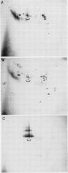
Proteins in intercellular washing fluid (IWF) from wheat (Triticum aestivum) and barley (Hordeum vulgare) leaves were separated by two-dimensional isoelectric focusing-polyacrylamide gel electrophoresis and stained with Coomassie brilliant blue (CBB) or silver. Intracellular protein from the cut ends of leaves accounted for only a small proportion of total protein in IWF from wheat leaves. When these were heavily infected with the stem rust fungus (Puccinia graminis f. sp. tritici) and grown at 19°C, four infection-related CBB-stainable proteins were detected in IWF.
To compare IWF proteins from wheat and barley leaves infected with the same pathogen, conditions were established that permitted luxuriant growth of stem rust of wheat in barley (exposure to chloroform before inoculation and maintenance at 25°C thereafter). Under these conditions, at least 10 infection-related silver-stainable proteins were detected in IWF from infected wheat in addition to the more than 50 that were of host origin. The electrophoretic properties of 8 of the infection-related proteins were the same as those of 8 infection-related proteins in IWF from barley.
IWF from wheat and barley grown under these conditions was analyzed for Concanavalin A-binding glycoproteins immobilized on nitrocellulose membrane replicas made from gels. Of the many infection-related glycoproteins that were detected in IWF from stem rust-affected wheat, approximately 20 occupied the same positions as those from stem rust-affected barley. The glycoprotein pattern of IWF prepared from wheat leaves grown at 19°C and infected with the leaf rust fungus (P. recondita f. sp. tritici) was markedly different to that of IWF from the same host infected with the stem rust fungus. We conclude that IWF from rust-affected cereal leaves may be a useful source of surface or extracellular proteins from the parasitic mycelium.
Full text
PDF


Images in this article


Selected References
These references are in PubMed. This may not be the complete list of references from this article.
- Bradford M. M. A rapid and sensitive method for the quantitation of microgram quantities of protein utilizing the principle of protein-dye binding. Anal Biochem. 1976 May 7;72:248–254. doi: 10.1016/0003-2697(76)90527-3. [DOI] [PubMed] [Google Scholar]
- Clegg J. C. Glycoprotein detection in nitrocellulose transfers of electrophoretically separated protein mixtures using concanavalin A and peroxidase: application to arenavirus and flavivirus proteins. Anal Biochem. 1982 Dec;127(2):389–394. doi: 10.1016/0003-2697(82)90192-0. [DOI] [PubMed] [Google Scholar]
- Gander J. E. Fungal cell wall glycoproteins and peptido-polysaccharides. Annu Rev Microbiol. 1974;28(0):103–119. doi: 10.1146/annurev.mi.28.100174.000535. [DOI] [PubMed] [Google Scholar]
- Goldstein I. J., Hayes C. E. The lectins: carbohydrate-binding proteins of plants and animals. Adv Carbohydr Chem Biochem. 1978;35:127–340. doi: 10.1016/s0065-2318(08)60220-6. [DOI] [PubMed] [Google Scholar]
- Hawkes R. Identification of concanavalin A-binding proteins after sodium dodecyl sulfate--gel electrophoresis and protein blotting. Anal Biochem. 1982 Jun;123(1):143–146. doi: 10.1016/0003-2697(82)90634-0. [DOI] [PubMed] [Google Scholar]
- Keen N. T., Yoshikawa M., Wang M. C. Phytoalexin Elicitor Activity of Carbohydrates from Phytophthora megasperma f.sp. glycinea and Other Sources. Plant Physiol. 1983 Mar;71(3):466–471. doi: 10.1104/pp.71.3.466. [DOI] [PMC free article] [PubMed] [Google Scholar]
- O'Farrell P. H. High resolution two-dimensional electrophoresis of proteins. J Biol Chem. 1975 May 25;250(10):4007–4021. [PMC free article] [PubMed] [Google Scholar]
- Ragster L. V., Chrispeels M. J. Azocoll-digesting Proteinases in Soybean Leaves: Characteristics and Changes during Leaf Maturation and Senescence. Plant Physiol. 1979 Nov;64(5):857–862. doi: 10.1104/pp.64.5.857. [DOI] [PMC free article] [PubMed] [Google Scholar]
- Rohringer R., Holden D. W. Protein blotting: detection of proteins with colloidal gold, and of glycoproteins and lectins with biotin-conjugated and enzyme probes. Anal Biochem. 1985 Jan;144(1):118–127. doi: 10.1016/0003-2697(85)90092-2. [DOI] [PubMed] [Google Scholar]
- Trocha P., Daly J. M. Cell Walls of Germinating Uredospores: II. Carbohydrate Polymers. Plant Physiol. 1974 Apr;53(4):527–532. doi: 10.1104/pp.53.4.527. [DOI] [PMC free article] [PubMed] [Google Scholar]
- Trocha P., Daly J. M., Langenbach R. J. Cell walls of germinating uredospores: I. Amino Acid and carbohydrate constituents. Plant Physiol. 1974 Apr;53(4):519–526. doi: 10.1104/pp.53.4.519. [DOI] [PMC free article] [PubMed] [Google Scholar]
- Vu C. V., Allen L. H., Bowes G. Effects of Light and Elevated Atmospheric CO(2) on the Ribulose Bisphosphate Carboxylase Activity and Ribulose Bisphosphate Level of Soybean Leaves. Plant Physiol. 1983 Nov;73(3):729–734. doi: 10.1104/pp.73.3.729. [DOI] [PMC free article] [PubMed] [Google Scholar]
- Wray W., Boulikas T., Wray V. P., Hancock R. Silver staining of proteins in polyacrylamide gels. Anal Biochem. 1981 Nov 15;118(1):197–203. doi: 10.1016/0003-2697(81)90179-2. [DOI] [PubMed] [Google Scholar]
- van der Wilden W., Segers J. H., Chrispeels M. J. Cell Walls of Phaseolus vulgaris Leaves Contain the Azocoll-Digesting Proteinase. Plant Physiol. 1983 Nov;73(3):576–578. doi: 10.1104/pp.73.3.576. [DOI] [PMC free article] [PubMed] [Google Scholar]