Abstract
In enteropathogenic and enterohemorraghic Escherichia coli (EPEC and EHEC), two members of the SlyA family of transcriptional regulators have been identified as SlyA. Western blot analysis of the wild type and the corresponding hosA and slyA deletion mutants indicated that SlyA and HosA are distinct proteins whose expression is not interdependent. Of 27 different E. coli strains (EPEC, EHEC, enteroinvasive, enteroaggregative, uropathogenic, and commensal) examined, 14 were positive for both genes and proteins. To investigate hosA expression, a hosA::luxCDABE reporter gene fusion was constructed. hosA expression was significantly reduced in the hosA but not the slyA mutant and was influenced by temperature, salt, and pH. In contrast to SlyA, HosA did not activate the cryptic E. coli K-12 hemolysin ClyA. Mutation of hosA did not influence type III secretion, the regulation of the LEE1 and LEE4 operons, or the ability of E2348/69 to form attaching-and-effacing lesions on intestinal epithelial cells. HosA is, however, involved in the temperature-dependent positive control of motility on swim plates and regulates fliC expression and FliC protein levels. In electrophoretic mobility shift assays, purified HosA protein bound specifically to the fliC promoter, indicating that HosA directly modulates flagellin expression. While direct examination of flagellar structure and the motile behavior of individual hosA cells grown in broth culture at 30°C did not reveal any obvious differences, hosA mutants, unlike the wild type, clumped together, forming nonmotile aggregates which could account for the markedly reduced motility of the hosA mutant on swim plates at 30°C. We conclude that SlyA and HosA are independent transcriptional regulators that respond to different physicochemical cues to facilitate the environmental adaptation of E. coli.
The SlyA family of transcriptional regulator proteins is found in both bacteria and archaea, where they facilitate adaptation to a variety of different growth environments. To date more than 120 SlyA family proteins have been identified, including 16 homologues in Escherichia coli alone (34). In Salmonella enterica serovar Typhimurium, slyA was initially presumed to encode a hemolytic virulence factor (salmolysin) which was required for survival in macrophages and for virulence in mice (15). Subsequently, SlyA was reclassified as a positively acting transcriptional regulator capable of inducing the expression of a cryptic hemolysin in E. coli K-12 (16, 22). Salmonella serovar Typhimurium slyA mutants exhibit altered survival in Tris-containing minimal medium as well as reduced production of flagellin and other outer membrane proteins, data which collectively suggest a role for SlyA in stationary-phase survival (28). Similarly, in enteroinvasive E. coli (EIEC), SlyA regulates the expression of a number of genes required for resistance to heat and acid stress and is considered important for survival and/or replication within phagocytic cells (27).
In other members of the Enterobacteriaceae, closely related SlyA family proteins regulate invasin, the primary invasion factor for both Yersinia enterocolitica and Yersinia pseudotuberculosis (RovA) (20, 23); the production of a carbapenem antibiotic and the red pigment prodigiosin in Serratia marcescens (Rap) (31); and exoenzymes, carbapenem, and virulence in the plant pathogen Erwinia carotovora (Hor) (31). More distantly related proteins include MarR and EmrR (E. coli) (18), which regulate genes involved in multiantibiotic resistance; PecS (Erwinia chrysanthemi), which controls plant cell wall-degrading exoenzymes (pectinases and cellulases) (24); and ScoC, a negative regulator of sporulation and exoprotease in Bacillus subtilis (13).
This diverse group of SlyA homologues shares a common winged-helix DNA-binding motif (2, 34). The overall protein structure is very similar, consisting of two domains, one for protein dimerization and one for DNA interaction. The DNA-binding domain includes two helices and several regions of β-sheet that associate to form a winged-helix structure, with an exposed wing region that contacts the DNA (2). It is likely that sequence variability in this wing region accounts for differences in DNA sequence recognition and the variety of modes of DNA interactions observed within this family (34).
Herbelin et al. (9) have shown that both enterohemorrhagic E. coli (EHEC) and enteropathogenic E. coli (EPEC) contain a novel 2.9-kb insertion sequence located at the 3′ terminus of rpoS that is not present in K-12 or ECOR group A strains of E. coli. Within this region they identified several open reading frames, including one that was identified as slyA. The inference from this finding was that EHEC and EPEC contain two slyA genes (9, 10). The first slyA gene, located at 36.7 to 37.1 min on the chromosome, is flanked by sodC and slyB, which code for a superoxide dismutase and an outer membrane lipoprotein, respectively. The second slyA-homologous gene, ECs3594 (GenBank accession no. AP002562), whose initial identification was based on an annotation of the rpoS-mutS region sequence from E. coli O157:H7, is located at 61.5 to 61.7 min, adjacent to rpoS (GenBank accession no. AJ006210) (9, 32). This second gene was reported to be present in some EPEC strains but not in E. coli K-12 lineage strains. Heroven and Dersch (10) suggested that although the two slyA genes were related, they were sufficiently different to warrant the renaming of the slyA gene identified by Herbelin et al. (9). Here we present data to show that this gene, which we have termed hosA (for homologue of slyA), is expressed in EPEC and other pathogenic E. coli strains and that, in contrast to SlyA, HosA is involved in the temperature-dependent control of swimming motility and cellular aggregation.
MATERIALS AND METHODS
Bacterial strains, plasmids, and growth media.
The bacterial strains and plasmids used in this study are detailed in Table 1. L broth (LB) used in this study consisted of 1% (wt/vol) tryptone, 0.5% (wt/vol) yeast extract, and 0.5% (wt/vol) NaCl, unless otherwise indicated. Tryptone broth used for motility studies consisted of 1% (wt/vol) tryptone and 0.5% NaCl. Agar plates were solidified with Bacteriological Agar no. 1 (Oxoid) to 1.5% (wt/vol) for general use. One-percent (weight-to-volume) tryptone swim plates containing 0.5% (wt/vol) NaCl were solidified with 0.3% (wt/vol) Eiken agar (Tanabe Seiyaku Co. Ltd., Osaka, Japan). Minimal M9 medium (25) was used to select for E2348/69 and against auxotrophic E. coli strains. Antibiotic, 100 mM morpholinepropanesulfonic acid (MOPS), and isopropyl-β-d-thiogalactopyranoside (IPTG) supplements were added as required. Unless otherwise stated, broth cultures were incubated in 50-ml disposable Falcon tubes at 37°C with shaking at 200 rpm.
TABLE 1.
Bacterial strains and plasmids used in this study
| Strain or plasmid | Genotype or phenotype | Source or reference |
|---|---|---|
| Strains | ||
| E2348/69 | O127:H6 EPEC isolate | 14 |
| II104 | E2348/69 slyA | This laboratory |
| II150 | E2348/69 hosA | This study |
| II151 | E2348/69 slyA hosA | This study |
| II152 | E2348/69 fliC | This study |
| DH5α | F−endA hsdR17 (rK− mk+) supE44 thi-1 recA1 gyrA96 φ80 lacZ | 7 |
| MG1655 | E. coli K-12 | 3 |
| S17-1 λpir | λpir lysogen thi pro HsdR−hsdM+recA RP4 2-Tc::Mu-Km::Tn7 (Tpr Smr) | 26 |
| Plasmids | ||
| pBAD24 | Expression vector with pBAD promoter and araC negative regulator | 6 |
| pDM4 | pir-dependent suicide vector, sacB double-cross over selection; Cmr | 19 |
| pUC57/T | T-cloning vector; Apr | Clontech |
| pII19 | pUC57/T containing slyA | This laboratory |
| pII30 | ptrc99A containing slyA, inducible by IPTG | This laboratory |
| pMJFA10 | pDM4 suicide plasmid containing hosA deletion (from nucleotide 4 to 375) | This study |
| pMJFA12 | pBAD24 containing hosA, inducible by arabinose | This study |
| pMJFA18 | PCR-amplified hosA promoter and gfp cloned into pSB377; Ampr | This study |
| pKB100 | PCR-amplified fliC promoter cloned into pSB377; Ampr | This study |
| pMJFA21 | PCR-amplified hosA cloned into pPROEX; Ampr | This study |
| pMJFA22 | PCR-amplified LEE1 operon promoter fused to luxCDABE in pSB377; Apr | This study |
| pHW100 | PCR-amplified LEE4 operon promoter fused to luxCDABE in pSB377; Apr | This study |
| pPROEX HT | IPTG-inducible expression vector, trc promoter, His, lacIq, f1 intergenic region; Apr | Invitrogen |
| pSB377 | luxCDABE reporter operon, rop, ColE1 ori; Apr | 33 |
| ptrc99A | ITPG-inducible expression vector, trc promoter; lacIqrrnBt1rrnBt2; Apr | Pharmacia |
DNA manipulations.
Standard methods for the manipulation of DNA were followed (25). A histidine tag was fused to the amino terminus of HosA by ligation of a PCR product into pPROEX-HTNcoI/Klenow/HindIII. hosA was ampli-fied with primers HOSA7 (5′-GCGTTACGAAATAAAGCGTTCCATCAG-3′) and HOSA10 (5′-GACGTCAGCCGTAAGCTTCGTCTTAAC-3′), containing a HindIII restriction site (underlined), by using genomic E2348/69 DNA as a template. The resulting plasmid, pMJFA21 (Table 1), contained hosA lacking the first ATG with an N-terminal six-histidine tag (HT) and an rTEV protease cleavage recognition site. An arabinose-inducible hosA plasmid was constructed by cloning a PCR product generated from E2348/69 genomic DNA by using primers HOSA7 and HOSA10 into pBAD24. This plasmid was named pMJFA12 (Table 1). PCR was used to amplify slyA from E2348/69 by using primers SLYF (5′-CTTAGCAAGCGAATTCTAAGGAGATG-3′) and SLYR (5′-CCGCGCTAAATAAGCTTGCGTGTGGTC-3′). The product was cloned into pUC57/T to generate pII19. To obtain IPTG-controlled expression of SlyA, the slyA gene from pII19 was cloned as an EcoRI-HindIII fragment into ptrc99A to produce pII30. A luxCDABE reporter fusion to the hosA promoter was constructed by cloning a PCR product generated from E2348/69 genomic DNA by using primers HOSA8 (5′-GTAAACTCCGGGCAAACTATC-3′) and HOSA9 (5′-GGAAGGAATTCGAGATGGATGGTGCTAA-3′), containing an EcoRI site (underlined), into pSB377 (30, 33) digested with SmaI and EcoRI. The resulting plasmid was named pMJFA18 (Table 1). Similarly, a fliC′::luxCDABE reporter gene fusion was constructed by cloning a PCR product generated from E2348/69 genomic DNA by using primers FLICP1 (5′-GTGGGAATTCTGGTGGTGCTGGTGGCG-3′), containing an EcoRI site (underlined), and FLICP2 (5′-GATTCGTTATCCTATATTGCAAGTC-3′) into pSB377 (30, 33). The resulting plasmid was called pKB100 (Table 1). The LEE1 and LEE4 luxCDABE reporter plasmids pMJFA22 and pHW100 (Table 1) were also constructed by cloning the LEE1 or LEE4 operon promoter region from E2348/69 into pSB377 as described above but using primers LEE101 (5′-CCTCGAATTCCCTTTTCAAGTTGCCTT-3′), containing an EcoRI site (underlined), and LEE102R (5′-TTAGCGACCTTATCCTTATC-3′) for LEE1 and primers LEE4-1 (5′-GTGAATTCGGTATCCAGAAGATCAAGAAGC-3′), containing an EcoRI site (underlined), and LEE4-2 (5′-GTGAATTCAAGTACTCGCATTCGCAC-3′) for LEE4 (30, 33).
Disruption of hosA.
Overlap extension PCR (11) was used to generate an in-frame deletion of hosA in E2348/69 to create the hosA mutant II150. Two PCR fragments were generated from the template genomic DNA with primer pairs HOSA1-HOSA2 (5′-CGAGATGGATGGTGCTAAGCGGCGTTT-3′ and 5′-CTTGCGCACCAACTGCATCATGTAAACTCCGGGCAAACTA-3′) and HOSA3-HOSA4 (5′-ATGCAGTTGGTGCGCAAGATGATGAACACATAG-3′ and 5′-GACGTCAGCCGCATGCTT CGTCTTAAC-3′). (The SphI restriction enzyme site, used for cloning into the suicide vector pDM4 [Table 1], is underlined. Complementary primer regions for overlap extension PCR are boldfaced.) The resulting products contained a 561-bp fragment containing the 5′ end of hosA and a 524-bp fragment containing the 3′ end of hosA, respectively. An 18-bp overlap in their sequences permitted amplification of a 1,067-bp product during a second PCR with primers HOSA1 and HOSA4. The resulting PCR product contained a deletion from nucleotides 4 to 375 of hosA, corresponding to HosA amino acid residues 2 to 126, and was cloned into pDM4 to create pMJFA10 (Table 1). Conjugation from E. coli S17-1 λpir was used to introduce pMJFA10 into E2348/69, and cells containing single-crossover events were isolated on minimal M9 agar containing chloramphenicol at 30 μg/ml. Double-crossover events were selected on LB-sucrose agar as described by Milton et al. (19). Chloramphenicol-sensitive colonies were screened by PCR using primers HOSA5 and HOSA6 (5′-CGTGGTGGACAATGTCATCTAC-3′ and 5′-CCCATAAACTGGACCACGAACC-3′), which are external to hosA. A PCR product of 1,603 bp indicated that hosA was intact, and a product of 1,232 bp indicated a hosA deletion. The 1,232-bp product was sequenced to confirm the hosA deletion. An E2348/69 hosA slyA double-mutant strain, II151 (Table 1), was constructed by introducing pMJFA10 into the slyA mutant II104 (Table 1) (to be described in detail elsewhere). The presence of both mutations in II151 was confirmed using PCR and DNA sequencing.
Hemolysin release assays.
Cell lysates were prepared from cultures grown in LB. Equal volumes of cell lysate were mixed with phosphate buffered saline (PBS), pH 7.4. An equal volume of washed sheep erythrocytes was added. After a 60-min incubation at 37°C, remaining whole cells were removed by centrifugation. The release of hemoglobin was measured at A540.
Swimming motility assays.
Tryptone motility plates were inoculated from bacterial cultures grown overnight by using toothpicks. Plates were incubated at 37 and 30°C and at room temperature (22°C) for as long as 48 h, and the diameters of migration on at least two axes were measured. A more detailed analysis of swimming motility of individual bacterial cells was undertaken by using a Hobson Tracker (Hobson Vision Ltd., Baslow, Derbyshire, United Kingdom), which simultaneously tracks moving bacterial cells in real time in a microscope field of view. For these experiments, bacteria were grown in LB at 30°C with shaking overnight. A 1-in-1,000 dilution into prewarmed LB was made, and the cultures were incubated for a further 1 to 2 h at 30°C. Optical density (OD) was monitored, and at an OD at 600 nm (OD600) of ∼0.05 to 0.1, samples were taken for analysis. Ten microliters of sample was placed on a microscope slide and covered. Analysis of each slide was carried out for a maximum period of 30 s, with two sets of data from each coverslip. A minimum of 20 samples were taken from each culture, and two independent cultures were analyzed per Hobson Tracker experiment.
Bioluminescence assays.
Expression of the lux-based reporter gene fusions was determined by measuring bioluminescence as a function of bacterial growth by using a combined, automated luminometer-spectrometer (the Anthos Labtech LUCY 1). Single bacterial colonies were resuspended in 100 μl of LB and then diluted 103-fold into the appropriate medium. Two hundred microliters of this suspension was pipetted into a Krystal 2000 96-well microplate (Thermo Life Sciences, Basingstoke, United Kingdom), which was placed inside the LUCY 1, and growth (measured as OD495) and light emission were recorded for at least 17 h.
Preparation of secreted proteins.
Bacteria from a 10-ml overnight LB culture were harvested by centrifugation for 7 min at 7,000 × g and 24°C. The pellet was resuspended in 10 ml of fresh, prewarmed (37°C) LB or Dulbecco's modified Eagle medium (DMEM) broth. A 0.5-ml volume was used to inoculate a 10-ml culture of the same medium containing the appropriate levels of antibiotic and arabinose. Cultures were incubated for 3 h, a 1.5-ml aliquot was taken, and the OD600 was recorded. Bacteria were pelleted in a microcentrifuge for 2 min, and either 1 ml or 65 μl of supernatant was taken. Proteins were concentrated by trichloroacetic acid precipitation as described by Swift et al. (29) and were resuspended in 65 μl of sterile distilled water.
Preparation of flagellar protein.
Bacteria from 50 ml of culture were pelleted by centrifugation and resuspended in 5 ml of PBS. Flagella were removed from the cell by vortexing for 15 to 30 s using a vortex shearing device (http://www.nottingham.ac.uk/iii/Methods/Bacteria_and_Molecular/Flagellinpreparations2.pdf). To remove intact bacterial cells, the resulting mixture was centrifuged twice at 10,000 × g for 10 min at room temperature. To pellet the flagellin protein, the supernatant was centrifuged for 1 h at 50,000 × g and 4°C, and the resulting pellet was resuspended in 100 μl of sterile distilled water.
Sodium dodecyl sulfate-polyacrylamide gel electrophoresis (SDS-PAGE).
Proteins were separated by using either Tricine-SDS-16% polyacrylamide gels (Novex) or SDS-11.5% polyacrylamide gels. For Western blotting, proteins were transferred to a 0.2-μm-pore-size nitrocellulose membrane (Protan; Schleicher & Schuell, London, United Kingdom) by electroblotting. Membranes were blocked for 1 h and probed overnight with the specified antibody. Rabbit polyclonal antibodies to the purified SlyA, HosA, and FliC proteins and to a mixture of EPEC secreted proteins were raised according to a standard protocol (8) and used at a 1:200 dilution. The primary antibody was visualized via a protein A-alkaline phosphatase conjugate (Sigma) detected by using 5-bromo-4-chloro-3-indolylphosphate (BCIP)-nitroblue tetrazolium (NBT) (Sigma).
A/E lesion formation on HT29 cells.
The capacity of EPEC strains to form attaching-and-effacing (A/E) lesions on the human colorectal cell line HT29 (American Type Culture Collection, Manassas, Va.) was investigated as follows. Ten milliliters of LB overnight cultures of EPEC was pelleted and gently resuspended in 10 ml of fresh, prewarmed DMEM. A 0.5-ml volume of this suspension was added to 10 ml of prewarmed DMEM. Ampicillin at 50 μg/ml was added where appropriate. Arabinose was added as 100 μl of a 20% solution to give a final concentration of 0.2% where appropriate. Bacteria were grown for 3 h at 37°C with shaking at 200 rpm. Bacteria were diluted 1:1 with fresh prewarmed DMEM, and 1 ml was added to HT29 cells grown on coverslips in 3 ml of DMEM in a six-well tray. Ampicillin and arabinose were added as appropriate to maintain culture concentrations. After infection (3 h), nonadherent bacteria were removed by washing (three times, for 5 min each time) with PBS and cells were formaldehyde fixed (3% in PBS for 20 min). Cells were permeabilized with 0.1% (vol/vol) Triton X-100 in PBS for 20 min and were washed (twice, for 5 min each time) with PBS. Actin filaments were stained by flooding the coverslip surface with 5 μg of phalloidin-fluorescein isothiocyanate (Sigma) ml−1 for 1 h. Coverslips were washed first with 0.1% (vol/vol) Triton X-100 in PBS (twice, for 5 min each time) and then with PBS (twice, for 5 min each time) and were mounted on glass slides for analysis. To quantify the proportion of HT29 cells carrying A/E lesions, at least 100 HT29 cells per coverslip were examined.
Purification of His-tagged HosA.
E. coli DH5α harboring plasmid pMJFA21 (Table 1) was grown in LB supplemented with 100 μg of carbenicillin/ml at 37°C to mid-log phase, IPTG (1 mM) was added to the culture, and incubation was continued for a further 2 h. Bacteria were harvested and resuspended in binding buffer (5 mM imidazole, 500 mM NaCl, 20 mM Tris-HCl [pH 7.9]) supplemented with 0.1% (vol/vol) Triton X-100. Cells were lysed by sonication, and after centrifugation, the soluble fraction was loaded onto a nickel-nitrilotriacetic acid His · Bind resin column (Novagen) equilibrated with binding buffer. The His-tagged HosA protein (>98% purity) was recovered by eluting with binding buffer containing 250 mM imidazole. The protein concentration was estimated by using Bradford reagent (Sigma) with bovine serum albumin as a standard. Protein purity was assessed by SDS-PAGE.
Electrophoretic mobility shift assays (EMSA).
A 494-bp DNA fragment containing the fliC promoter and upstream region was amplified by PCR using genomic DNA from E2348/69 as a template and primers FLICP1 and FLICP2. The fliC promoter DNA was labeled at 3′-OH ends by the addition of 0.5 U of Klenow enzyme and 50 μM digoxigenin-labeled deoxynucleoside triphosphates. Ten nanograms of labeled DNA was incubated with increasing amounts of purified His-tagged HosA (from 0.2 to 5 μM) for 5 min at room temperature in 10 mM Tris-HCl (pH 7.5) containing 50 mM KCl and 1 mM dithiothreitol. Prior to use, the HosA protein sample was dialyzed in incubation buffer. Lambda control DNA was prepared by PCR using primers Lambda1 (5′-GCGGAATTCAAACAGGTGCTGGGG-3′) and Lambda2R (5′-CCGCTCTAGACGGATGGAAACGCCTTA-3′). Samples were electrophoresed on precast 6% DNA retardation gels (Invitrogen) according to the manufacturer's instructions and were transferred to a nylon membrane. A 104-fold dilution of alkaline phosphatase-conjugated anti-digoxigenin (Roche) was used to detect the labeled DNA.
RESULTS
HosA and SlyA are distinct proteins.
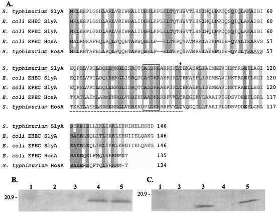
Amino acid sequence comparisons of SlyA and HosA from the archetypal EPEC strain E2348/69 suggest that HosA is a member of the MarR-SlyA family of regulators (Fig. 1A). Overall, HosA shares 24% identity and 47% similarity with SlyA, with 30% identity and 60% similarity over the region proposed to be involved in DNA binding. Interestingly, the similarity between SlyA and HosA increases to 68% over the region required to form the winged-helix fold, and the 5 amino acids that form the single wing region in SlyA, proposed to locate within the DNA groove, have 100% similarity with HosA. Furthermore, tertiary structural analysis using 3D-pssm (http://www.sbg.bio.ic.ac.uk/∼3dpssm/) predicted that HosA would form a tertiary structure similar to those of E. coli MarR and Enterococcus faecalis SlyA (2, 34).
FIG. 1.
Comparison of SlyA and HosA. (A) Multiple amino acid sequence alignment of SlyA from Salmonella serovar Typhimurium LT2 (NP460407), EHEC (NP288078), and EPEC (AAF97817) and HosA from EPEC (AAG14991) and Salmonella serovar Typhimurium LT2 (NP461841). Dark shading, highly conserved amino acids; boldfaced letters, fully conserved amino acids; light shading, three of five amino acids conserved. Box indicates the amino acids that form the single wing region; dashed underline indicates the proposed DNA-binding domain. Asterisk indicates the threonine that is conserved in the MarR family. (B and C) Western blot analysis for the presence of SlyA (B) and HosA (C) in cell extracts from the hosA slyA mutant II151 (lane 2), the slyA mutant II104 (lane 3), the hosA mutant II150 (lane 4), and the wild-type strain E2348/69 (lane 5). Lane 1, 20.9-kDa molecular mass marker.
To investigate the antigenic relationship between SlyA and HosA, the conservation of HosA, and the relative levels of the two transcriptional-regulator proteins, we used PCR to amplify both genes from E2348/69. The genes were then cloned into the overexpression vectors ptrc99A and pBAD24 to produce pII30 and pMJFA12, respectively, as described in Materials and Methods. Both proteins were purified from SDS-PAGE gels and used to raise rabbit monospecific polyclonal antibodies. These were used to probe Western blots of whole-cell lysates prepared from the parent EPEC strain, E2348/69, and from strains carrying slyA (II104) hosA (II150), or slyA hosA (II151) mutations, constructed as described in Materials and Methods. Figure 1B and C show that both proteins are expressed in the parent EPEC strain and that they are antigenically distinct. Antibodies raised against SlyA reacted only to a single 24-kDa protein present in the lysates prepared from E2348/69 and the hosA mutant (Fig. 1B), while those raised against HosA recognized a 21-kDa protein in the parent and the slyA mutant only (Fig. 1C). These data suggest that the absence of SlyA does not affect the expression of HosA or vice versa, indicating that their expression is not interdependent. Neither antibody recognized any proteins present in the lysate prepared from the double hosA slyA mutant (Fig. 1B and C, lanes 2).
HosA is expressed in a diverse range of E. coli strains.
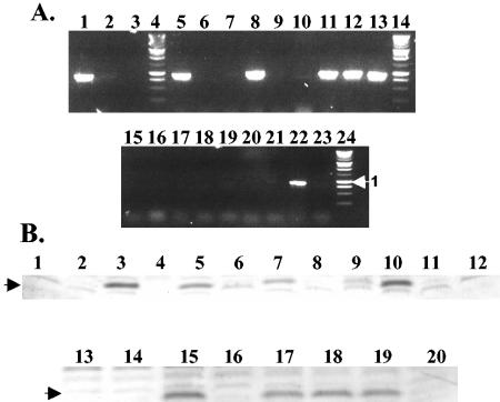
Since hosA is absent from E. coli K-12 strains, we sought to determine the prevalence of hosA in a selected group of enterovirulent, uropathogenic (UPEC), and commensal E. coli strains. PCR and Southern blot analysis of these strains indicated that hosA was present in many but not all E. coli types. The data summarized in Fig. 2A and Table 2 show, for example, that while hosA is conserved in EPEC (7 of 10) and EHEC (5 of 5) strains, it was absent from UPEC (n = 4) strains. To determine whether hosA was expressed, Western blot analysis was performed on cell lysates prepared from a selection of hosA-positive and -negative strains identified by PCR (Fig. 2B). In each case, strains which were positive for the hosA gene by PCR contained the HosA protein, while all those that were negative by PCR did not.
FIG. 2.
Conservation of HosA in E. coli. (A) PCR was used to confirm the presence of hosA in different E. coli strains. Lane 1, E2348/69 (EPEC); lane 2, hosA mutant II150 (EPEC); lane 3, MG1655 (K-12); lane 5, BJ4 (commensal); lane 6, 6601 (commensal); lane 7, 160DA (EPEC); lane 8, F630C (EPEC); lane 9, O55:H7 (EPEC); lane 10, O119:H6 (EPEC); lane 11, EDL933 O157:H7 (EHEC); lane 12, Sakai O157:H7 (EHEC); lane 13, NCTC12900 (EHEC); lane 15, 536 (UPEC); lane 16, DS17 (UPEC); lane 17, NU14 (UPEC); lane 18, O6:K13 (UPEC); lane 19, 1457-75 (EIEC); lane 20, 8271-77 (EIEC); lane 21, 5898-71 (EIEC); lane 22, JPN10 (EaggEC); lane 23, O111:H12 (EaggEC). Lanes 4, 14, and 24 contain a 1-kb ladder (Promega). (B) Total-cell proteins were isolated from exponentially growing cultures of different E. coli strains and pathotypes. Western blot analysis was carried out to confirm the expression of HosA. Lane 1, MG1655 (K-12); lane 2, II150 (EPEC); lane 3, E2348/69 (EPEC); lane 4, broad-range markers (Bio-Rad); lane 5, BJ4 (commensal); lane 6, 6601 (commensal); lane 7, JPN10 (EaggEC); lane 8, O111:H12 (EaggEC); lane 9, 29962 (EHEC); lane 10, BK33 (EHEC); lane 11, 1457-75 (EIEC); lane 12, 8271-77 (EIEC); lane 13, 536 (UPEC); lane 14, NU14 (UPEC); lane 15, F630C (EPEC); lane 16, O55:H7 (EPEC); lane 17, 259 (EPEC); lane 18, 260 (EPEC); lane 19, 237 (EPEC); lane 20, 272 (EPEC).
TABLE 2.
Conservation of hosA in E. coli
| Strain classification | No. of strains analyzed | No. of strains in which hosA is present | % of strains with hosA |
|---|---|---|---|
| Nondiarrheagenic | |||
| UPEC | 4 | 0 | 0 |
| Commensal | 2 | 1 | 50 |
| Laboratory | 1 | 0 | 0 |
| Diarrheagenic | |||
| EIEC | 3 | 0 | 0 |
| EaggEC | 2 | 1 | 50 |
| EPEC | 10 | 7 | 70 |
| EHEC | 5 | 5 | 100 |
| Total | 27 | 14 | 52 |
HosA does not confer hemolytic activity on E. coli MG1655.
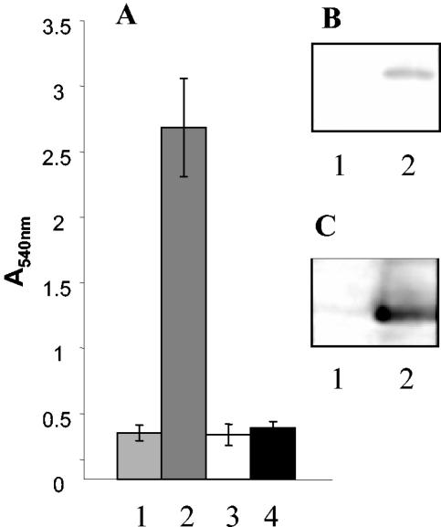
SlyA was originally identified as a transcriptional regulator by its ability to activate a cryptic E. coli K-12 hemolysin termed ClyA (15). If HosA and SlyA are functionally equivalent, we hypothesized that HosA should be capable of activating clyA. To investigate this possibility, we transformed pII30 and pMJFA12 into E. coli MG1655. Hemoglobin release assays showed a fivefold increase in the level of hemoglobin in the supernatant under conditions of SlyA induction but no change following the induction of HosA (Fig. 3). These data suggest that SlyA and HosA are distinct regulators, and as such they are likely to control different promoters.
FIG. 3.
HosA does not activate hemolytic activity in E. coli MG1655. (A) MG1655 carrying a plasmid (pII30 and pMJFA12) with either hosA or slyA, respectively, cloned downstream of an inducible promoter were grown in LB with either 0.1 mM IPTG or 0.2% arabinose, respectively. Cell lysates were prepared by sonication and mixed with sheep erythrocytes. After 60 min, the hemoglobin released was measured at A540. Column 1, E. coli MG1655(ptrc99A); column 2, E. coli MG1655(pII30); column 3, E. coli MG1655(pBAD24); column 4, E. coli MG1655(pMJFA12). (B) Western blot of SlyA in cell lysates prepared from E. coli MG1655(ptrc99A) (lane 1) and E. coli MG1655(pII30) (lane 2). (C) Western blot of HosA in cell lysates from E. coli MG1655(pBAD24) (lane 1) and E. coli MG1655(pMJFA12) (lane 2).
HosA does not influence A/E lesion formation.
Since hosA is primarily associated with pathogenic strains of E. coli, we hypothesized that HosA may have a role in the regulation of proteins that are important for the establishment of infection. Both EPEC and EHEC employ tightly regulated type III protein secretion systems to deliver effector proteins into mammalian cells. These translocated proteins direct the intimate attachment of the bacteria to the host cell surface and subvert host cell signaling pathways to induce the formation of an actin pedestal beneath the bacteria. The formation of these characteristic A/E lesions relies on the gene products encoded at a pathogenicity island termed the locus of enterocyte effacement (LEE) (4, 21). Genetic analysis of LEE has identified the components of the type III secretion system, which are transcribed from three polycistronic operons designated LEE1, LEE2, and LEE3 (17), while the secreted effector (Esp) proteins are encoded by a fourth polycistronic operon designated LEE4 (17).
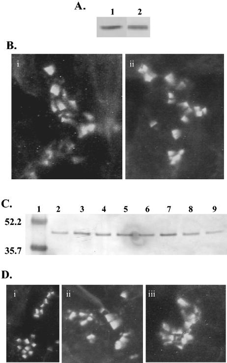
To determine whether HosA might regulate the expression of either LEE1 or LEE4 genes, transcriptional luxCDABE reporter gene fusions containing either the LEE1 (pMFJA22) or the LEE4 (pHW100) promoter were transformed into wild-type E2348/69 and the E2348/69 hosA mutant. These fusions contained promoter sequences and at least 400 bp of upstream sequence to allow the binding of regulatory proteins. No difference in the expression of either promoter was observed (data not shown). To determine whether any regulation may be occurring at the posttranscriptional level, antibodies were raised against EspB, a type III effector protein encoded within the LEE4 operon, and were used to monitor EspB levels in the presence or absence of HosA. Secreted proteins were prepared from E2348/69, E2348/69 hosA, E2348/69(pMJFA12), and E2348/69(pBAD24) grown in LB at 37°C. Western blot analysis revealed no obvious changes in EspB levels (Fig. 4A and C), indicating that HosA does not influence either the expression or the translocation of the type III secreted proteins. Furthermore, mutation of hosA did not affect the capacity of E2348/69 to form microcolonies or A/E lesions on an intestinal epithelial cell line (HT29) (Fig. 4B and D). Indeed, similar clusters of A/E lesions and similar numbers of lesions within clusters were observed.
FIG. 4.
HosA does not influence the expression of EPEC type III secreted proteins or the ability to infect HT29 cells. (A) Secreted proteins were purified from supernatants of exponentially growing cultures of E2348/69 and the hosA mutant II150 and were normalized for culture density. Western blot analysis was carried out to confirm the expression and secretion of EspB, a key type III secreted effector protein. Lane 1, E2348/69; lane 2, II150. (B) A/E lesion formation on HT29 cells by wild-type EPEC and the hosA mutant. Micrographs show typical fields of E2348/69 (i) and the hosA mutant II150 (ii) after incubation with HT29 colorectal cells. (C) Secreted proteins were purified from supernatants of exponentially growing cultures of E2348/69 or the hosA mutant II150 complemented with either pBAD24 (vector control) or pMJFA12, respectively, and were normalized for culture density. Western blot analysis was carried out to confirm the expression and secretion of EspB as a representative type III secreted protein. Lane 1, broad-range prestained 52.2- and 35.7-kDa markers (Bio-Rad); lane 2, E2348/69(pBAD24) with no induction; lane 3, E2348/69(pBAD24) with 0.2% (wt/vol) arabinose; lane 4, E2348/69(pMJFA12) with no induction; lane 5, E2348/69 (pMJFA12) with 0.2% (wt/vol) arabinose; lane 6, the hosA mutant II150 transformed with pBAD24 with no induction; lane 7, II150(pBAD24) with 0.2% (wt/vol) arabinose; lane 8, II150(pMJFA12) with no induction; lane 9, II150(pMJFA12) with 0.2% (wt/vol) arabinose. (D) A/E lesion formation on HT29 cells induced by EPEC overexpressing hosA. Micrographs show typical fields of E2348/69 containing either pBAD24 or pMJFA12 after incubation with HT29 colorectal cells. (i) E2348/69(pBAD24) with 0.2% (wt/vol) arabinose; (ii) E2348/69(pMJFA12) with no induction; (iii) E2348/69(pMJFA12) with 0.2% (wt/vol) arabinose.
HosA regulates motility in EPEC.
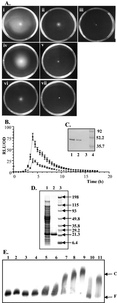
Since flagellum-driven swimming motility may contribute to the establishment of infection, we examined the motility of the hosA mutant. On swim plates incubated at 37°C, both E2348/69 and the hosA mutant II150 were motile and able to swim to the edge of the plate within 16 h (data not shown). However, at 30°C, the absence of HosA severely reduced motility (Fig. 5A). The hosA mutant exhibited a fourfold reduction in swimming motility after a 24-h incubation at 30°C and failed to swim at room temperature (approximately 22°C). Growth was observed around the inoculum site after 24 h, confirming cell viability. At room temperature, the parent strain, E2348/69, was able to swim but required 48 h to reach the edge of the plate (data not shown). To determine whether the motility of the hosA mutant on tryptone plates containing IPTG could be restored by a plasmid-borne copy of hosA, pMJFA21 was introduced into both the hosA mutant II150 and wild type E2348/69. Although no complementation of the mutant was observed (Fig. 5A, panels vi and vii), overexpression of hosA in the parent strain resulted in the inhibition of motility (Fig. 5A, panels iv and v), providing further evidence of a role for HosA in modulating flagellum-mediated motility.
FIG. 5.
HosA modulates swimming motility in EPEC strain E2348/69. (A) Swim plates incubated at 30°C. (i) E2348/69; (ii) hosA mutant (II150); (iii) fliC mutant (II152); (iv) E2348/69(pMJFA21); (v) E2348/69(pMJFA21) plus 1 mM IPTG; (vi) II150(pMJFA21); (vii) II150(pMJFA21) plus 1 mM IPTG. (B) Light emission from strains E2348/69 (▪) and II150 (▴) containing a fliC′::luxCDABE reporter fusion was measured during growth at 30°C in tryptone broth swim medium. Relative light units (RLU) are calculated as light emitted normalized to the OD495 at each time point. This experiment was performed in triplicate. (C) Western blot analysis of the major flagellin protein FliCin E2348/69 (lane 1), the hosA mutant (lane 2), and the fliC mutant (lane 3). Lane 4, molecular mass markers. (D) His-tagged HosA was purified by using nickel-nitrilotriacetic acid His · Bind resin. The purity of the protein was confirmed by SDS-PAGE. Lane 1, crude extract; lane 2, 10 μg of purified HosA; lane 3, broad-range protein markers (Bio-Rad). (E) Band shift assay of the fliC promoter region by His-tagged HosA. A labeled fragment (F) and DNA-protein complexes (C) are indicated. Lane 1, no protein; lane 2, 0.2 μM HosA; lane 3, 0.4 μM HosA; lane 4, 0.8 μM HosA; lane 5, 1.6 μM HosA; lane 6, 2 μM HosA; lane 7, 3 μM HosA; lane 8, 4 μM HosA; lane 9, 5 μM HosA; lane 10, 5 μM HosA with a 100-fold molar excess of unlabeled fliC DNA; lane 11, 5 μM HosA with a 100-fold molar excess of unlabeled λcIII DNA.
To determine whether HosA influenced motility through control of the flagellin structural gene, fliC, we constructed a fliC′::luxCDABE reporter gene fusion which was transformed into both E2348/69 and II150. When these strains were grown in tryptone motility broth at 30°C, there was a two to threefold decrease in fliC promoter activity in the hosA mutant, suggesting that HosA has a role in regulating fliC expression (Fig. 5B). Immunoblot analysis of flagellin preparations also indicated (Fig. 5C) that FliC protein levels were reduced in the hosA mutant, in agreement with the reporter gene fusion data. However, a decrease in fliC′::luxCDABE expression was also noted after growth in tryptone motility broth at 37°C (data not shown), even though the motility of the hosA mutant at this growth temperature was wild type.
To determine whether the decrease in fliC expression occurred via a direct interaction with the fliC promoter, the HosA protein was His tagged, overexpressed, and purified (Fig. 5D) for use in band shift assays. Figure 5E shows that HosA is able to bind to a 494-bp fragment containing the fliC promoter and upstream sequences. The specificity of binding was confirmed, since the band shift was abolished by the addition of excess unlabeled fliC DNA but was retained in the presence of DNA fragments generated from λ DNA.
Examination by transmission electron microscopy of individual E2348/69 hosA mutant cells grown at 30°C revealed the presence of normal flagella on most bacteria, suggesting that HosA may influence flagellar rotation rather than flagellum formation per se (data not shown). To investigate this possibility, we employed a Hobson Tracker to examine the movement of individual E2348/69 and hosA mutant cells. This investigation included analysis of the length of continuous directed movement, the time taken for this movement, and the frequency and duration of tumbling for an individual bacterium as it moved across the field of view. Comparison of E2348/69 and the hosA mutant, II150, grown in LB or tryptone motility broth at 37°C revealed no obvious differences in motility. Table 3 summarizes the data obtained for the two strains grown in LB at 30°C to an OD of ∼0.1. Again, very few obvious differences were observed in the motility of individual bacterial cells. However, microscopic examination of the cultures grown to an OD600 of >0.2 revealed that, unlike E2348/69, the majority of the hosA mutant cells had clumped together, forming large aggregates that were no longer able to swim (data not shown). During this analysis of motility using the Hobson Tracker, it was possible to see movement of individual cells within some of the smaller clumps, which was most likely caused by flagellar movement. For example, where two or three cells clumped together, the group of cells could be seen to spin rapidly, with no directional motion. Furthermore, cells which were observed to dissociate from the aggregates and the few remaining single cells in the culture were swimming with motile parameters that did not differ from those observed in samples prepared from cultures at lower cell densities.
TABLE 3.
Summary of motility characteristics of E2348/69 and the hosA mutant, II150, grown in LB at 30°C
| Characteristic | Strain
|
|
|---|---|---|
| E2348/69 | II150 | |
| Length of run (μm) | 22.2 ± 5.5 | 21.6 ± 4.5 |
| 20.6 ± 3.5 | 19.8 ± 3.4 | |
| 17.3 ± 2.8 | 19.7 ± 4.0 | |
| Run time(s) | 1.0 ± 0.2 | 1.0 ± 0.2 |
| 1.0 ± 0.2 | 0.9 ± 0.2 | |
| 0.8 ± 0.1 | 0.9 ± 0.2 | |
| Speed (μm s−1) | 23.4 ± 1.1 | 21.4 ± 1.3 |
| 21.3 ± 1.3 | 21.0 ± 1.1 | |
| 22.9 ± 1.4 | 21.0 ± 1.3 | |
Expression of hosA as a function of the growth environment.
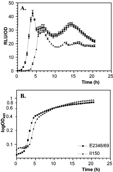
To investigate the expression of hosA, we constructed pMJFA18, a hosA′::luxCDABE reporter gene fusion. The reporter plasmid was introduced into E2348/69 and the hosA mutant II150, and light output and growth were determined over 18 h by using 96-well plates in a combined luminometer-photometer. Figure 6 shows that in E2348/69 at 37°C in LB, hosA was expressed early in the exponential phase at low cell population densities and reached a maximum around the onset of stationary phase, after which expression levels fell to about 50% of maximum. In the hosA mutant, hosA gene expression was delayed by approximately 2 h and expression failed to reach wild-type levels in stationary phase (Fig. 6A). These data indicate that HosA is autoregulated to some extent, although the presence of a functional HosA protein is not an absolute requirement for hosA expression. To determine whether SlyA also controls the timing of hosA expression, pMJFA18 was introduced into the E2348/69 slyA mutant, II104. No detectable differences in hosA expression over the growth curve were observed when II104 and E2348/69 were compared (data not shown). Indeed, the converse was also true: the absence of HosA did not affect the expression of a slyA reporter (data not shown).
FIG. 6.
The hosA promoter is positively autoregulated by HosA. (A) Light emission from strain E2348/69 and the hosA mutant II150 containing pMJFA18 (hosA′::luxCDABE) was determined as a function of growth. Relative light units (RLU) are calculated as the light emitted normalized to the OD495 at each time point. Each experiment was performed twice in triplicate. (B) Growth of E2348/69 and the hosA mutant II150 containing pMJFA18 (hosA′::luxCDABE).
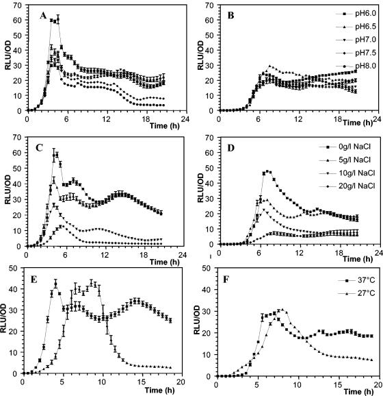
Since pathogenic E. coli strains need to survive and adapt to different environmental conditions during passage from the external environment into the host gastrointestinal tract (1, 12), we examined the expression of hosA′::luxCDABE in E2348/69 and the hosA mutant, II150, as a function of pH, osmolarity, and temperature. Figure 7A shows that hosA expression in E2348/69 was influenced by pH, since maximal light output in LB was observed at a pH of 6.0. Compared with that in the parent strain, hosA expression in the hosA mutant was both delayed and reduced at pH 6.0; increasing the pH above 6.0 had little effect on hosA expression in the hosA mutant (Fig. 7B). hosA expression in II150 was also delayed, relative to that in E2348/69 (Fig. 7C), in LB containing a range of NaCl concentrations (Fig. 7D), although increasing osmolarity reduced hosA expression only slightly. Figure 7E shows that hosA expression in E2348/69 was delayed at 27°C in a manner similar to that noted at 37°C for the hosA mutant (Fig. 7F), although in the latter, hosA expression followed a similar pattern irrespective of temperature. These data indicate that the timing and level of hosA expression are influenced primarily by the presence of an intact hosA gene, by temperature, and by pH.
FIG. 7.
Environmental regulation of hosA as a function of pH, osmolarity, and temperature. Light emission from strains E2348/69 (A, C, and E) and the hosA mutant II150 (B, D, and F) containing pMJFA18 (hosA′::luxCDABE) was determined as a function of pH (A and B), osmolarity (C and D), and temperature (E and F). Relative light units (RLU) are calculated as the light emitted normalized to the OD495 at each time point. Each experiment was performed twice in triplicate.
DISCUSSION
A major aim of this study was to resolve the confusion over the identity of the gene located at 61.5 to 61.7 min in the EPEC and EHEC chromosomes, which had been incorrectly identified as slyA (9). It is important to acknowledge that this error was identified by Heroven and Dersch (10) and noted by Whittam (32). However, the gene was not renamed. To avoid further confusion, we have named this gene hosA, since the data presented in this study clearly demonstrate that although HosA shares a number of the structural features associated with the SlyA protein family, HosA is antigenically and functionally distinct from SlyA. The low overall protein identity (25%) between SlyA and HosA increases to 60% over the specific region identified by comparison with the crystal structure as important for interacting with target DNA sequences and to 100% with respect to the five key amino acids making contact with the DNA. Variation in the winged-helix fold has been hypothesized to be important for recognition of different DNA target sequences. Analysis of the crystal structures of SlyA and MarR has revealed a potential binding site for a small cofactor (2, 34). However, the nature of the cofactor is unclear but it is likely to be different for each regulator, since this region is not well conserved. It is possible that binding of a cofactor or different cofactors may lead to alterations in target recognition, thus making this family of regulators highly responsive to environmental changes.
The amino acid sequence differences between SlyA and HosA suggested that these proteins are likely to recognize different elements within target DNAs and therefore to regulate the expression of different subsets of genes. This appears to be the case in that HosA, in contrast to SlyA, is unable to activate the cryptic hemolysin in E. coli K-12 strains (Fig. 3) (16, 22). A consensus for the binding site (TTAGCAAGCTAA) for SlyA in Salmonella has been defined by Stapleton et al. (28). This consensus sequence is found in both the slyA and clyA promoter regions but is absent from both the hosA and fliC promoters of E2348/69, both of which we have shown to be regulated by HosA. We have shown that the purified HosA protein is able to bind to the fliC promoter region, suggesting that HosA recognizes a sequence distinct from that recognized by SlyA (Fig. 5). This possibility is further highlighted by the lack of regulation of slyA by HosA or vice versa, although lux reporter gene fusion studies indicate that the expression profiles of hosA (Fig. 6) and slyA (unpublished data) in E2348/69 are similar throughout the growth curve.
Examination of the sequenced genomes of E. coli revealed the presence of hosA only in the pathogenic strains. Herbelin et al. (9) reported that HosA was not present in K-12 or ECOR group A strains of E. coli. To determine whether HosA is conserved among pathogenic E. coli isolates, we examined a variety of different strains by PCR, Southern blot analysis, and immunoblot analysis. This included screening of enteroaggregative (EaggEC), EIEC, EPEC, EHEC, UPEC, and commensal strains. Surprisingly, we found that while the EPEC and EHEC groups contained the highest number of hosA-positive strains, none of the EIEC strains contained hosA. Thus, HosA is unlikely to be essential for intracellular survival within host phagocytic cells. These data also suggested that hosA was not essential for bacterial growth, a finding confirmed by the ease of construction of a hosA deletion mutant. However, the link between HosA and EPEC and EHEC strains did suggest to us that HosA could be involved either in modulating virulence gene expression or in facilitating adaptation to changing environments. Using E2348/69, a well-characterized member of the EPEC group, we examined the ability of a hosA deletion mutant to form A/E lesions on the gastrointestinal cell line HT29. However, we were unable to detect any changes in the nature of the A/E lesions formed or the kinetics of A/E lesion formation. Furthermore, there were no differences in the levels of LEE1 and LEE4 operon expression when the wild type and the hosA mutant were compared. Interestingly, however, we have noted that although a slyA mutation does not affect A/E lesion formation, overexpression of SlyA in EPEC has a marked effect on A/E lesion formation, which is almost completely lost (unpublished data). This is not the case for HosA, however; overexpression of hosA had no effect on A/E lesion formation.
Although flagella are important extracellular structures required for swimming and swarming motility, in EPEC they have also been reported to mediate adherence to host cells (5). The presence of HosA was discovered to be important for motility as judged by swim plate assays and was dependent on temperature. A reduction in temperature from 37 to 22°C resulted in the reduction and subsequent loss of swimming motility. This suggested that HosA functions as a positive control element in the regulation of flagellum-driven motility and may modulate gene expression in response to changes in temperature and other, as yet unidentified environmental signals. Indeed, we have shown that hosA expression is modulated by temperature, growth phase, and pH. Other members of the SlyA family of regulators have also been reported to be regulated by changes in environmental conditions. For example, Y. pseudotuberculosis rovA is expressed maximally in stationary-phase cells grown at 25°C, with significantly lower levels of expression observed at 37°C and in exponentially growing cells (20). Furthermore, rovA expression is also influenced by osmolarity, pH, and nutrient availability. Interestingly, although common environmental signals have different effects on rovA and hosA expression, it is clear that, in common with slyA, both genes are positively autoregulated, although HosA is not an absolute requirement for hosA expression.
Although swim plate assays revealed no obvious differences in motility when the wild type was compared with the hosA mutant at 37°C, the motility of the hosA mutant was reduced as the incubation temperature was lowered to 30°C or less, suggesting that HosA may directly regulate flagellar expression. However, we were unable to complement the hosA mutant with a plasmid-borne copy of the intact gene, perhaps as a consequence of our inability to match the timing and level of HosA required by using an inducible expression system. Nevertheless, mutation of hosA also resulted in reductions in fliC expression and FliC protein levels. Furthermore, EMSA band shift assays demonstrated that HosA recognized and bound directly to DNA sequences located upstream of the fliC promoter. However, electron microscopy showed that most hosA mutant cells grown at 30°C possessed flagella. Taken together, these data suggest that the inability of the hosA mutant to swim on motility agar at low temperatures is due not to a lack of functional flagella but perhaps to a change in their movement. Using a Hobson Tracker, it is possible to monitor individual bacterial cells as they pass through a field of view. This technique revealed that although single bacterial cells appeared to be capable of swimming at 30°C, the level of cellular aggregation increased as the culture cell density increased, reducing the capacity for effectual swimming. It is therefore likely that this increased cellular aggregation inhibited the migration of the hosA mutant to the edge of the swim plate. This would suggest that the surface properties of the hosA mutant are different from those of the parent when the strains are grown at 30°C. In Salmonella, SlyA regulates the expression of a number of surface proteins (28), and thus the physicochemical properties of the cell surface are likely to be modified. Further work will be required to determine whether HosA regulates the expression of surface proteins in addition to FliC, which together may account for the cell aggregation phenotype and the temperature-dependent loss of motility observed.
In conclusion, it is clear that in EPEC, HosA is functionally distinct from SlyA; although the mechanisms by which these two related transcriptional regulators interact with DNA are likely to be similar, they clearly have different regulatory functions.
Acknowledgments
We thank Simon Swift for helpful discussions and the construction of the E2348/69 slyA mutant and pII30.
This work was supported by a Medical Research Council (MRC) United Kingdom Career Development Fellowship (to H.W.) and by research grants (to P.W.) from the MRC and the Biotechnology and Biological Sciences Research Council of the United Kingdom, which are gratefully acknowledged.
Editor: J. B. Bliska
REFERENCES
- 1.Abe, H., I. Tatsuno, T. Tobe, A. Okutani, and C. Sasakawa. 2002. Bicarbonate ion stimulates the expression of locus of enterocyte effacement-encoded genes in enterohemorrhagic Escherichia coli O157:H7. Infect. Immun. 70:3500-3509. [DOI] [PMC free article] [PubMed] [Google Scholar]
- 2.Alekshun, M. N., S. B. Levy, T. R. Mealy, B. A. Seaton, and J. F. Head. 2001. The crystal structure of MarR, a regulator of multiple antibiotic resistance, at 2.3 Å resolution. Nat. Struct. Biol. 8:710-714. [DOI] [PubMed] [Google Scholar]
- 3.Bachmann, B. J. 1987. Derivations and genotypes of some mutant derivatives of Escherichia coli K-12, p. 1197-1219. In F. C. Neidhardt, J. L. Ingraham, K. B. Low, B. Magasanik, M. Schaechter, and H. E. Umbarger (ed.), Escherichia coli and Salmonella typhimurium: cellular and molecular biology. American Society for Microbiology, Washington, D.C.
- 4.Donnenberg, M. S., and T. S. Whittam. 2001. Pathogenesis and evolution of virulence in enteropathogenic and enterohemorrhagic Escherichia coli. J. Clin. Investig. 107:539-548. [DOI] [PMC free article] [PubMed] [Google Scholar]
- 5.Girón, J. A., A. G. Torres, E. Freer, and J. B. Kaper. 2002. The flagella of enteropathogenic Escherichia coli mediate adherence to epithelial cells. Mol. Microbiol. 44:361-379. [DOI] [PubMed] [Google Scholar]
- 6.Guzman, L.-M., D. Belin, M. J. Carson, and J. Beckwith. 1995. Tight regulation, modulation, and high-level expression by vectors containing the arabinose pBAD promoter. J. Bacteriol. 177:4121-4130. [DOI] [PMC free article] [PubMed] [Google Scholar]
- 7.Hanahan, D. 1983. Studies on transformation of Escherichia coli with plasmids. J. Mol. Biol. 166:557-580. [DOI] [PubMed] [Google Scholar]
- 8.Harlow, E., and D. P. Lane. 1988. Antibodies: a laboratory manual. Cold Spring Harbor Laboratory Press, Cold Spring Harbor, N.Y.
- 9.Herbelin, C. J., S. C. Chirillo, K. A. Melnick, and T. S. Whittam. 2000. Gene conservation and loss in the mutS-rpoS genomic region of pathogenic Escherichia coli. J. Bacteriol. 182:5381-5390. [DOI] [PMC free article] [PubMed] [Google Scholar]
- 10.Heroven, A. K., and P. Dersch. 2002. Two different open reading frames named slyA in the Escherichia coli sequence databases. Trends Microbiol. 10:267-268. [DOI] [PubMed] [Google Scholar]
- 11.Ho, S. N., H. D. Hunt, R. M. Horton, J. K. Pullen, and L. R. Pease. 1989. Site-directed mutagenesis by overlap extension using the polymerase chain reaction. Gene 77:51-59. [DOI] [PubMed] [Google Scholar]
- 12.Kenny, B., A. Abe, M. Stein, and B. B. Finlay. 1997. Enteropathogenic Escherichia coli protein secretion is induced in response to conditions similar to those in the gastrointestinal tract. Infect. Immun. 65:2606-2612. [DOI] [PMC free article] [PubMed] [Google Scholar]
- 13.Koide, A., M. Perego, and J. A. Hoch. 1999. ScoC regulates peptide transport and sporulation initiation in Bacillus subtilis. J. Bacteriol. 181:4114-4117. [DOI] [PMC free article] [PubMed] [Google Scholar]
- 14.Levine, M. M., J. P. Nataro, H. Karch, M. M. Baldini, J. B. Kaper, R. E. Black, M. L. Clements, and A. D. O'Brien. 1985. The diarrheal response of humans to some classic serotypes of enteropathogenic Escherichia coli is dependent on a plasmid encoding an enteroadhesiveness factor. J. Infect. Dis. 152:550-559. [DOI] [PubMed] [Google Scholar]
- 15.Libby, S. J., W. Goebel, A. Ludwig, N. Buchmeier, F. Bowe, F. C. Fang, D. G. Guiney, J. G. Songer, and F. Heffron. 1994. A cytolysin encoded by Salmonella is required for survival within macrophages. Proc. Natl. Acad. Sci. USA 91:489-493. [DOI] [PMC free article] [PubMed] [Google Scholar]
- 16.Ludwig, A., C. Tengel, S. Bauer, A. Bubert, R. Benz, H. J. Mollenkopf, and W. Goebel. 1995. SlyA, a regulatory protein from Salmonella typhimurium, induces a haemolytic and pore-forming protein in Escherichia coli. Mol. Gen. Genet. 249:474-486. [DOI] [PubMed] [Google Scholar]
- 17.Mellies, J. L., S. J. Elliot, V. Sperandio, M. S. Donnenberg, and J. B. Kaper. 1999. The Per regulon of enteropathogenic Escherichia coli: identification of a regulatory cascade and a novel transcriptional activator, the locus of enterocyte effacement (LEE)-encoded regulator (Ler). Mol. Microbiol. 33:296-306. [DOI] [PubMed] [Google Scholar]
- 18.Miller, P. F., and M. C. Sulavik. 1996. Overlaps and parallels in the regulation of intrinsic multiple-antibiotic resistance in Escherichia coli. Mol. Microbiol. 21:441-448. [DOI] [PubMed] [Google Scholar]
- 19.Milton, D. L., R. O'Toole, P. Horstedt, and H. Wolf-Watz. 1996. Flagellin A is essential for the virulence of Vibrio anguillarum. J. Bacteriol. 178:1310-1319. [DOI] [PMC free article] [PubMed] [Google Scholar]
- 20.Nagel, G., A. Lahrz, and P. Dersch. 2001. Environmental control of invasin expression in Yersinia pseudotuberculosis is mediated by regulation of RovA, a transcriptional activator of the SlyA/Hor family. Mol. Microbiol. 41:1249-1269. [DOI] [PubMed] [Google Scholar]
- 21.Nataro, J. P., and J. B. Kaper. 1998. Diarrheagenic Escherichia coli. Clin. Microbiol. Rev. 11:142-201. [DOI] [PMC free article] [PubMed] [Google Scholar]
- 22.Oscarsson, J., Y. Minzunoe, B. E. Uhlin, and D. J. Haydon. 1996. Induction of haemolytic activity in Escherichia coli by the slyA gene product. Mol. Microbiol. 20:191-199. [DOI] [PubMed] [Google Scholar]
- 23.Revell, P. A., and V. L. Miller. 2000. A chromosomally encoded regulator is required for expression of the Yersinia enterocolitica inv gene and for virulence. Mol. Microbiol. 35:677-685. [DOI] [PubMed] [Google Scholar]
- 24.Reverchon, S., W. Nasser, and J. Robert-Baudouy. 1994. pecS: a locus controlling pectinase, cellulase and blue pigment production in Erwinia chrysanthemi. Mol. Microbiol. 11:1127-1139. [DOI] [PubMed] [Google Scholar]
- 25.Sambrook, J., E. F. Fritsch, and T. Maniatis. 1989. Molecular cloning: a laboratory manual, 2nd ed. Cold Spring Harbor Laboratory Press, Cold Spring Harbor, N.Y.
- 26.Simon, R., J. Quandt, and W. Klipp. 1989. New derivatives of transposon Tn5 suitable for mobilization of replicons, generation of operon fusions and induction of genes in Gram-negative bacteria. Gene 80:161-169. [DOI] [PubMed] [Google Scholar]
- 27.Spory, A., A. Bosserhoff, C. von Rhein, W. Goebel, and A. Ludwig. 2002. Differential regulation of multiple proteins of Escherichia coli and Salmonella enterica serovar Typhimurium by the transcriptional regulator SlyA. J. Bacteriol. 184:3549-3559. [DOI] [PMC free article] [PubMed] [Google Scholar]
- 28.Stapleton, M. R., V. Norte, R. C. Read, and J. Green. 2002. Interaction of the Salmonella typhimurium transcription and virulence factor SlyA with target DNA and identification of members of the SlyA regulon. J. Biol. Chem. 277:17630-17637. [DOI] [PubMed] [Google Scholar]
- 29.Swift, S., M. J. Lynch, L. Fish, D. F. Kirke, J. M. Tomás, G. S. A. B. Stewart, and P. Williams. 1999. Quorum sensing dependent regulation and blockade of exoprotease production in Aeromonas hydrophila. Infect. Immun. 67:5192-5199. [DOI] [PMC free article] [PubMed] [Google Scholar]
- 30.Swift, S., M. K. Winson, P. F. Chan, N. J. Bainton, M. Birdsall, P. J. Reeves, C. E. D. Rees, S. R. Chhabra, P. J. Hill, J. P. Throup, B. W. Bycroft, G. P. C. Salmond, P. Williams, and G. S. A. B. Stewart. 1993. A novel strategy for the isolation of luxI homologues: evidence for the widespread distribution of a LuxR:LuxI superfamily in enteric bacteria. Mol. Microbiol. 10:511-520. [DOI] [PubMed] [Google Scholar]
- 31.Thomson, N. R., A. Cox, B. W. Bycroft, G. S. A. B. Stewart, P. Williams, and G. P. C. Salmond. 1997. The rap and hor proteins of Erwinia, Serratia and Yersinia: a novel subgroup in a growing superfamily of proteins regulating diverse physiological processes in bacterial pathogens. Mol. Microbiol. 26:531-544. [DOI] [PubMed] [Google Scholar]
- 32.Whittam, T. S. 2002. Two different open reading frames named slyA in the E. coli sequence databases. Trends Microbiol. 10:268. [DOI] [PubMed] [Google Scholar]
- 33.Winson, M. K., S. Swift, P. J. Hill, C. M. Sims, G. Griesmayr, B. W. Bycroft, P. Williams, and G. S. A. B. Stewart. 1998. Engineering the luxCDABE genes from Photorhabdus luminescens to provide a bioluminescent reporter for constitutive and promoter probe plasmids and mini-Tn5 constructs. FEMS Microbiol. Lett. 163:193-202. [DOI] [PubMed] [Google Scholar]
- 34.Wu, R., R. Zhang, O. Zagnitko, I. Dementieva, N. Maltzev, J. D. Watson, R. Laskowski, P. Gornicki, and A. Joachimiak. 2003. Crystal structure of Enterococcus faecalis SlyA-like transcriptional factor. J. Biol. Chem. 278:20240-20244. [DOI] [PMC free article] [PubMed] [Google Scholar]


