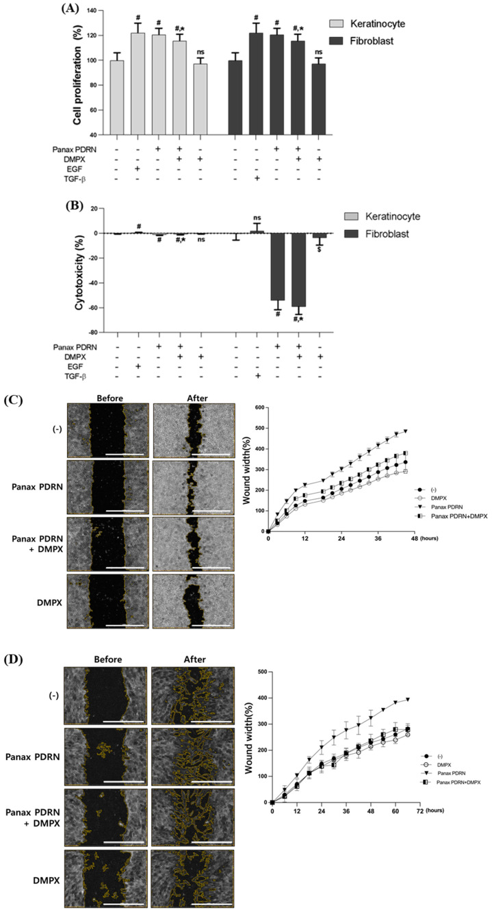
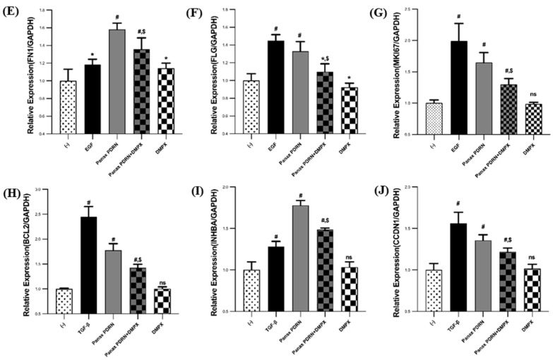
Figure 2.
Analysis of cell proliferation and regeneration-promoting effects of Panax PDRN on keratinocytes and fibroblasts. (A) Cell proliferation and (B) cytotoxicity were analyzed for Panax PDRN in keratinocytes (HaCaT) and fibroblasts (HDF). (C,D) A wound-healing assay was performed to confirm cell proliferation and regeneration ability in HaCaT and HDF (scale bar represents 1000 μm). (E–J) The expression of each gene related to cell proliferation and regeneration (fibronectin, filaggrin, Ki-67, Bcl-2, inhibin A, and cyclin D1) was confirmed via qRT-PCR in HaCaT and HDF. mRNA levels were normalized to GAPDH expression. Expression levels for each gene are indicated as fold-changes compared with the control levels. # p < 0.01 compared with basal levels; * p < 0.05; $ p < 0.01 compared with Panax PDRN levels; ns means statistically not significant; DMPX: 3,7-Dimethyl-1-propargylxanthine.


