Abstract
Background
The presence of T cells and suppressive myeloid cells in epithelial ovarian cancer (EOC) correlate with good and bad clinical outcome, respectively. This suggests that EOC may be sensitive to adoptive cell therapy with autologous tumor-infiltrating lymphocytes (TIL), provided that immunosuppression by myeloid-derived suppressor cells and M2 macrophages is reduced. Platinum-based chemotherapy can alleviate such immunosuppression, potentially creating a window of opportunity for T cell-based immunotherapy.
Methods
We initiated a phase I/II trial (NCT04072263) in patients with recurrent platinum-sensitive EOC receiving TIL during platinum-based chemotherapy. TILs were administered 2 weeks after the second, third and fourth chemotherapy course. Patients were treated in two cohorts with or without interferon-α (IFNa), as conditioning and TIL support regimen. The primary endpoint was to evaluate the feasibility and safety according to CTCAE V.4.03 criteria and the clinical response and immune modulatory effects of this treatment were evaluated as secondary endpoints.
Results
Sixteen patients were enrolled. TIL could be successfully expanded for all patients. TIL treatment during chemotherapy without IFNa (n=13) was safe but the combination with IFNa added to the chemotherapy-induced toxicity with 2 out of 3 patients developing thrombocytopenia as dose-limiting toxicity. Fourteen patients completed treatment with a full TIL cycle and were further evaluated for clinical and immunological response. Platinum-based chemotherapy resulted in reduction of circulating myeloid cell numbers and IL-6 plasma levels, confirming its immunosuppression-alleviating effect. Three complete (CR), nine partial responses and two stable diseases were recorded, resulting in an objective response rate of 86% (Response Evaluation Criteria In Solid Tumors V.1.1). Interestingly, progression free survival that exceeded the previous platinum-free interval was detected in two patients, including an exceptionally long and ongoing CR in one patient that coincided with sustained alleviation of immune suppression.
Conclusion
TIL therapy can be safely combined with platinum-based chemotherapy but not in combination with IFNa. The chemotherapy-mediated reduction in immunosuppression and the increase in platinum-free interval for two patients warrants further exploration of properly-timed TIL infusions during platinum-based chemotherapy, possibly further benefiting from IL-2 support, as a novel treatment option for EOC patients.
Keywords: Immunotherapy, Adoptive; Lymphocytes, Tumor-Infiltrating; Combined Modality Therapy; Myeloid-Derived Suppressor Cells; Tumor Microenvironment
WHAT IS ALREADY KNOWN ON THIS TOPIC
The clinical efficacy of adoptive transfer of tumor infiltrating lymphocytes (TIL) in epithelial ovarian cancer (EOC) is moderate when used as monotherapy, probably due to its immunosuppressive tumor environment. This warrants exploration of combined treatment to improve efficacy of TIL in EOC.
WHAT THIS STUDY ADDS
We showed that chemotherapy alleviates immunosuppression in EOC patients and that the combined treatment of TIL during chemotherapy was feasible and safe and resulted in an increased secondary platinum-free interval in some patients.
HOW THIS STUDY MIGHT AFFECT RESEARCH, PRACTICE OR POLICY
This study shows that combined chemoimmunotherapy provides a treatment window for a potentially more effective TIL therapy and may directly impact development of T cell-based treatment options for EOC patients with recurrent disease.
Background
The 5-year survival rate for epithelial ovarian cancer (EOC) patients is 40%–50%, due to the frequent diagnosis at an advanced stage and because most patients do not, or temporarily, benefit from treatment. Although survival for patients with BRCA-mutated EOC will increase after registration of olaparib, disease recurrence still occurs in approximately 70% of all patients.1 Relapsed advanced ovarian cancer is considered incurable, underscoring the unmet need for new treatment options, preferably before treatment-resistance develops.
The rationale to use immunotherapy in EOC patients is based on it being an immunogenic tumor, as indicated by the clear correlation between strong CD8+ T-cell infiltration and improved overall survival (OS) in EOC.2–4 The antitumor activity of CD8+ tumor-infiltrating lymphocyte T cells (TIL) is dramatically impaired by various immunosuppressive mechanisms and cells, including CD163+ myeloid cells, myeloid-derived suppressor cells (MDSCs) and regulatory T cells (Treg). These cells are major constituents of EOC tumors and their presence has been associated with disease progression and worse prognosis.5–7 In addition, accumulation of MDSC in the peripheral blood and ascites of patients has been associated with worse clinical outcome.8–10 Furthermore, tumor-induced elevation of circulating myeloid-cell frequencies have also been associated with bad prognosis,11–13 but this depends on the type of myeloid cells as reflected by the association of a high dendritic cell (DC) to MDSC ratio with a better OS in EOC patients.10
Platinum-based carboplatin-paclitaxel (CP) chemotherapy has been shown to significantly improve systemic immunity in EOC patients by reduction of circulating Treg.14 We observed a similar reduction in Treg and myeloid cell frequencies after CP-chemotherapy in cervical cancer, coinciding with enhanced vaccine-induced T-cell immunity and better survival.15 16 In a mouse model the CP-chemotherapy-mediated normalization of myeloid cells was observed in blood as well as in tumors, indicating that changes in the composition of circulating cells reflected the situation in the tumor.15 Optimal normalization of lymphocyte to myeloid cell ratios were obtained 2 weeks after the second chemotherapy in cervical cancer patients.15 Since Treg and immune-suppressive myeloid cells play a prominent role in EOC, a pronounced beneficial effect of CP-chemotherapy on immune-suppression is expected in EOC. Indeed, CP-chemotherapy in EOC patients resulted in a reduction of circulating Treg frequencies which coincided with an enhanced IFN-gamma response upon stimulation of peripheral blood T cells with autologous tumor antigens.14 This underscores that CP-chemotherapy potentially creates a window of opportunity for effective T cell-based immunotherapy in EOC, similar to what has been observed in cervical cancer.15 16 Treatment with autologous TIL has proven to induce durable responses in several solid tumors.17–20 Only few reports are available of TIL therapy in EOC showing clinical potential especially when TIL are given in combination with chemotherapy, while efficacy of TIL monotherapy is moderate.20–22 This limited clinical efficacy may be attributed to the relatively strong immunosuppressive TME in EOC compared with melanoma, where adoptive T cell therapy (ACT) is effective. We hypothesized that normalization of this immunosuppression by chemotherapy would enhance the clinical benefit of TIL infusions. We have shown that the generally used lymphodepleting preconditioning regimens and administration of high-dose IL-2 postinfusion23 24 can be replaced by a milder preconditioning regimen based on IFNa, with 29% clinical benefit in stage IV metastatic melanoma, including patients refractory to CTLA-4 and/or PD-1 checkpoint-blockade therapy.25
Altogether, this provided the rationale for a clinical trial in which recurrent platinum-sensitive EOC patients were treated with TIL infusions during standard platinum-based chemotherapy with or without additional IFNa. The primary objective was to evaluate feasibility and safety and the secondary objectives were to evaluate the clinical response and to study underlying mechanisms and immune parameters and contextualize these with clinical outcome.
Materials and methods
Patient selection
Patients were eligible if 18 years or older with histologically proven, recurrent, platinum-sensitive EOC and amenable to platinum-based chemotherapy. All patients had measurable progressive disease (PD) according to Response Evaluation Criteria In Solid Tumors (RECIST V.1.1) or confirmed elevated CA125>2 times the upper limit of normal (ULN) within 3 months. Patients had a life-expectancy of at least 3 months and a WHO performance status 0–2. Prior treatment, including immunotherapy, was allowed but had to be discontinued for at least 2 weeks before study entry. At least one resectable lesion was required for the isolation and expansion of TIL. Preferably at least one additional measurable target lesion was required for response evaluation. Alternatively, CA125 was used to assess the clinical response. Patients with brain metastasis, clinically significant heart disease (New York Heart Association class III or IV), active immunodeficiency or autoimmune disease, with other malignancy within 2 years prior to entry into the study, a known allergy to penicillin or streptomycin, or seropositivity for hepatitis B/C, HIV, human T cell lymphotropic virus (HTLV) or Treponema pallidum, were excluded.
Study design
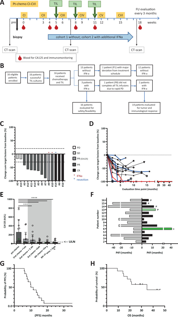
The combined treatment is depicted in figure 1A and consisted of six cycles of standard platinum-based chemotherapy (carboplatin area under the curve 5 or 6 plus paclitaxel 175 mg/m2 or cisplatin 50 mg/m2 in case of hypersensitivity to carboplatin), once every 3 weeks. TILs were given as three infusions at 3 weeks intervals without IFNa in cohort 1, starting 14 days after the second course of chemotherapy (ie, week 0) and also 2 weeks after the third and fourth chemotherapy cycle. TILs were given intravenously at a dose of 1–7.5×10⁸ T cells per infusion over a time period of 30–60 min during day care. Multiple TIL infusions are given to provide higher levels of circulating TIL for a prolonged time. The administered TIL dose is based on our experience in advanced melanoma where this dose range was shown to be safe and effective.25 After completion of three and six cycles of chemotherapy the clinical response was assessed by physical examination and imaging studies (CT scan) according to the RECIST V.1.1 and immune-related response criteria (irRC). If no progression of disease was observed and if TILs were available another cycle of three TIL infusions without chemotherapy was administered. During the follow-up, imaging was repeated every 3 months. Patients were regularly monitored for chemical and immunological parameters including CA125 levels and white blood cell counts. Heparinized venous blood was collected for immunological monitoring at several time points after infusion. Peripheral blood mononuclear cells (PBMCs) as well as serum/plasma samples were cryopreserved. In the second cohort, TIL infusions during chemotherapy were combined with pegylated-IFNa (Pegasys, Intron-A, 1 µg/kg/week s.c. with a maximum of 90 µg/week) starting 1 week before the first TIL infusion and for 12 weeks in total. The primary objective was to evaluate the safety of TIL with or without low dose IFNa during platinum-based chemotherapy, as assessed by the NIH Common Terminology Criteria for Adverse Events (CTCAE) V.4.03. As secondary objectives the clinical response including the best overall response (BOR) according to RECIST V.1.1 and irRC, the disease control rate (DCR) defined as complete response (CR)+partial response (PR)+stable disease (SD) at 6 months, the progression-free survival (PFS), OS and platinum-free interval (PtFI) pretreatment and post-treatment were evaluated. The PFS is defined as the duration from start of platinum-based chemotherapy until first observation of radiologically confirmed PD. The PtFI is defined as the duration from the last platinum-based chemotherapy cycle until the first observation of radiologically confirmed PD. In addition, hypothesis-related immune parameters, including immune-modulation by chemotherapy, immune modulation by TIL with or without IFNa and phenotypic and functional characteristics of TIL were evaluated.
Figure 1.
Study design and patient inclusion and clinical response evaluation. (A) Treatment schedule given in weeks. Platinum-based-chemotherapy is given as six consecutive cycles (CI thru CVI) starting form week 0. Three TIL infusions are given two weeks after the second, third and fourth chemotherapy cycle. (B) Patient disposition; 16 patient were enrolled and received chemotherapy+TIL and three of them also received IFNa. All these patients were evaluated for safety and feasibility. Two patients were not evaluated for tumor and immunological response due to gross deviation from the treatments schedule or rapid PD precluding completion of TIL infusions. (C–H) Clinical response was evaluated according to RECIST V.1.1. in patients (n=14) that completed chemotherapy and TIL infusion cycles. (C) Waterfall plot showing the % change in sum of target lesion size from baseline. The best overall response is indicated in the color of the bars. The red asterisk indicates the two patients that received IFNa in addition to chemotherapy and TIL, the blue asterisk indicates the patient that obtained a CR after resection of the remaining target lesion. Patient #9 did not have an evaluable target lesion, but was evaluated based on reduction of CA125 level. (D) Spider plot showing the change in sum of target lesions from baseline in time. Blue lines show the response in patients that received PARP inhibitor after completion of the chemotherapy plus TIL cycles. In red the patient that obtained a durable CR after chemotherapy and TIL is depicted. (E) Change of tumor-marker CA125 measured in blood samples before, during and after therapy. Bars represent the mean CA125 levels (kU/L) and dots the values for individual patients. The gray area indicates the period when the 3 consecutive TIL infusions were administered. The dotted line indicates the upper level of normal (ULN). Significant change from baseline: ***p<0.001, ****p<0.0001 by Friedman with Dunn’s correction for multiple comparisons. (F) Platinum free interval (PtFI) prior to treatment (gray bars, to the left) and post-treatment (black and green bars, to the right), measured as time to progression according to RECIST V.1.1 from the time point of the last chemotherapy cycle (in months). The green bars indicate patients with a longer PtFI post-treatment than pretreatment. Patients that received PARPi after chemotherapy plus TIL are indicated by the p and ongoing response by the >behind the appropriate bars. (G) PFS as evaluated according to RECIST V.1.1 and (H) OS curves according to Kaplan-Meier. CR, complete response; OS, overall surviva; PD, progressive disease; PFS, progression-free survival; PR, partial response; RECIST, Response Evaluation Criteria In Solid Tumors; SD, stable disease.
Generation of TIL for infusion
TILs for infusion were cultured from a small resected tumor sample or biopsy, as previously described25 26 (online supplemental material).
jitc-2023-007697supp002.pdf (296.5KB, pdf)
Immunological response evaluation in blood samples
Cytokine analysis in serum/plasma
The serum/plasma concentrations of immune-modulating proinflammatory/anti-inflammatory cytokines IL-6 (Immunotools, 31670069) and IL-8 (Invitrogen, CNB0011IL-7) and homeostasis regulating-cytokines IL-7, IL-15 and IL-21 (R&D diagnostics; DY207; Biolegend, 435104; Mabtech; 3540–1 H-6, respectively) were analyzed using ELISA, according to the manufacturer’s instructions.
Immunophenotyping of PBMC
PBMC samples were immunophenotyped using a 40-marker panel and multispectral flow cytometry as described in online supplemental methods.
Lymphocyte proliferative and antigen presenting capacity
The proliferative potential of PBMC collected at the indicated time-points was evaluated using a proliferation assay and the same samples were used to assess the effect of treatment on the antigen-presenting capacity of PBMC using a mixed lymphocyte reaction (MLR), as previously described,15 with minor modifications (see online supplemental material).
Immunophenotyping of TIL batches
To assess the phenotypic characteristics of TIL batches used for infusion, cryopreserved reference vials of TIL were thawed and divided into multiple samples for staining with separate antibody panels for activation/inhibitory, memory, homing and regulatory T cell markers (see online supplemental table 2), respectively, as we previously described25 (online supplemental material).
Functional analysis of the infused TIL
Tumor reactivity: The antigen-specificity of the infusion product was tested against a broad panel of EOC cell lines that were (partially) matched for at least one HLA-class I allele with the corresponding patient using IFNg release as a read-out, as previously described25 (online supplemental material). IFNg secretion in response to CD3/CD28 activation beads (Dynabeads, Thermofisher ratio 1:4 beads to T cells) was used to assess the activation and cytokine release potential of the TIL batches.
Statistical analysis
Descriptive statistics were used to summarize patient baseline characteristics at start of treatment. Survival and PFS were estimated according to the Kaplan-Meier’s method using GraphPad Prism version V.9.3.1. 1 for Windows (GraphPad Software, La Jolla, California, USA). Although group comparisons based on <6 months or >6 months PFS is more generally accepted, only one of the patients evaluable for clinical and immunological responses experienced a PFS<6 months. Therefore, we categorized patients based on < or >median PFS for comparisons and this corresponds grosso modo with a PtFI>6 months. Statistics used for analysis of immune parameters are described in online supplemental methods and figure legends.
Results
Patient characteristics and TIL expansion
In total, 16 patients were included in our phase I/II clinical trial (NCT04072263) between November 2018 and March 2021. Patient characteristics are given in table 1.
Table 1.
Patient characteristics
| All patients (n=16) |
Chemo+TIL (n=13) |
Chemo+TIL+IFNa (n=3) | |
| Age | |||
| Median age and range (year) | 61 (29–77) | 62 (29–77) | 58 (53–73) |
| WHO status; n (%) | |||
| 0 | 6 (38) | 6 (46) | 0 (0) |
| 1 | 10 (62) | 7 (54) | 3 (100) |
| 2 | 0 (0) | 0 (0) | 0 (0) |
| Tumor type | |||
| HGSOC | 15 (94) | 12 (92) | 3 (100) |
| LGMOC | 1 (6) | 1 (8) | 0 (0) |
| Platinum sensitivity | |||
| >6 and <12 months | 6 (38) | 5 (38) | 1 (33) |
| >12 months | 10 (62) | 8 (62) | 2 (67) |
| Mutation status; n (%) | |||
| BRCA mutation | 2 (13) | 2 (15) | 0 (0) |
| HR status; n (%) | |||
| ER− and PrR− | 4 (25) | 3 (23) | 1 (33) |
| ER+ or PrR+ | 7 (44) | 6 (46) | 1 (33) |
| Unknown | 5 (31) | 4 (31) | 1 (33) |
| Previous lines of chemotherapy; n (%) | |||
| 1 | 12 (75) | 10 (77) | 2 (67) |
| 2 | 3 (19) | 2 (15) | 1 (33) |
| >3 | 1 (6) | 1 (8) | 0 (0) |
| Other previous treatments; n (%) | |||
| Bevacizumab | 2 (13) | 1 (8) | 1 (33) |
| Endocrine therapy | 2 (13) | 1 (8) | 1 (33) |
| PARPi | 2 (13) | 1 (8) | 1 (33) |
ER, estrogen receptor; HGSOC, high grade serous ovarian cancer; HR, hormone receptor; LGMOC, low-grade mucinous ovarian cancer; PrR, progesterone receptor.
All but one of the enrolled patients had a high-grade serous ovarian carcinoma histology type ovarian cancer. All patients were treated with platinum-based chemotherapy and started TIL infusions. Two patients (P1 and P8) were not evaluable for the toxicity and efficacy of TIL with (P8) or without IFNa (P1) during chemotherapy, respectively (figure 1B). For one of these patients (P1), additional toxicity of the standard chemotherapy required such an adjustment (dose reductions and delay) of the treatment schedule that proper evaluation of the safety and effect of TIL infusions was no longer possible. In the case of patient P8 not all TIL infusions of the first cycle could be given due to the rapid disease progression. The remaining fourteen patients completed treatment with at least one full cycle of three TIL infusions and were further evaluated for tumor and immunological response parameters. The median age of all 16 patients was 61 years (range 29–77) with no (significant) difference between the patients that received additional IFNa (n=3) and those that did not (n=13). All patients had WHO status 0–1 and recurrent platinum-sensitive disease with a PtFI varying between 6 and 12 months, that is, a partial platinum-sensitive recurrence, in 38% (6/16), and a PtFI of more than 12 months (range 12–55 months) in 62% (10/16) of the patients. CA125 levels before start of treatment ranged between 14 and 928 kU/L (median 219 kU/L). BRCA mutation was detected in 13% (2/16) of the patients. Twenty-five percent of the patients received more than one line of prior systemic (chemo)therapies.
All patients were discharged the same day of obtaining an excisional biopsy for TIL collection. TIL could be successfully cultured from tumor samples or biopsies for all included patients, although for some patients (P8, P10, P13 and P16) this required an extra biopsy from another lesion, possibly reflecting intrapatient heterogeneity in immune infiltrate between lesions.27 A median initial culture time of 16 days (range 9–30 days) followed by rapid expansion during 14 days, resulting in a 1516-fold (range 106–2769) expansion, was required to obtain enough cells for infusion.
Safety of TIL with or without additional IFNa during platinum-based chemotherapy
The first cohort of six patients was treated with TIL and platinum-based chemotherapy without IFNa which did not result in toxicity other than what is known from platinum-based chemotherapy alone (table 2). In the majority of patients, all observed side effects were present before TIL infusions and, therefore, definitely not related to TIL infusions.
Table 2.
Adverse events ≥grade 3 and their relation to treatment for individual patients
| Patient | Related to | ||||
| ID | (S)AE term | Grade | Chemo | TIL | IFNa |
| Cohort 1 without IFNa | |||||
| P1* | Neutrophil count decreased | 3 | YES | y/n | – |
| Platelet count decreased | 3 | YES | NO | – | |
| White cell count decreased (pan cytopenia) | 3 | YES | NO | – | |
| Lymphocyte count decreased | 3 | YES | y/n | – | |
| Anemia | 3 | y/n | y/n | – | |
| Nausea | 3 | y/n | NO | – | |
| P2 | Neutrophil count decreased | 3 | YES | NO | – |
| P3 | Neutrophil count decreased | 4 | YES | NO | – |
| White cell count decreased | 3 | YES | NO | – | |
| P4 | Neutrophil count decreased | 3 | YES | NO | – |
| P5 | Neutrophil count decreased | 3 | YES | NO | – |
| Upper respiratory infection | 3 | NO | NO | . | |
| P6 | Vomiting | 3 | y/n | NO | – |
| Diarrhea | 3 | YES | NO | – | |
| P7 | None | – | – | – | – |
| P11 | None | – | – | – | – |
| P12 | Neutrophil count decreased | 4 | YES | NO | – |
| P13 | Neutrophil count decreased | 3 | YES | NO | – |
| P14 | Neutrophil count decreased | 3 | YES | NO | – |
| P15 | Neutrophil count decreased | 3 | YES | NO | – |
| P16 | Neutrophil count decreased | 4 | YES | NO | – |
| Cohort two with IFNa | |||||
| P8* | None | – | – | – | – |
| P9 | Platelet count decreased | 3 | YES | y/n | y/n |
| White cell count decreased (pancytopenia) | 3 | YES | y/n | NO | |
| Neutrophil count decreased | 4 | YES | y/n | y/n | |
| P10 | Neutrophil count decreased | 4 | YES | y/n | y/n |
| White cell count decreased | 3 | YES | NO | y/n | |
| Platelet count decreased | 3 | y/n | NO | y/n | |
| Other general; malaise with PD | 3 | NO | NO | NO | |
All grade 3 or more (S)AEs and their relation to therapy according to CTCAE V.4.03 are given. (S)AEs grade 4 are in bold. Relatedness to therapy: YES; definitely related, NO; definitely not related and y/n; possibly related. ‘–‘=not applicable.
*P1 and P8 grossly deviated from treatment schedule and were only evaluated for safety/feasibility.
CTCAE, Common Terminology Criteria for Adverse Events; PD, progressive disease; (S)AE, (serious) adverse events.
In the second cohort, IFNa was added to the treatment. An aggravation of chemotherapy-induced pancytopenia and especially neutropenia (grade 4) and thrombocytopenia (grade 3) was observed in two out of three patients resulting in DLT. Therefore, the addition of IFNa to TIL during chemotherapy was discontinued. The remaining patients (P11 thru P16) were treated according to the first cohort, resulting in a total of 13 patients treated with TIL during chemotherapy without IFNa.
Clinical evaluation
The clinical response was evaluated in 14 patients who completed at least one full cycle of three TIL infusions and without gross deviation from the treatment schedule at the data cut-off point of April 2023. The obtained BOR was recorded as three CR, nine PR and two SD; resulting in an objective response rate (ORR) of 86% (figure 1C) and a 100% DCR at 6 months. Response recorded as a more than 30% decrease in the sum of the target lesions (or reduction of CA125 cancer marker in P9) was reached at the first evaluation point in 12/14 patients (figure 1D). In all patients, a drop in tumor-marker CA125 level was observed after treatment, that decreased below the ULN threshold of 35 kU/mL in 12/14 patients (figure 1E). The PtFI observed after platinum-based chemotherapy and TIL exceeded the PtFI obtained after the last platinum-based treatment prior to enrolment in patient P13 and patient P6 (green bars in figure 1F). The PtFI post-treatment ranged from 2 to 35 months (median 6.5 months). Interestingly, for P6 the ongoing PFS has already increased fivefold after combined TIL and platinum-based chemotherapy. In general, the response duration varied and resulted in a median PFS of 10.7 months (range 6–39+ months), corresponding to a median platinum-free survival of 6.5 months (figure 1F and figure 1G). At the data cut-off point (April 2023), median probability of OS was 34.7 months according to Kaplan-Meier analysis (figure 1H).
Translational studies
Detailed characterization of infused TIL
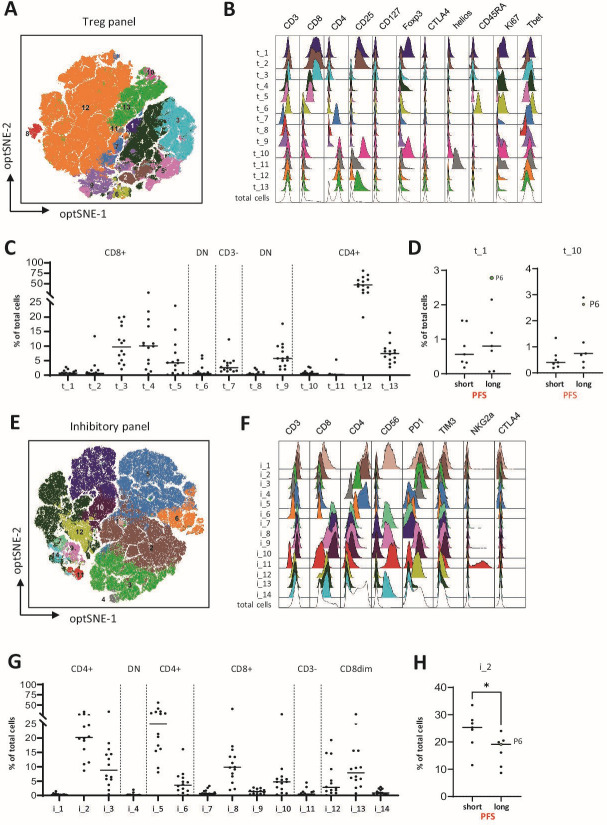
TIL consisted of a mix of on average two-thirds CD4 and one-third CD8 T cells (figure 2), which did not differ between patients with a shorter or longer than median PFS. T-distributed stochastic neighbor embedding (tSNE) was used to cluster similar cells in 2D-plots. In TIL stained with a Treg marker panel we defined 13 different populations including a CD4+ cell population (t_1), reflecting CD4+Treg as determined using the consensus marker expression profile,28 as well as a CD8+ Treg-like cell population (t_10), both highly expressing CD25 and FoxP3 (figure 2A–D). However, populations t_1 and t_10 made up 1% or less of the final expanded TIL product. None of the T-cell populations identified by the Treg marker panel distinguished patients with a shorter or longer than median PFS (figure 2D) or OS (online supplemental figure S1).
Figure 2.
Immunophenotyping of infused TIL. Administered TILs were stained with antibody panels specific for regulatory T cells (t) and inhibitory/activation (i) T cell markers, as indicated in online supplemental table 2. High-dimensional single cell data analysis of the stained TIL were performed by opt-Distributed Stochastic Neighbor Embedding (optSNE) and FLOWSOM using OMIQ software (A) Overlay of 13 different FLOWSOM clusters (t_1 thru t_13) for all TIL batches stained with the Treg marker Ab-panel plotted on the optSNE map. (B) Expression levels of each of the indicated regulatory T cell markers are depicted for the individual populations t_1 thru t_13 and for the total TIL population. (C) Frequencies of the identified populations in the total TIL populations are shown and grouped on major phenotypic characteristics, shown above the data. (D) Frequencies of CD8+ (t_1) and CD4+ (t_10) T cell populations with a regulatory T cell phenotype are shown as percentage of total TIL for patients with a shorter (short) or longer (long) than median PFS, respectively. (E) Overlay of 14 different FLOWSOM clusters (i_1 thru i_14) for all TIL batches stained with the inhibitory/activation marker Ab-panel plotted on the optSNE map. (F) Expression levels of the indicated inhibitory/activation T cell markers is depicted for these individual populations i_1 thru i_14 and for the total TIL population. (G) Frequencies of the identified populations in the total TIL populations are shown and grouped on major phenotypic characteristics, shown above the data. (H) Frequencies of the CD4+PD-1+Tim-3+ (i_2) T cell population is shown as percentage of total TIL for patients with a shorter (short) or longer (long) than median PFS, respectively. *p<0.05 by Mann-Whitney U test, and P6 indicates the patient with the durable CR after treatment. CR, complete response; PFS, progression-free survival; TIL, tumor-infiltrating lymphocytes.
jitc-2023-007697supp001.pdf (2.5MB, pdf)
Based on expression of inhibitory/activation markers, we distinguished 14 different cell clusters in tSNE plots (figure 2E–H and online supplemental figure S2). Especially within the CD4+ T cells, populations i_1, i_2 and i_3 show clear expression of the inhibitory/activation marker PD-1. While populations i_1, and i_3 moderately expressed any of the other inhibitory/exhaustion markers, population i_2 coexpresses Tim-3, possibly reflecting a more exhausted phenotype. Patients with a longer than median PFS were treated with TIL containing significantly less of these i_2 CD4+PD-1+Tim-3+ T cells (figure 2H). Within the different CD8+ T cell populations, populations i_9 and i_10, expressed PD-1 but little to no other exhaustion markers, reflecting an activated, but not exhausted phenotype. Furthermore, staining for memory and homing markers shows that most TIL express a T cell effector phenotype (Teff, CD45RO+CCR7-CD62L-CD27-CD28-)) and less than 5% express CCR7 and a low level of CD62L representing a central memory-like (Tcm) phenotype (population m_4 in online supplemental figure S3). Moderate expression of CXCR3, CCR4 and CCR6, representing activated Th1 and effector T cells is observed on (a subfraction of) the largest CD4+ (h_2) and CD8+ (h_10) T cell populations, respectively (online supplemental figure S4). Differences in memory or homing markers were not associated with shorter or longer than median PFS or OS (online supplemental figure S3 and S4).
To assess tumor cell recognition, TILs were cocultured with a panel of ovarian cancer cell lines, partially matched for HLA class I. In 1 of the tested TIL batches (P16) modest recognition of one or more of the matched cell lines was detected (online supplemental figure S4), suggesting that in most cases tumor-reactivity may be present against private antigens but not against shared tumor antigens, similar to what we found in melanoma.25 All TIL batches were able to secrete the type 1 cytokine IFNg when stimulated with Staphylococcus enterotoxin B (SEB) and anti-CD3/CD28 but differences in IFNg production, as well as the proliferation rate during rapid expansion of TIL and the total administered dose and percentage CD8+ in infused TIL, were not related to PFS or OS (online supplemental figure S6).
Therapy-induced changes in blood counts of myeloid cells but not lymphocytes
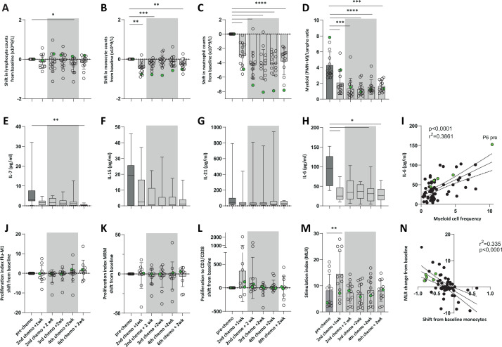
The effect of platinum-based chemotherapy on the absolute numbers of circulating leukocytes, in particular neutrophils, monocytes and lymphocytes, was analyzed by standard white blood cell counts in all 14 evaluable patients. While absolute numbers of monocytes and neutrophils were reduced, the absolute lymphocyte counts were hardly affected (figure 3A–C). In general, the monocyte counts returned to baseline levels at 2 weeks after the third chemotherapy cycle, while the reduction in neutrophils was retained. Interestingly, the absolute monocyte and neutrophil numbers remained reduced throughout all TIL infusion time points in the two patients with increased PtFl but the strongest reduction was observed in the patient with the most durable PtFl (P6; green dots in figure 3B–C). As a result of the chemotherapy-induced changes observed in the total patient group, the myeloid cell (monocytes plus neutrophils) to lymphocyte ratio is drastically reduced. Maximal reduction is reached 2 weeks after the second chemotherapy cycle and retained until 2 weeks after the sixth chemotherapy cycle (figure 3D).
Figure 3.
Therapy-induced changes in the circulation. (A–D) White blood cell counts were performed on blood samples obtained at the indicated time points before and during treatment. Shifts from baseline in absolute (A) lymphocyte, (B) monocyte, and (C) neutrophil cell counts (×109/L) are shown. (D) Changes in the ratio of myeloid (neutrophils (PMN) and monocytes (M)) cells to lymphocytes are depicted. (E–I) Plasma samples were collected at the indicated time points before and during treatment to assess changes in circulating cytokine levels. The levels of (E) IL-7, (F) IL-15, (G) IL-21, and (H) IL-6 cytokines were analyzed by ELISA. (I) The correlation between circulating myeloid cell frequencies and IL-6 cytokine levels is shown by linear regression. P6-pre, indicates the high IL-6 level corresponding to high myeloid cell frequency observed in P6 prior to treatment. (J–N) Treatment effect on circulating lymphocyte and antigen presenting cell function was assessed in peripheral blood mononuclear cells isolated from venous blood samples collected at the indicated time-points before and during treatment. The proliferative response of lymphocytes to (J) influenza M1 (influenza) antigen, (K) memory response mix (MRM) and (L) CD3/CD28 activation beads, was measured by 3H-thymidine incorporation. The changes are depicted as shift from baseline of the proliferation index. (M) The APC function was evaluated using a mixed lymphocyte reaction (MLR). (N) The correlation between APC function (MLR change from baseline) and reduction in circulating monocyte numbers (shift from baseline) is shown by linear regression. The gray areas indicate the period when the three consecutive TIL infusions were administered. Results for the patient with a durable CR (P6) are depicted in green. *p<0.05, **p<0.01, ***p<0.001, ****p<0.0001 indicate significant changes from baseline by Kruskal-Wallis with Dunn’s correction for multiple comparisons. CR, complete response.
Therapy-induced changes in serum/plasma factors
The effect of platinum-based chemotherapy and subsequent TIL infusions did not overtly affect the levels of the T-cell supportive circulating cytokines IL-7, IL-15 and IL-21 (figure 3E–G).
In addition, we analyzed changes in IL-6 and IL-8 levels, since these cytokines were described to promote the attraction of and differentiation toward immune suppressive myeloid cells and to correlate with disease progression and worse prognosis in EOC.6 8 The IL-6 serum level was clearly reduced by chemotherapy (figure 4H) starting from the first evaluation point onward and throughout the period that TIL infusions were given. IL-6 levels were correlated to the total number of myeloid cells as determined by differential white cell counts (figure 4I). IL-8 serum levels were low and hardly altered (not shown).
Figure 4.
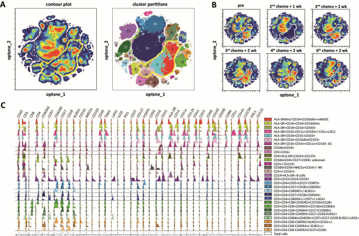
Detailed evaluation of treatment effect on circulating leukocyte populations. PBMCs were isolated from venous blood samples collected at the indicated time points before and during treatment and stained with a panel of 40 markers indicated in online supplemental table 1. High-dimensional single cell data analysis of the stained PBMC was performed by opt-Distributed Stochastic Neighbor Embedding (optSNE) and FLOWSOM using OMIQ software (A) OptSNE plots visualizing a contour plot (left) and cluster partitions by FLOWSOM (right) for all patient samples. Populations representing myeloid cells are indicted by the yellow bordered area. (B) Changes of the cell clusters visualized in contour plots are shown for the different time points and show clear reduction of the myeloid cell populations, especially at 1 week after the second chemotherapy cycle. (C) The major and/or specific combination of markers distinguishing the defined cell populations are indicated (on the right) and the complete expression profile for each individual marker (indicated above the graph) is shown for each cell population (in different colors) and for the total cell population (in white, lower panels) in histogram plots. PBMC, peripheral blood mononuclear cell.
Transient therapy-induced increase in T-cell stimulatory capacity of antigen presenting cells
Next, we studied whether the platinum-based-chemotherapy influenced the function of immune cells. PBMCs were tested in a lymphocyte stimulation test to assess their reactivity to common recall antigens, such as influenza antigen (influenza) or memory response mix (MRM). No overt changes were observed. A slightly improved reactivity was detected when lymphocytes were stimulated with CD3/CD28 beads (figure 3J–L). Importantly, all PBMC samples showed a very potent proliferative response to CD3/CD28-bead stimulation and the stimulation index in response to MRM was comparable to what we previously observed in healthy donors and in cervical cancer patients after chemotherapy-induced normalization of circulating myeloid cell frequencies (online supplemental figure S7).15 These data demonstrate that lymphocyte function is not negatively affected by the chemotherapy treatment in EOC.
A significantly improved APC function was detected by MLR 1 week after the second cycle of chemotherapy (1 week before TIL infusion, figure 3M). This effect was transient and correlated with reduced monocyte numbers (figure 3N) and returned to baseline during the remainder of the chemotherapy cycles (figure 3M). Notably, the improved APC function of PBMC from the patient with a long PtFl after therapy was sustained throughout the complete treatment (green dots in figure 3M and online supplemental figure S7).
Therapy-induced changes in specific cell populations in the circulation
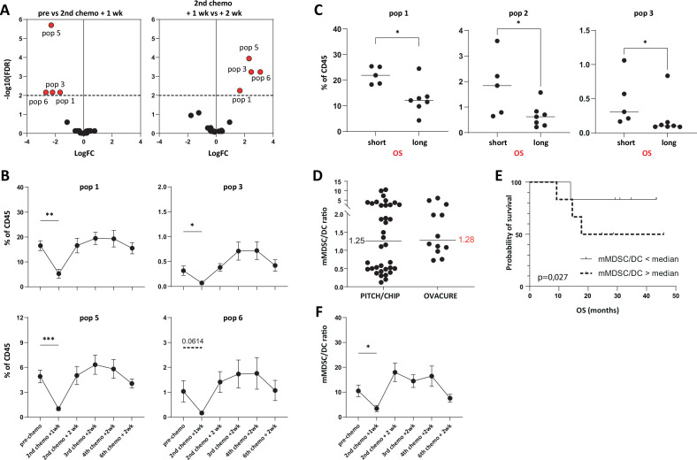
Next, in-depth analysis of therapy-induced changes in PBMC and their pre-treatment frequencies in relation to PFS and OS was performed. From 12 of the 14 evaluable patients samples of all time points were available. Immunophenotyping using a 40-marker panel and optSNE-FlowSOM cluster-segmentation revealed 29 different subpopulations of immune cells (figure 4A). The density plots of the PBMC samples at different time points show a very clear change, mainly in populations 1, 3, 5, and 6 and most pronounced at 1 week after the second chemotherapy cycle (figure 4B and figure 5A). The largest of the therapy-altered cell populations represents classical mMDSC (population 1). Population 3 comprises non-classical HLA-DR+CD14+CD16+CD163+ monocytes and populations 5 and 6 HLA-DR+CD14+CD163+M2 macrophage-like cells (figure 4C and online supplemental figure S8). The observed reduction at 1 week after the second chemotherapy cycle is transient and bounces back 2 weeks after the second chemotherapy cycle (figures 4B and 5B). This is also apparent when the most significant changes in populations between pretreatment vs week 1 and week 1 vs week 0 values are shown in a volcano plot (figure 5A) or at the quantitative level (figure 5B). Pretreatment frequencies of populations 1, 3 and 2 (CD163dim M2 macrophage-like cells) correlated with shorter OS (figure 5C). Pretreatment frequencies of all other subpopulations neither correlate to OS nor significantly changed during therapy (online supplemental figure S9 and S10). Clearly the lymphocytes (figures 4 and 5A), including several subpopulations of CD4 and CD8 T cells (populations 17–21 and 23–25, respectively, in online supplemental figure S10), were not altered during therapy.
Figure 5.
Treatment affects circulating myeloid cell numbers and the mMDSC/DC ratio. (A) Treatment-induced changes in specific cell populations, defined and described in figure 4, were further analyzed. Populations 1, 3, 5, and 6 showed the largest and most significant change when frequencies between pretreatment versus second chemotherapy+1 week samples (left panel), and also when samples from 1 vs 2 weeks after the second chemotherapy (right panel), were compared using OMIQ and edgeR. (B) Frequencies of sequential blood samples show a transient and significant decrease in populations 1, 3, 5, and a near significant decrease in population 6 from baseline, (C) Baseline frequencies of population 1, 2 and 3 were depicted for patients with a shorter and longer than median OS, respectively. Given p values according to Mann-Whitney U test. (D) Baseline mMDCS/DC ratios were calculated and compared with ratios previously determined and described for ovarian cancer patients included in the PITCH or CHIP trial.10 In brief, CD3−CD19−CD56-HLA-DR−/lowCD14+CD15− (mMDSC) and CD3−CD19−CD56−HLA-DR+CD14−CD11c+ (DC) were determined as percentage of total CD45+ cells after multicolor flowcytometry (as described in figure 4) and ratios of these cell populations are plotted for evaluated patients in the PITCH-CHIP trial (n=36) and OVACURE trial (n=12). (E) Kaplan-Meier plots show the survival of patients divided in patients with an mMDSC/DC ratio above (dotted line) and below (solid line) median. (F) mMDSC and DC frequencies were determined in sequential blood samples obtained at baseline and during treatment and show a significant but transient, treatment-induced reduction in mMDSC/DC ratio at 1 week after the second chemotherapy cycle.*p<0.05, **p<0.01, ***p<0.001 by Kruskal-Wallis with Dunn’s correction for multiple comparisons. DC, dendritic cell; MDSC, myeloid-derived suppressor cells; OS, overall survival.
We reported that the baseline mMDSC/DC ratio is an independent predictive factor for OS in two different cohorts of EOC.10 Therefore, the pretreatment mMDSC/DC ratio was determined as described and compared with this earlier cohort,10 showing high similarity (median 1.28 vs 1.25) in its distribution among patients (figure 5D). Furthermore, also in the current patient cohort, a baseline mMDSC/DC-ratio below median is associated with a better OS, although not significant due to the limited patient number (figure 5E). Interestingly, platinum-based chemotherapy resulted in a significant but transient reduction of the mMDSC/DC ratio (figure 5F).
Summarizing translational data of the patient experiencing an unexpected long CR and PtFI after treatment
Patient P6 experienced a very long lasting CR after therapy resulting in a much longer PtFI after therapy than observed prior to inclusion in this trial. The translational data indicate that this patients stands out from all other patients and to fully comprehend their potential impact, these results are summarized here.
Compared with TIL given to the other patients, TIL administered to this patient show lower frequencies of population i_2, which was significantly associated with a longer PFS (figure 2H), and these TIL produced the highest level of IFNg when stimulated with CD3/CD28 activation beads (online supplemental figure S6).
At baseline, this patient had the highest level of circulating myeloid cell frequencies corresponding to the highest myeloid cell/lymphocyte ratio (figure 3D), mMDSC/DC ratio (figure 5D) and circulating IL-6 level (figure 3I), all depicted in green dots. The platinum-based chemotherapy induced reduction of monocytes, neutrophils and myeloid cell/lymphocyte ratio (figure 3B–D, respectively) was most pronounced and retained for the longest period in this patient. These chemotherapy induced changes associated with a reduction of circulating IL-6 levels (figure 3H,I) and a slight but more prolonged improvement of APC function than observed for all other patients (figure 3M).
Discussion
In this study, we show that treatment of patients with recurrent EOC with the combination of TIL during platinum-based chemotherapy is feasible and safe but that addition of IFNa resulted in aggravation of chemotherapy induced neutropenia and thrombocytopenia. It was feasible to obtain TIL in a timely fashion for treatment of all patients. Platinum-based chemotherapy resulted in a reduction of immunosuppressive myeloid cells, and in combination with TIL a DCR of 100% at 6 months and an ORR of 86% was obtained. The median PFS of 10.7 months in the current trial is comparable to the median PFS of 9 months reported in the Calypso trial (n=507)29 and a median PFS of 13 months and ORR of 66% in the ICON4 trial (n=392)30 for patients with a recurrent platinum-sensitive EOC. Importantly, the clinical response to retreatment with platinum-based chemotherapy depends on the PtFI after previous treatment and a progressively shorter PtFI is reported after each subsequent line of platinum-based chemotherapy.31 Therefore, the PtFI observed for patient P6 after similar platinum-based chemotherapy with TIL far exceeds that what could be expected based on the PtFI after the previous line of platinum-based treatment (>42 vs 8 months). Such a durable response after retreatment with a similar line of platinum-based chemotherapy potentially can be attributed to the concomitant administration of TIL and holds promise for future studies on TIL infusions during platinum-based-chemotherapy.
The potential of adoptively transferred TIL as treatment for EOC has thus far only been investigated in a limited number of clinical trials. ACT after commonly used non-myeloablative conditioning and IL-2 support resulted in disease stabilization for 3–5 months without severe side effects in a small cohort (n=6) of platinum-resistant EOC patients.20 Combined TIL infusions with cisplatin-based chemotherapy in 10 patients with EOC after a single cyclophosphamide conditioning cycle, without IL-2 support, resulted in an ORR of 90%. However, 8/10 of these patients did not have any prior treatment and most likely responded very well to platinum-based chemotherapy, which has a general first-line response rate of 60%–90%. This precludes distinguishing clinical benefit of chemotherapy from that of chemotherapy with TIL.22 However, the reported early signs of clinical activity by the combined treatment, together with the earlier observed window of opportunity created by chemotherapy15 16 and association of immune infiltration with recurrence and OS2 provided a rationale for further exploration of the clinical effect and additional investigation of underlying mechanisms of action of the combined treatment, as performed in our trial.
We did not use non-myeloablative conditioning or IL-2 support,25 but standard chemotherapy and IFNa as TIL-support regimen, aiming to reduce treatment-related toxicity and thereby increase the number of patients eligible for TIL treatment. Unfortunately, IFNa added to chemotherapy-induced toxicity and could not be combined in subsequently treated patients. Platinum-based chemotherapy induced a transient reduction of the absolute number of monocytes and a more sustained reduction of total leukocytes and neutrophils. This is in line with previous reports showing that EOC patients present with high neutrophil to lymphocyte ratio (NLR), and that a platinum-based chemotherapy-induced reduction thereof correlates with clinical response to therapy.32 33 We show that absolute lymphocyte counts nor lymphocyte function were affected by the used chemotherapy. In addition, we showed that the myeloid cell/lymphocyte ratio was strongly reduced, reaching a maximal effect at 2 weeks after second chemotherapy. Moreover, we showed that chemotherapy preserved and even transiently improved antigen presenting potential of circulating immune cells, coinciding with the reduction of several CD14+ myeloid cell populations, including the classical mMDSC and CD163+ cells. These results confirm our previous findings in cervical cancer patients where platinum-based chemotherapy (CP) normalized the aberrant tumor-induced immune cell composition, and in particular, the number of circulating CD14+myeloid cells resulting in an improved T cell response to recall antigens.15 16 It also confirms our notion that in order to infuse TILs when the excessive numbers of myeloid cells are reduced, one to 2 weeks after the second chemotherapy cycle is the optimal timing for combined immunochemotherapy.
EOC patients presenting with increased tumor-induced systemic inflammation, including increased NLR and IL-6 levels, may exhibit an impaired lymphocyte-mediated immune response to tumors, underlying their worse prognosis and increased potential for tumor recurrence.33 Here, we show that platinum-based chemotherapy may alleviate immunosuppression via the reduction of several circulating myeloid cell populations (neutrophils, monocytes and mMDSCs) and the plasma IL-6 level. Increased IL-6 levels have been correlated to the abundance of mMDSC and worse prognosis in EOC,8 and is described to be involved in expansion, accumulation and promotion of the immunosuppressive phenotype of MDSCs via activation of the STAT3-signaling pathway.8 34 Our previous studies showing that EOC with high epithelial IL-6 expression display a dense infiltration of mature myeloid cells,6 and that ovarian cancer cell-derived IL-6 can polarize differentiation of monocytes into the suppressive CD163+M2 like macrophages,35 further adds to the notion that platinum-based chemotherapy may change the immunosuppressive TME in favor of the antitumor immune response. However, the chemotherapy-driven reduction of mMDSC in EOC is transient in most of the patients, except the one with an exceptionally good clinical response. Hence, it would be of interest to investigate if this effect could be prolonged. Other chemotherapeutics used in EOC, such as doxorubicin and gemcitabine, do not alter the numbers of circulating MDSC.10 One possible option is to use tocilizumab (anti-IL-6 receptor). We previously reported a clinical trial showing that at a functionally effective dose of tocilizumab, the myeloid cells expressed less STAT3 and produced more IL-12, and this coincided with the production of higher amounts of the effector T cell cytokines IFNg and TNFa in EOC patients.36 Alternatively, one may add MDSC-specific targeting compounds to alleviate immune suppression.37
The tumor-induced increase of mMDSC results in an increased mMDSC/DC ratio at baseline. Similar to our earlier findings in two different cohorts of patients with EOC10 a trend for better OS was observed in patients with a lower than median mMDSC/DC ratio. Interestingly, the patient displaying the highest mMDSC/DC ratio at baseline but also the strongest and most retained treatment-induced reduction of the mMDSC/DC ratio, responded best to treatment with an ongoing CR of more than 3 years at data closure. This patient also displayed a high circulating IL-6 level and the highest myeloid cell frequency and myeloid cell/lymphocyte ratio at baseline and a very clear reduction of all of these parameters after platinum-based chemotherapy, suggesting that the efficient alleviation of immunosuppression in this specific patient may have contributed to the exceptional clinical response. This is further supported by the most sustained and pronounced improved APC function observed in PBMC from this specific patient.
The infused TIL consist of a heterogenous population of CD4+ and CD8+ T cells with a CD45RO+phenotype. The majority of the T cells express PD-1, but not any of the other inhibitory/exhaustion markers, which may reflect an activated but not exhausted phenotype. However, the largest population of CD4+ T cells in the TIL population did coexpress PD-1 as well as Tim-3 and may reflect a more exhausted phenotype.38 Interestingly, patients with a shorter than median PFS were treated with TIL that contained significantly more of such PD-1+Tim-3+ double positive CD4+ T cells.
This study has a number of limitations. Due to the design of the study the number of patients is limited and no control group is available, hence all references to the efficacy of the treatment tested should be interpreted with caution. Furthermore, while EOC-derived TILs were used as treatment modality, we have not established if the infused cells persisted in the patients or whether they were able to reach the tumors. This is part of future translational studies. Finally, we have made use of partly HLA-matched tumor cell lines to assess tumor recognition by TIL products. Although tumor cell-specific cytokine production as result of allorecognition is not likely to occur in these extensively cultured T cell populations, it cannot be formally excluded.
In conclusion, our results show that it is feasible to obtain TIL for treatment and that it is safe to treat patient with TIL during platinum-based chemotherapy, when given without additional IFNa. The platinum-based chemotherapy induced reduction of myeloid cells is maximal at 1–2 weeks after the second chemotherapy and, therefore, considered the optimal timing for TIL infusions. Since the combination of platinum-based chemotherapy with IFNa resulted in dose-limiting toxicity, less toxic IL-2 formulations, not aggravating chemotherapy-related toxicities, may be necessary to provide additional T cell support and improve efficacy. This warrants further investigation of combined chemotherapy and TIL therapy with optimal T cell support in EOC patients.
Acknowledgments
We thank all patients for their participation in this trial.
Footnotes
SHVdB and JRK contributed equally.
Contributors: Conceptualization and funding acquisition: EMEV, JRK and SHVdB; investigations: EMEV, SJS, LdB, MV, CEvdM, PMdK, NML and SB; ATMP release: EMEV, IMW and PM; data analysis: EMEV, SJS and MJPW; manuscript writing: EMEV, SJS, SHvdB and JRK; manuscript review: SJS, MJPW, SHvdB and JRK; guarantor: EMEV.
Funding: The study was financially supported by Ovacure.org and the Dutch Cancer Society grant 2021-13676 to EMEV, JRK and SHVdB.
Competing interests: None declared.
Provenance and peer review: Not commissioned; externally peer reviewed.
Supplemental material: This content has been supplied by the author(s). It has not been vetted by BMJ Publishing Group Limited (BMJ) and may not have been peer-reviewed. Any opinions or recommendations discussed are solely those of the author(s) and are not endorsed by BMJ. BMJ disclaims all liability and responsibility arising from any reliance placed on the content. Where the content includes any translated material, BMJ does not warrant the accuracy and reliability of the translations (including but not limited to local regulations, clinical guidelines, terminology, drug names and drug dosages), and is not responsible for any error and/or omissions arising from translation and adaptation or otherwise.
Data availability statement
All data relevant to the study are included in the article or uploaded as online supplemental information.
Ethics statements
Patient consent for publication
Not applicable.
Ethics approval
All patients gave written informed consent before inclusion. This phase I/II study was approved by the Central Committee on Research involving Human Subjects (CCMO), and extension by hospital exemption approved by the Health and Youth Care Inspectorate (IGJ) and the Medical Ethics Committee of the Leiden University Medical Center (OVACUre study number L18.012, NCT04072263). The study was conducted in accordance with the Declaration of Helsinki.
References
- 1. DiSilvestro P, Banerjee S, Colombo N, et al. Overall survival with maintenance Olaparib at a 7-year follow-up in patients with newly diagnosed advanced ovarian cancer and a BRCA mutation: the Solo1/GOG 3004 trial. J Clin Oncol 2023;41:609–17. 10.1200/JCO.22.01549 [DOI] [PMC free article] [PubMed] [Google Scholar]
- 2. Zhang L, Conejo-Garcia JR, Katsaros D, et al. Intratumoral T cells, recurrence, and survival in epithelial ovarian cancer. N Engl J Med 2003;348:203–13. 10.1056/NEJMoa020177 [DOI] [PubMed] [Google Scholar]
- 3. Vermeij R, de Bock GH, Leffers N, et al. Tumor-infiltrating cytotoxic T lymphocytes as independent prognostic factor in epithelial ovarian cancer with Wilms tumor protein 1 overexpression. J Immunother 2011;34:516–23. 10.1097/CJI.0b013e31821e012f [DOI] [PubMed] [Google Scholar]
- 4. Hwang WT, Adams SF, Tahirovic E, et al. Prognostic significance of tumor-infiltrating T cells in ovarian cancer: a meta-analysis. Gynecol Oncol 2012;124:192–8. 10.1016/j.ygyno.2011.09.039 [DOI] [PMC free article] [PubMed] [Google Scholar]
- 5. Curiel TJ, Coukos G, Zou L, et al. Specific recruitment of regulatory T cells in ovarian carcinoma fosters immune privilege and predicts reduced survival. Nat Med 2004;10:942–9. 10.1038/nm1093 [DOI] [PubMed] [Google Scholar]
- 6. Wouters M, Dijkgraaf EM, Kuijjer ML, et al. Interleukin-6 receptor and its ligand Interleukin-6 are opposite markers for survival and infiltration with mature myeloid cells in ovarian cancer. Oncoimmunology 2014;3:e962397. 10.4161/21624011.2014.962397 [DOI] [PMC free article] [PubMed] [Google Scholar]
- 7. Cui TX, Kryczek I, Zhao L, et al. Myeloid-derived Suppressor cells enhance stemness of cancer cells by inducing microrna101 and suppressing the Corepressor Ctbp2. Immunity 2013;39:611–21. 10.1016/j.immuni.2013.08.025 [DOI] [PMC free article] [PubMed] [Google Scholar]
- 8. Wu L, Deng Z, Peng Y, et al. Ascites-derived IL-6 and IL-10 synergistically expand Cd14(+)HLA-DR(-/Low) myeloid-derived Suppressor cells in ovarian cancer patients. Oncotarget 2017;8:76843–56. 10.18632/oncotarget.20164 [DOI] [PMC free article] [PubMed] [Google Scholar]
- 9. Obermajer N, Muthuswamy R, Odunsi K, et al. PGE(2)-Induced Cxcl12 production and Cxcr4 expression controls the accumulation of human Mdscs in ovarian cancer environment. Cancer Res 2011;71:7463–70. 10.1158/0008-5472.CAN-11-2449 [DOI] [PMC free article] [PubMed] [Google Scholar]
- 10. Santegoets SJAM, de Groot AF, Dijkgraaf EM, et al. The blood mMDSC to DC ratio is a sensitive and easy to assess independent predictive factor for epithelial ovarian cancer survival. Oncoimmunology 2018;7:e1465166. 10.1080/2162402X.2018.1465166 [DOI] [PMC free article] [PubMed] [Google Scholar]
- 11. Bishara S, Griffin M, Cargill A, et al. Pre-treatment white blood cell subtypes as prognostic indicators in ovarian cancer. Eur J Obstet Gynecol Reprod Biol 2008;138:71–5. 10.1016/j.ejogrb.2007.05.012 [DOI] [PubMed] [Google Scholar]
- 12. Eo WK, Kwon S, Koh SB, et al. The lymphocyte-monocyte ratio predicts patient survival and aggressiveness of ovarian cancer. J Cancer 2016;7:538–45. 10.7150/jca.14206 [DOI] [PMC free article] [PubMed] [Google Scholar]
- 13. Xiang J, Zhou L, Li X, et al. Preoperative monocyte-to-lymphocyte ratio in peripheral blood predicts stages, metastasis, and histological grades in patients with ovarian cancer. Transl Oncol 2017;10:33–9. 10.1016/j.tranon.2016.10.006 [DOI] [PMC free article] [PubMed] [Google Scholar]
- 14. Wu X, Feng QM, Wang Y, et al. The immunologic aspects in advanced ovarian cancer patients treated with paclitaxel and carboplatin chemotherapy. Cancer Immunol Immunother 2010;59:279–91. 10.1007/s00262-009-0749-9 [DOI] [PMC free article] [PubMed] [Google Scholar]
- 15. Welters MJ, van der Sluis TC, van Meir H, et al. Vaccination during myeloid cell depletion by cancer chemotherapy fosters robust T cell responses. Sci Transl Med 2016;8:334ra52. 10.1126/scitranslmed.aad8307 [DOI] [PubMed] [Google Scholar]
- 16. Melief CJM, Welters MJP, Vergote I, et al. Strong vaccine responses during chemotherapy are associated with prolonged cancer survival. Sci Transl Med 2020;12:535. 10.1126/scitranslmed.aaz8235 [DOI] [PubMed] [Google Scholar]
- 17. Rosenberg SA, Yang JC, Sherry RM, et al. Durable complete responses in heavily pretreated patients with metastatic melanoma using T-cell transfer immunotherapy. Clin Cancer Res 2011;17:4550–7. 10.1158/1078-0432.CCR-11-0116 [DOI] [PMC free article] [PubMed] [Google Scholar]
- 18. Ellebaek E, Iversen TZ, Junker N, et al. Adoptive cell therapy with Autologous tumor infiltrating lymphocytes and low-dose Interleukin-2 in metastatic melanoma patients. J Transl Med 2012;10:169. 10.1186/1479-5876-10-169 [DOI] [PMC free article] [PubMed] [Google Scholar]
- 19. Creelan BC, Wang C, Teer JK, et al. Tumor-infiltrating lymphocyte treatment for anti-PD-1-resistant metastatic lung cancer: a phase 1 trial. Nat Med 2021;27:1410–8. 10.1038/s41591-021-01462-y [DOI] [PMC free article] [PubMed] [Google Scholar]
- 20. Pedersen M, Westergaard MCW, Milne K, et al. Adoptive cell therapy with tumor-infiltrating lymphocytes in patients with metastatic ovarian cancer: a pilot study. Oncoimmunology 2018;7:e1502905. 10.1080/2162402X.2018.1502905 [DOI] [PMC free article] [PubMed] [Google Scholar]
- 21. Fujita K, Ikarashi H, Takakuwa K, et al. Prolonged disease-free period in patients with advanced epithelial ovarian cancer after adoptive transfer of tumor-infiltrating lymphocytes. Clin Cancer Res 1995;1:501–7. [PubMed] [Google Scholar]
- 22. Aoki Y, Takakuwa K, Kodama S, et al. Use of adoptive transfer of tumor-infiltrating lymphocytes alone or in combination with cisplatin-containing chemotherapy in patients with epithelial ovarian cancer. Cancer Res 1991;51:1934–9. [PubMed] [Google Scholar]
- 23. Dudley ME, Wunderlich JR, Yang JC, et al. Adoptive cell transfer therapy following non-myeloablative but Lymphodepleting chemotherapy for the treatment of patients with refractory metastatic Melanoma. JCO 2005;23:2346–57. 10.1200/JCO.2005.00.240 [DOI] [PMC free article] [PubMed] [Google Scholar]
- 24. Dudley ME, Yang JC, Sherry R, et al. Adoptive cell therapy for patients with metastatic melanoma: evaluation of intensive myeloablative chemoradiation preparative regimens. J Clin Oncol 2008;26:5233–9. 10.1200/JCO.2008.16.5449 [DOI] [PMC free article] [PubMed] [Google Scholar]
- 25. Verdegaal E, van der Kooij MK, Visser M, et al. Low-dose interferon-alpha preconditioning and adoptive cell therapy in patients with metastatic melanoma refractory to standard (immune) therapies: a phase I/II study. J Immunother Cancer 2020;8:e000166. 10.1136/jitc-2019-000166 [DOI] [PMC free article] [PubMed] [Google Scholar]
- 26. Dudley ME, Wunderlich JR, Robbins PF, et al. Cancer regression and autoimmunity in patients after clonal repopulation with antitumor lymphocytes. Science 2002;298:850–4. 10.1126/science.1076514 [DOI] [PMC free article] [PubMed] [Google Scholar]
- 27. Burdett NL, Willis MO, Alsop K, et al. Multiomic analysis of homologous recombination-deficient end-stage high-grade serous ovarian cancer. Nat Genet 2023;55:437–50. 10.1038/s41588-023-01320-2 [DOI] [PubMed] [Google Scholar]
- 28. Santegoets SJAM, Dijkgraaf EM, Battaglia A, et al. Monitoring regulatory T cells in clinical samples: consensus on an essential marker set and gating strategy for regulatory T cell analysis by flow cytometry. Cancer Immunol Immunother 2015;64:1271–86. 10.1007/s00262-015-1729-x [DOI] [PMC free article] [PubMed] [Google Scholar]
- 29. Pujade-Lauraine E, Wagner U, Aavall-Lundqvist E, et al. Pegylated liposomal doxorubicin and carboplatin compared with paclitaxel and carboplatin for patients with platinum-sensitive ovarian cancer in late relapse. J Clin Oncol 2010;28:3323–9. 10.1200/JCO.2009.25.7519 [DOI] [PubMed] [Google Scholar]
- 30. Parmar MKB, Ledermann JA, Colombo N, et al. Paclitaxel plus platinum-based chemotherapy versus conventional platinum-based chemotherapy in women with relapsed ovarian cancer: the Icon4/AGO-OVAR-2.2 trial. Lancet 2003;361:2099–106. 10.1016/s0140-6736(03)13718-x [DOI] [PubMed] [Google Scholar]
- 31. Markman M, Markman J, Webster K, et al. Duration of response to second-line, platinum-based chemotherapy for ovarian cancer: implications for patient management and clinical trial design. J Clin Oncol 2004;22:3120–5. 10.1200/JCO.2004.05.195 [DOI] [PubMed] [Google Scholar]
- 32. Milne K, Alexander C, Webb JR, et al. Absolute lymphocyte count is associated with survival in ovarian cancer independent of tumor-infiltrating lymphocytes. J Transl Med 2012;10. 10.1186/1479-5876-10-33 [DOI] [PMC free article] [PubMed] [Google Scholar]
- 33. Sanna E, Tanca L, Cherchi C, et al. Decrease in neutrophil-to-lymphocyte ratio during neoadjuvant chemotherapy as a predictive and prognostic marker in advanced ovarian cancer. Diagnostics (Basel) 2021;11:1298. 10.3390/diagnostics11071298 [DOI] [PMC free article] [PubMed] [Google Scholar]
- 34. Jiang M, Chen J, Zhang W, et al. Interleukin-6 Trans-signaling pathway promotes immunosuppressive myeloid-derived suppressor cells via suppression of suppressor of cytokine signaling 3 in breast cancer. Front Immunol 2017;8:1840. 10.3389/fimmu.2017.01840 [DOI] [PMC free article] [PubMed] [Google Scholar]
- 35. Dijkgraaf EM, Heusinkveld M, Tummers B, et al. Chemotherapy alters monocyte differentiation to favor generation of cancer-supporting M2 macrophages in the tumor microenvironment. Cancer Res 2013;73:2480–92. 10.1158/0008-5472.CAN-12-3542 [DOI] [PubMed] [Google Scholar]
- 36. Dijkgraaf EM, Santegoets SJAM, Reyners AKL, et al. A phase I trial combining carboplatin/doxorubicin with Tocilizumab, an anti-IL-6R monoclonal antibody, and interferon-Alpha2B in patients with recurrent epithelial ovarian cancer. Ann Oncol 2015;26:2141–9. 10.1093/annonc/mdv309 [DOI] [PubMed] [Google Scholar]
- 37. Sarkar OS, Donninger H, Al Rayyan N, et al. Monocytic Mdscs exhibit superior immune suppression via adenosine and depletion of adenosine improves efficacy of immunotherapy. Sci Adv 2023;9:eadg3736. 10.1126/sciadv.adg3736 [DOI] [PMC free article] [PubMed] [Google Scholar]
- 38. Rallón N, García M, García-Samaniego J, et al. Expression of PD-1 and Tim-3 markers of T-cell exhaustion is associated with Cd4 Dynamics during the course of untreated and treated HIV infection. PLoS One 2018;13:e0193829. 10.1371/journal.pone.0193829 [DOI] [PMC free article] [PubMed] [Google Scholar]
Associated Data
This section collects any data citations, data availability statements, or supplementary materials included in this article.
Supplementary Materials
jitc-2023-007697supp002.pdf (296.5KB, pdf)
jitc-2023-007697supp001.pdf (2.5MB, pdf)
Data Availability Statement
All data relevant to the study are included in the article or uploaded as online supplemental information.


