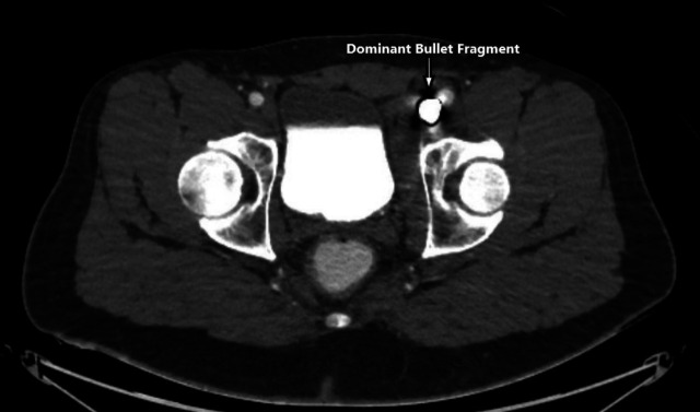
A man presented to a level I trauma center with three gunshot wounds, one each to the left shoulder, lower back, and posterior left thigh. His primary survey was intact. His secondary survey demonstrated palpable distal pulses and no hard signs of vascular injury. His ankle-brachial indices (ABIs) were normal. CT scan demonstrated metallic fragments in the spinal canal at L4 with an associated vertebral body fracture and a missile adjacent to the left common femoral artery. A CT angiography (CTA) was limited due to scatter but demonstrated no obvious injury to the artery and no surrounding hematoma (figure 1). Three-vessel run-off was present. The patient remained hemodynamically normal, and there was no groin hematoma on repeat evaluation.
Figure 1.
CT angiography cross-section showing dominant bullet fragment adjacent to left common femoral artery.
What would you do?
Go to the operating room for left groin exploration
Formal angiography
Observe in the intensive care unit (ICU) with plan for arterial duplex
What we did and why
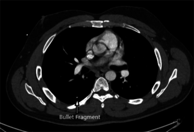
Because the patient was hemodynamically normal, had normal ABIs, and had no evidence of expanding hematoma, we observed the patient in the ICU and obtained an arterial duplex. This demonstrated no arterial injury, but femoral vein thrombosis was identified. The patient’s spinal cord injury and clinical examination were suspicious for cauda equina syndrome, and neurosurgery recommended against anticoagulation. Given the femoral vein thrombus and neurosurgical recommendation, an inferior vena cava (IVC) filter was placed. The patient’s pulse examination subsequently changed on day 3, and he was noted to have only Doppler signals on the left. A repeat CTA demonstrated an embolized metallic fragment in the right lung, presumably from the missile now located in the distal iliac vein (figure 2). There was no injury seen to the artery. However, it was again limited by artifact.
Figure 2.
CT angiography cross-section showing bullet fragment embolized to right lung.
What would you do?
Formal angiography and anticoagulate when able
Open exploration of the left femoral artery and vein
Formal angiography and bulletectomy
What we did and why
Using the hybrid room, we asked vascular surgery to perform angiography to rule out arterial injury. Ultimately, given the metallic fragment embolization to the right lung, we decided that a missile removal was warranted. As the missile had migrated on imaging, we also thought use of the c-arm would be helpful to determine the best operative exposure.
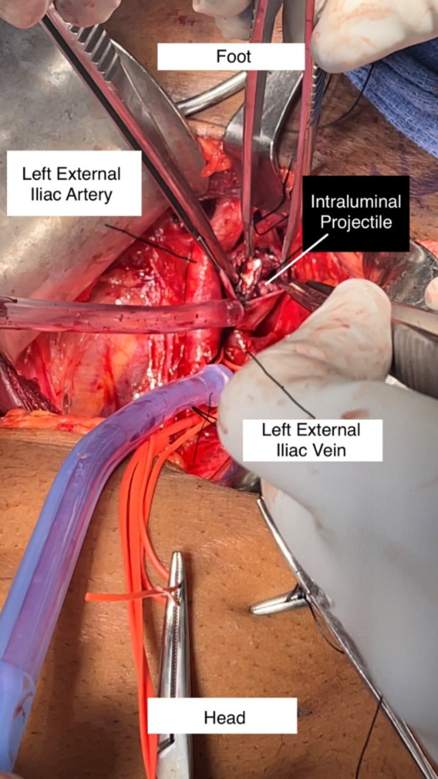
Vascular surgery performed an angiography demonstrating no arterial injury. Using the c-arm, we identified the missile just proximal to the inguinal ligament. Given this location, we elected to perform a left-sided retroperitoneal exposure. We isolated the external iliac vein and identified the missile within the vein (figure 3). There was no injury to the external iliac vein, confirming our suspicion that the bullet migrated proximally. There was extensive clot burden noted in the external iliac vein with extension into the femoral vein, and intraoperative Doppler demonstrated venous flow just proximal to the missile but no flow distally. Due to the concern for thrombus embolization, we decided to ligate the external iliac vein proximally and distally. A longitudinal venotomy was made, and missile fragments along with the associated thrombus were removed. Once deemed safe by neurosurgery, the patient was started on systemic anticoagulation. He was ultimately discharged to acute rehabilitation with a plan for outpatient removal of his IVC filter.
Figure 3.
Intraoperative view of dominant bullet fragment during extraction from left external iliac vein.
Footnotes
Contributors: PJP and CD were primarily responsible for drafting and creating figures for the article. AP, ZM, and JA were responsible for revising the article and its figures. JA was responsible for the final approval before submission.
Funding: The authors have not declared a specific grant for this research from any funding agency in the public, commercial or not-for-profit sectors.
Competing interests: None declared.
Provenance and peer review: Not commissioned; internally peer reviewed.
Ethics statements
Patient consent for publication
Not required.


